Download presentation
Presentation is loading. Please wait.

1
Procedure Guidelines and Practical Applications For PET \CT Imaging by Dr. H. Hawesa RAD 466-Lecture 7

2
Advantages of PET : 1- Better ( spatial resolution, quantitation). 2- Potential for labeling of biological compounds with positron emitters including C11, N13, O15, F18 3-F18 –flourodeoxyglucose (FDG) is the most commonly used radiopharmaceutical in all applications. F18 –flourodeoxyglucose (FDG): 1-F18 is acyclotron – produced radionuclide with a half-life of 110 minutes. 2-integration of F18 into the Glucose molecule in position 2= F18-fluro-2- deoxyglucose. 3-Behaves like a sugar 4-Measures tissue metabolism,which is enhanced in many tumors with significantly increased uptake compared to normal tissue
 is the most commonly used radiopharmaceutical in all applications. F18 –flourodeoxyglucose (FDG): 1-F18 is acyclotron – produced radionuclide with a half-life of 110 minutes. 2-integration of F18 into the Glucose molecule in position 2= F18-fluro-2- deoxyglucose. 3-Behaves like a sugar 4-Measures tissue metabolism,which is enhanced in many tumors with significantly increased uptake compared to normal tissue.")
3
5- F18- FDG is transported & phosphorlylated remains trappedin the cell 6- Unlike glucise, this tracer does not undergo further significant enzymatic reactions. 7- F18- FDG,the accumulation of this compound is pe=roportional to glycolytic rate.

4
Clinical Indications: 1-Differentiation between benign & malignant tumors. 2-Staging of cancer at the time of initial diagnose & after recurrence. 3-Evaluation of the effect of therapy on cancer. 4-Ptovided images of viable tumor distribution for target definition for optimal external beam radiation therapy. 5- Cardiology & Neurology 6- Whole body screening for cancer.

5
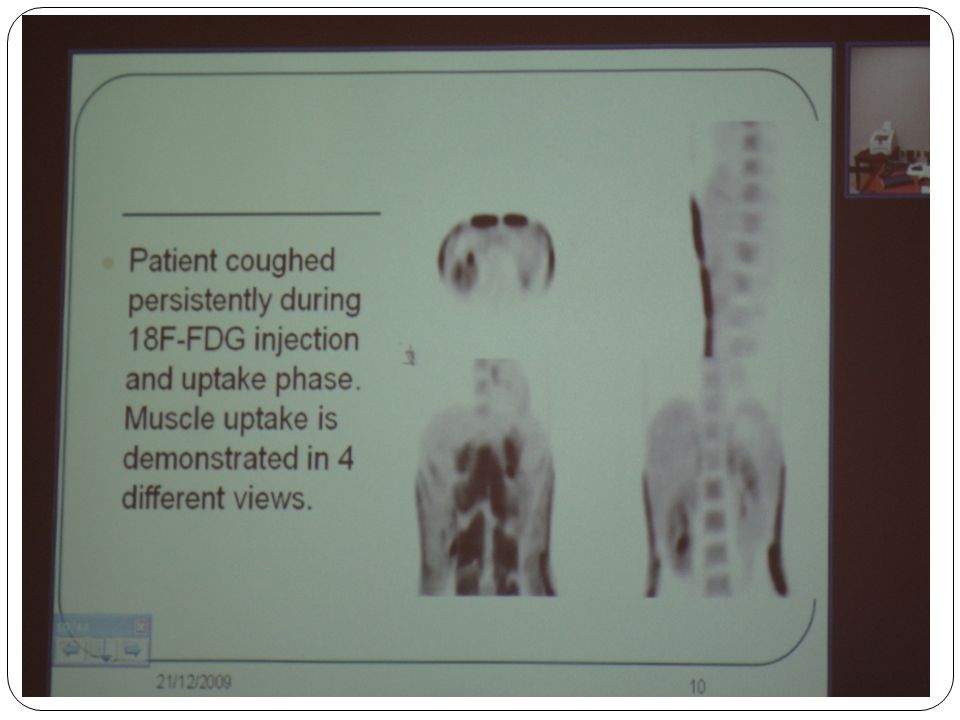
Patient preparation : 1-Give instruction to the patient..2-Remove any metallic object that can cause attenuation artifacts 3-NPO for 4-6 hr prior to scan (decrease insulin level) 4-blood sugar level should be within reference range. 5 -Non –diabetic patient ( start scanning the morning after an overnight fasting.) 6 -Diabetic patient can eat their usual breakfast & medication & they start scanning around noon. 7- Avoid vigorous exercise for 24hr prior to beginning the study. 8 - No interventions (surgery –chemotherapy) within the last 1-2 months. 9 -Muscle uptake should be controlled: A- Patient should lie still throughout the uptake period. B- Patient shouldn’t talk ( to minimize the uptake in the laryngeal muscle.) C- Use muscle relaxant
 6 -Diabetic patient can eat their usual breakfast & medication & they start scanning around noon. 7- Avoid vigorous exercise for 24hr prior to beginning the study. 8 - No interventions (surgery –chemotherapy) within the last 1-2 months. 9 -Muscle uptake should be controlled: A- Patient should lie still throughout the uptake period. B- Patient shouldn’t talk ( to minimize the uptake in the laryngeal muscle.) C- Use muscle relaxant.")
6
10 - Keep the patient warm during uptake period to minimize the uptake in brown adipose tissue (e.g: superclavicular & paraspinal regions) 11 - Hydrate the patient with 1-2 glass if water 12 - Sedation may be need for patient with claustrophobia. 13 - patient should be screened for a history of iodinated contrast material allergy (in CT study.) 14 - For brain imaging, patient should be in a quite & dimly lit room. 15 - Blood glucose should be checked before FDG administration. (tumor uptake of FDG is reduced in Hyperglycemic states) 16 - Arms should be elevated over head. 17- FDG should be injected at a site contra lateral to the site of concern.
 14 - For brain imaging, patient should be in a quite & dimly lit room Blood glucose should be checked before FDG administration. (tumor uptake of FDG is reduced in Hyperglycemic states) 16 - Arms should be elevated over head. 17- FDG should be injected at a site contra lateral to the site of concern..")
11
PET scan procedure : 1- Dose = 10-20 mci (370 - 740 MBq) of F18-FDG. 2- Begin image acquistion = 45-60 minutes later. 3-Scan acquisitions : A-Limited area scanning B-Dynamic imaging C- Whole body imaging D-Total body imaging 4- 2D or 3D. 5-Dedicated PET or PET/CT 6-Imaging time (E = 4-5 min/bed position)
.")
12
Technical consideration :.1- The dose prior to dispensing 2- The dose prior to injection(dispensing). A-Don’t invert the lead pot & vial to withdraw FDG. B-Use of a long flexible needle (angled away from the top of the vial). C-Shielded long flexible needle D- Dose drawing system.
. C-Shielded long flexible needle D- Dose drawing system..")
14
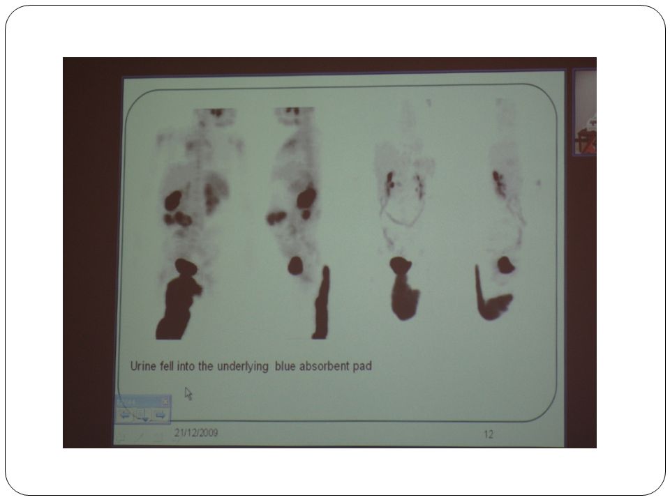
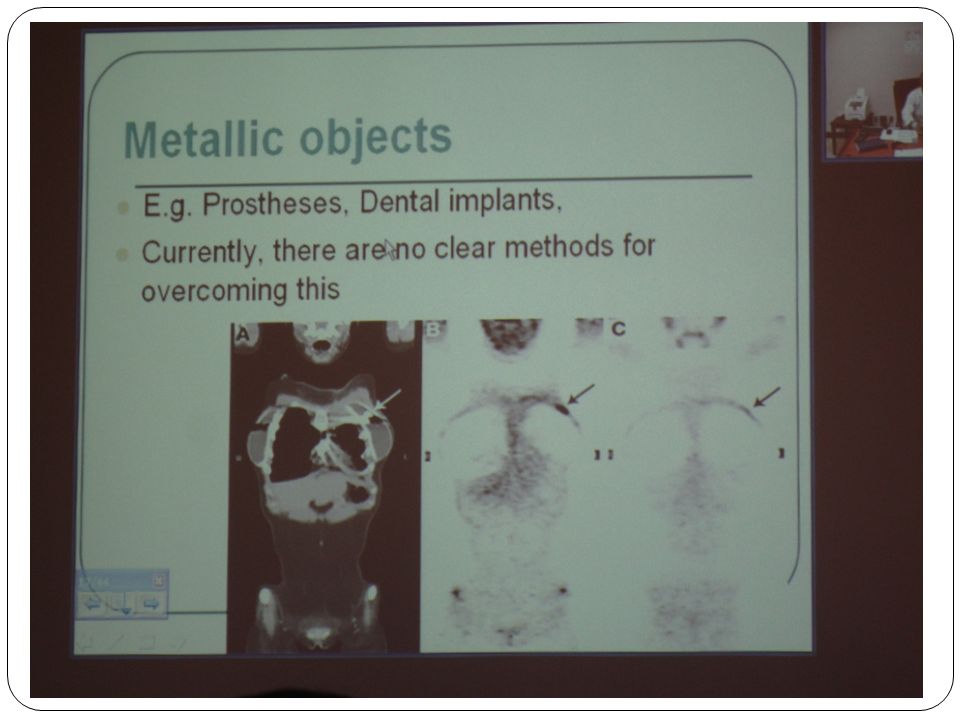
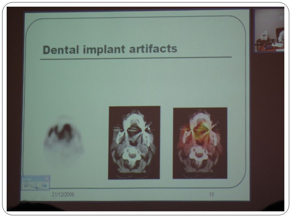
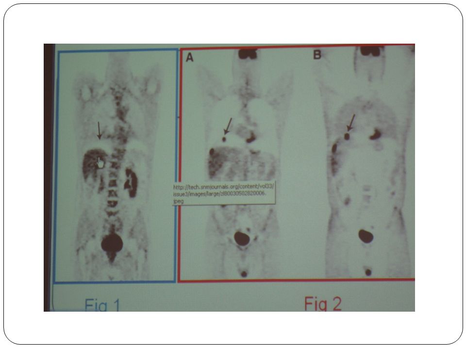
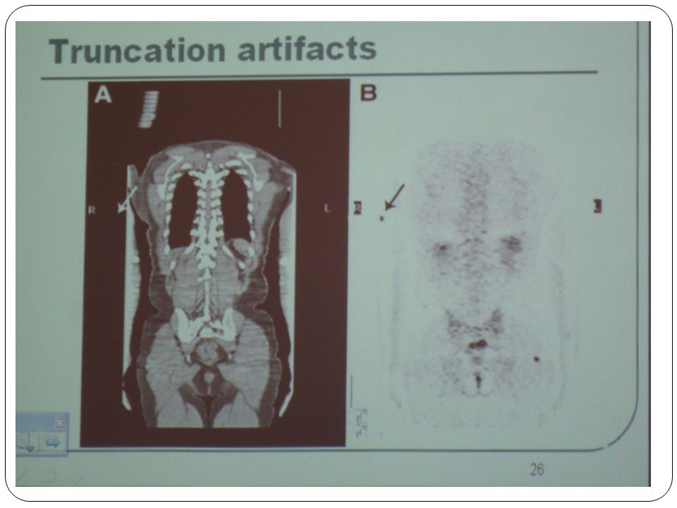
Normal FDG scan:.1- Brain.2- Heart activity (varible).3- Kidney & the Bladder Imaging Artifacts : Objects. 1- Metallic.2- Respiratory motion 3- Contrast medium. 4- Truncation. Metallic Object: -e.g: Prostheses,dental implants.- Currently, there are no clear methods for overcoming this

17
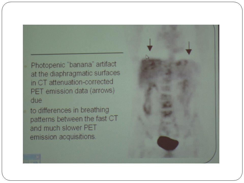
Respiratory Atrifacts : CT = with breath hold at full inspiration PET= breathing normally - The anatomy of CT should match with PET images (If not = artifacts ) Solution : 1-PET (no AC) & reviewed with PET (AC) 2- Use of multislice CT 3- Respiratory gating 4- Correction algorithm 5- Immobilization AC= Attenuation correction
 Solution : 1-PET (no AC) & reviewed with PET (AC) 2- Use of multislice CT 3- Respiratory gating 4- Correction algorithm 5- Immobilization AC= Attenuation correction")
20
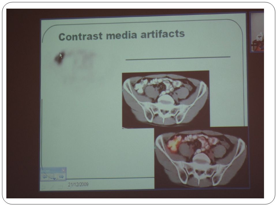
Contrast Media: - Iodinated contrast used in CT to enhance attenuation values in the vessels (IV) & gastrointestinal tract GIT (oral) Solution: -Perform two CT scans : 1-Clinical CT with CM. 2-CT for AC (non-contrast, low – dose ) -Use of algorithms to set the contrast - enhanced pixels to a tissue-equivalent value
 -Use of algorithms to set the contrast - enhanced pixels to a tissue-equivalent value.")
23
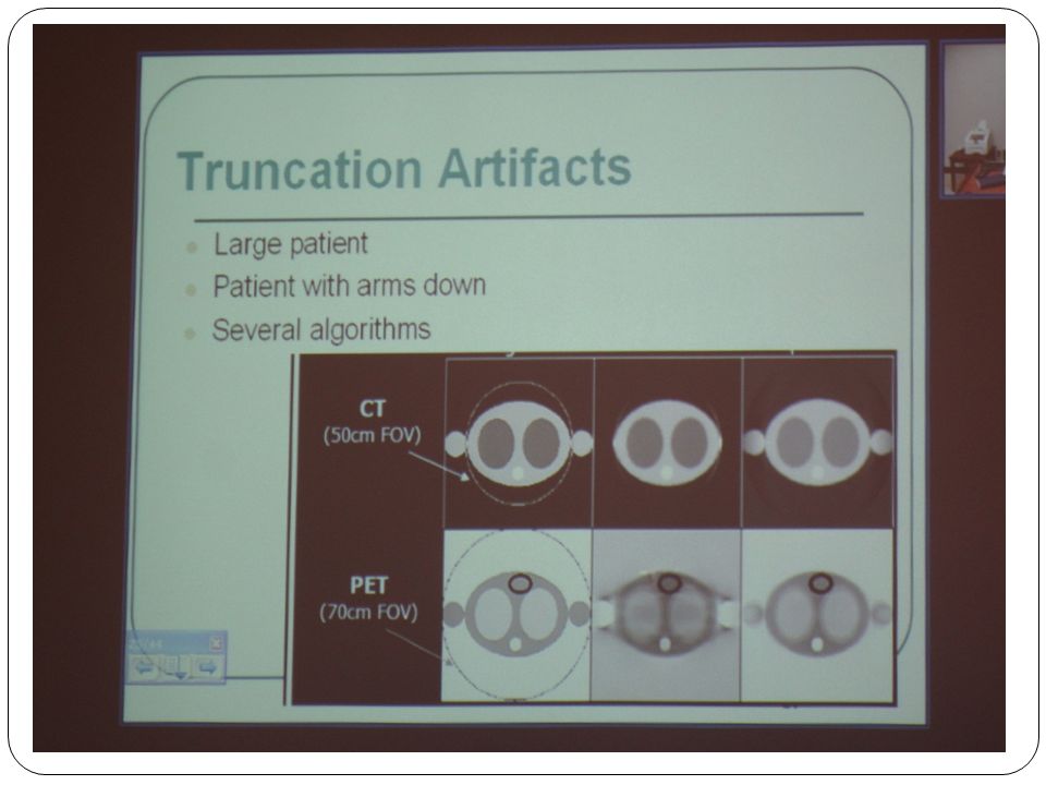
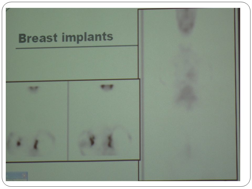
Truncation: 1- Large patient. 2-patient with arms down. 3-Several algorithms..4-Breast implant

29
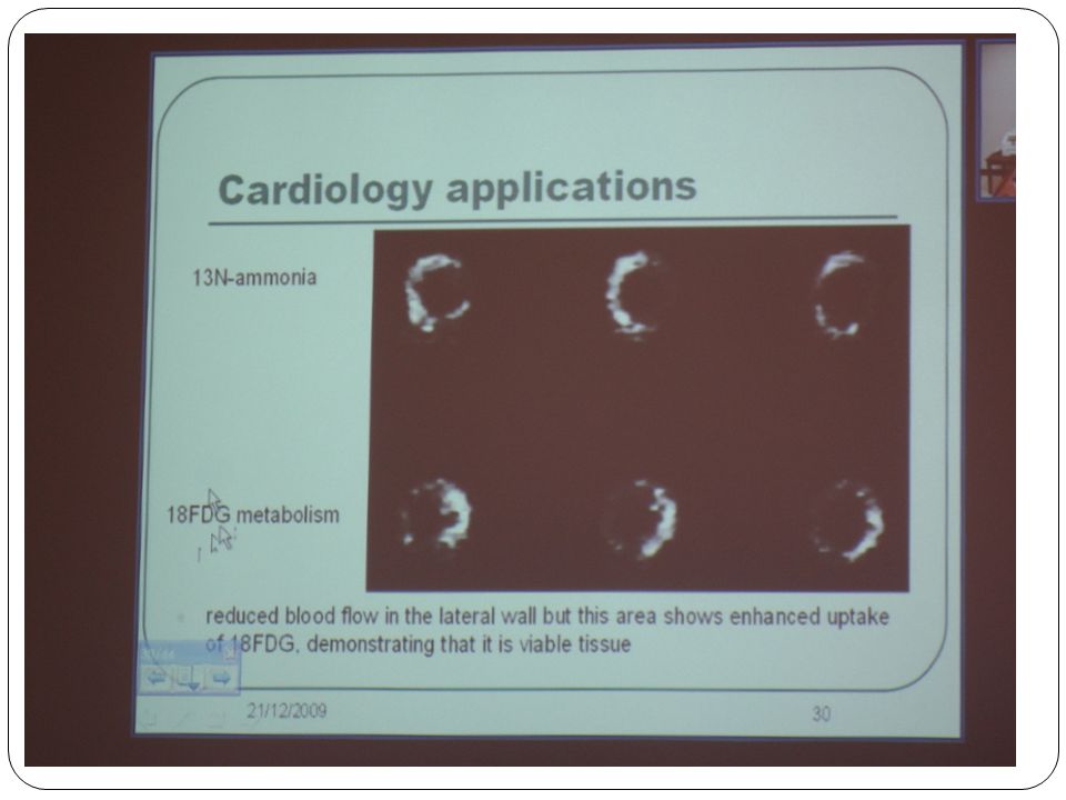
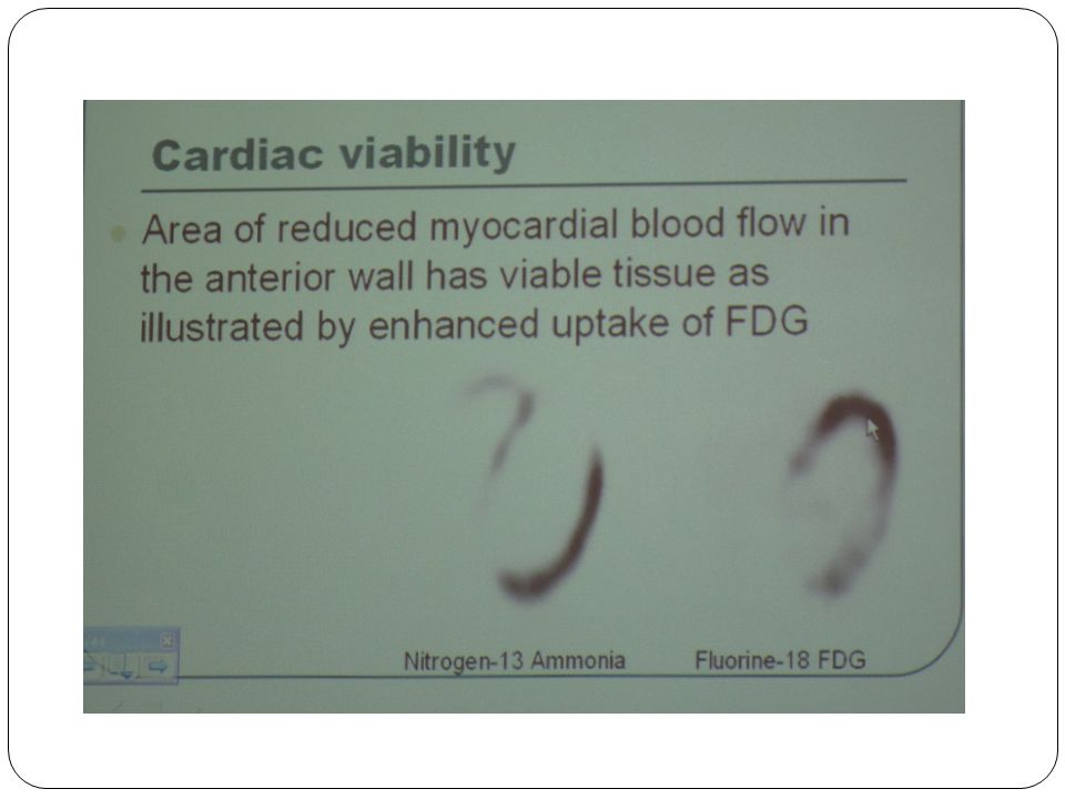
Cardiac viability.. Area of reduced myocardial blood flow in The anterior wall has visible tissue as illustrated by enhanced uptake of FDG.

31
Neurology application.. Dopamine receptor system is particularly useful in patients with movement disorders ( e.g. parkinson’s disease ).. Blood flow using O 15 labelled water.. MRI & CT. ( replaced much of the brain studies,. SPECT. ( recommended in the case of epilepsy)
.. Blood flow using O 15 labelled water.. MRI & CT. ( replaced much of the brain studies,. SPECT. ( recommended in the case of epilepsy).")
32
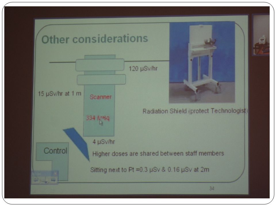
Radiation Protection. Fast & reliable technique.. L – shield.. Dose to technologist.. Use of unit doses.. Minimize the time in dispensing.. Minimize the time handling of syringe.. Minimize the time spent with injected patient.. Use of cart to transport the dose from the hot lab to injection room.. Use of additional shielding in the injection room & waiting area.. Using long flexible needle & long – handled forceps. Time distance e s shielding

34
Thank you

Similar presentations


 CT scanning or (CAT scanning) is using X-rays to create a 3D image of the inside of an object. CT stands for computed tomography.>")


