Download presentation
Presentation is loading. Please wait.

1
Family Pseudomonaceae Genus Pseudomonase
Pseudomonads and Acinetobacters are widely distributed in soil and water

2
Objective To be familiar with Pseudomonas species
To describe the characteristic To understand the pathogenesis To explain the diseases , Clinical finding, diagnosis , treatment and prevention

3
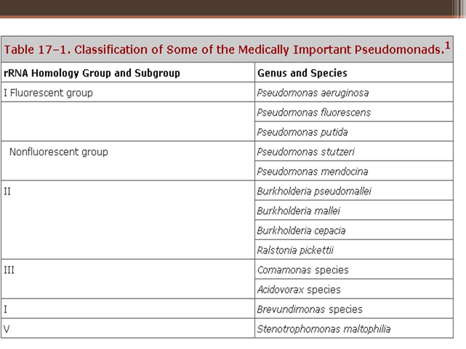
Pseudomonase Gram-negative motile aerobic rods
some of which produce water-soluble pigments The classification is based on rRNA/DNA homology and common culture characteristics.

5
P aeruginosa Widely distributed in nature, commonly in moist environments in hospitals. Colonize normal humans Causes disease in humans with abnormal host defenses.

6
Pseudomonas aeruginosa
Gram-negative bacteria Small numbers in the normal intestinal flora and on the skin of humans . The major human pathogen of the group. invasive Toxigenic Important nosocomial pathogen.

7
Pathogenesis Pathogenic only when introduced into areas devoid of normal defenses, Mucous membranes and skin are disrupted by direct tissue damage; Intravenous or urinary catheters are used; Neutropenia is present, as in cancer chemotherapy.

8
The bacterium attaches to and colonizes the mucous membranes or skin, invades locally, and produces systemic disease.

9
Clinical Findings Infection of wounds and burns, Meningitis,
Urinary tract infection, catheters , instruments or in irrigating solutions. Respiratory tract, contaminated respirators, results in necrotizing pneumonia. Mild otitis externa in swimmers. ,invasive (malignant) otitis externa in diabetic patients. Eye infection , lead to rapid destruction of the eye, most commonly after injury or surgical procedures. In infants or debilitated persons, invade the bloodstream and result in fatal sepsis; in leukemia, lymphom, antineoplastic drugs, radiation therapy, patients with severe burns.
 otitis externa in diabetic patients. Eye infection , lead to rapid destruction of the eye, most commonly after injury or surgical procedures. In infants or debilitated persons, invade the bloodstream and result in fatal sepsis; in leukemia, lymphom, antineoplastic drugs, radiation therapy, patients with severe burns.")
10
Symptoms & Signs Related to the organ .
verdoglobin (a breakdown product of hemoglobin) or fluorescent pigment can be detected in wounds, burns, or urine by ultraviolet fluorescence. Hemorrhagic necrosis of skin occurs often in sepsis due to P aeruginosa; the lesions, ecthyma gangrenosum, are surrounded by erythema and often do not contain pus. P aeruginosa can be seen on Gram-stained specimens from ecthyma lesions, and cultures are positive.
 or fluorescent pigment can be detected in wounds, burns, or urine by ultraviolet fluorescence. Hemorrhagic necrosis of skin occurs often in sepsis due to P aeruginosa; the lesions, ecthyma gangrenosum, are surrounded by erythema and often do not contain pus. P aeruginosa can be seen on Gram-stained specimens from ecthyma lesions, and cultures are positive.")
11
Sign and Symptoms Fever, Shock, Oliguria, Leukocytosis and leukopenia,
Disseminated intravascular coagulation, Adult respiratory distress syndrome.

12
Antigenic Structure & Toxins
Pili (fimbriae) attachment to host epithelial cells. Alginate exopolysaccharide mucoid colonies seen in cultures from patients with cystic fibrosis. Endotoxin, P. aeruginosa can be typed by lipopolysaccharide immunotype and by pyocin (bacteriocin) susceptibility. extracellular enzymes : Elastases, proteases, and two hemolysins: a heat-labile phospholipase C and a heat-stable glycolipid.
 attachment to host epithelial cells. Alginate exopolysaccharide mucoid colonies seen in cultures from patients with cystic fibrosis. Endotoxin, P. aeruginosa can be typed by lipopolysaccharide immunotype and by pyocin (bacteriocin) susceptibility. extracellular enzymes : Elastases, proteases, and two hemolysins: a heat-labile phospholipase C and a heat-stable glycolipid.")
13
Exotoxin Exotoxin A, tissue necrosis , lethal for animals when injected in purified form. The toxin blocks protein synthesis mechanism of action identical to diphtheria toxin, though the structures of the two toxins are not identical. Antitoxins to exotoxin A are found in some human sera, including those of patients who have recovered from serious P aeruginosa infections

14
Morphology & Identification
Motile Rod-shaped Gram-negative (single, pairs, and occasionally in short chains)
")
15
Culture Obligate aerobe Grows on many types of culture media,
Sweet or grape-like or corn taco-like odor. Some strains hemolyze blood. Smooth round colonies with a fluorescent greenish color : pyocyanin: Nonfluorescent bluish pigment Pyoverdin: fluorescent pigment, greenish color Pyorubin: dark red pigment pyomelanin : black pigment

16
Cultures from patients with cystic fibrosis
Form mucoid colonies as a result of overproduction of alginate, an exopolysaccharide. In cystic fibrosis patients, the exopolysaccharide appears to provide the matrix for the organisms to live in a biofilm

17
Diagnostic Laboratory Tests
Specimens Specimens from skin lesions, pus, urine, blood, spinal fluid, sputum, and other material should be obtained as indicated by the type of infection. Smears Gram-negative rods Biochemical tests ; Oxidase , growth at 42 °C, Pigments , oxidize glucose

18
Growth Characteristics
Does not ferment carbohydrates, but many strains oxidize glucose. Oxidase positivity, The presence of characteristic pigments, growth at 42 °C. Differentiation of P aeruginosa from other pseudomonads on the basis of biochemical activity requires testing with a large battery of substrates.

19
Culture blood agar , differential media commonly used to grow the enteric gram-negative rods. P aeruginosa does not ferment lactose and is easily differentiated from the lactose-fermenting bacteria. Culture is the specific test for diagnosis of P aeruginosa infection

20
P aeruginosa and other pseudomonads are resistant to many antimicrobial agents and therefore become dominant and important when more susceptible bacteria of the normal flora are suppressed

21
Treatment Rapidly develop resistance when single drugs are employed. So,Clinically significant infections with P aeruginosa should not be treated with single-drug therapy Ticarcillin or piperacillin—is used in combination with an aminoglycoside, usually tobramycin Aztreonam, imipenem, and the newer quinolones, including ciprofloxacin. Newer cephalosporins, ceftazidime and cefoperazone are active against P aeruginosa

22
Epidemiology and Control
Nosocomial pathogen Special attention should be paid to sinks, water baths, showers, hot tubs, and other wet areas Epidemiologic purposes, strains can be typed by pyocins and by lipopolysaccharide immunotypes. Vaccine from appropriate types administered to high-risk patients provides some protection against pseudomonas sepsis. Such treatment has been used experimentally in patients with leukemia, burns, cystic fibrosis, and immunosuppression.

23
Burkholderia pseudomallei
small, motile, aerobic gram-negative bacillus a natural saprophyte ; soil, fresh water, rice paddies, and vegetable . Human infection ; contamination of skin abrasions and possibly by ingestion or inhalation. Epizootic B pseudomallei infection occurs in sheep, goats, swine, horses, and other animals, though animals do not appear to be a primary reservoir for the organism.

24
Melioidosis of humans Melioidosis may manifest itself as acute, subacute, or chronic infection. IP 2–3 days, but latent periods of months to years also occur. A localized suppurative infection can occur at the inoculation site where there is a break in the skin. acute septicemic form of infection with involvement of many organs. The most common form of melioidosis is pulmonary infection, which may be a primary pneumonitis Melioidosis has a high mortality rate if untreated

25
Burkholderia mallei :glanders, a disease of horses, mules, and donkeys transmissible to humans
Burkholderia cepacia:is an environmental organism that is able to grow in water, soil, plants, animals, and decaying vegetable materials

26
Thank you ?

Similar presentations


 Prof. Dr. Ebtisam.F. El Ghazzawi Medical Research Institute (MRI) Alexandria University.>")


>")
 –Bacteria or their products may migrate to adjacent tissues or bloodstream.>")
SM MT 418 Clinical Microbiology Student Laboratory Session.>")
>")

>")
