Download presentation
Presentation is loading. Please wait.

1
Enterobacteriaceae

2
Enterobacteriaceae The most important bacterial family in human medicine Well-defined diseases with typical clinical symptoms: Typhoid fever, dysentery and plague Nosocomial infections: Urinary tract infections, pneumonias, wound infections and sepsis The most important bacterial family in human medicine is the Enterobacteriaceae. This family includes genera and species that cause well-defined diseases with typical clinical symptoms (typhoid fever, dysentery, plague) as well as many opportunists that cause mainly nosocomial infections (urinary tract infections, pneumonias, wound infections, sepsis). Enterobacteriaceae are Gram-negative, usually motile, facultatively anaerobic rod bacteria. The high levels of metabolic activity observed in them are made use of in identification procedures. The species are subdivided into epidemiologically
 as well as many opportunists that cause mainly nosocomial infections (urinary tract infections, pneumonias, wound infections, sepsis). Enterobacteriaceae are Gram-negative, usually motile, facultatively anaerobic rod bacteria. The high levels of metabolic activity observed in them are made use of in identification procedures. The species are subdivided into epidemiologically.")
3
Definition and significance
41 genera with hundreds of species Gram-negative, facultatively anaerobic rod Natural habitat: intestinal tract of humans and animals

4
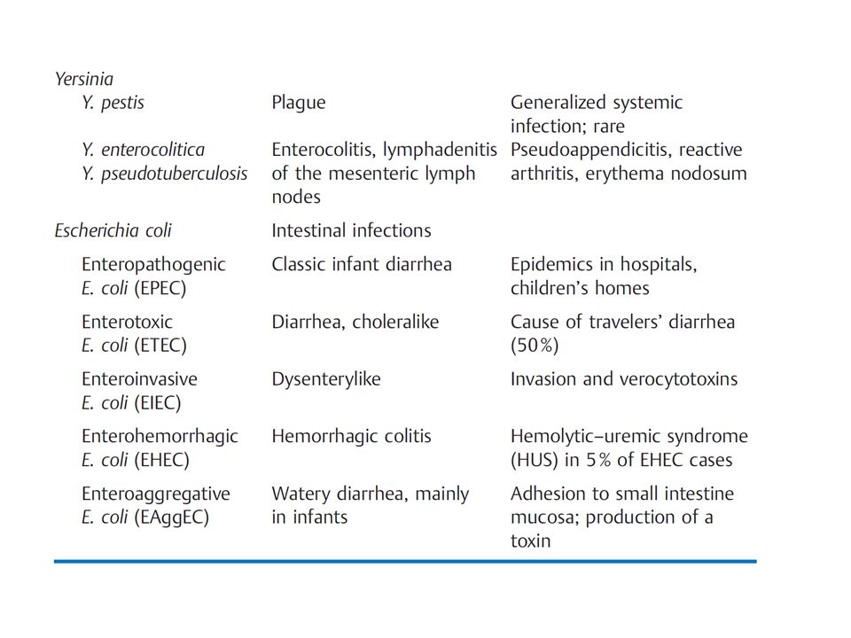
The Most Important Genera/Species/Vars of Enterobacteriaceae and the Corresponding Clinical Pictures

6
Virulence and pathogenicity
The most important pathogenicity factors: Colonizing factors Invasins Endotoxin Exotoxins Enterobacteriaceae are the most significant contributors to intestinal infections The most important pathogenicity factors of Enterobacteriaceae are colonizing factors, invasins, endotoxin, and various exotoxins. Enterobacteriaceae are the most significant contributors to intestinal infections, which are among the most frequent diseases of all among the developing world populace.

7
Identification of Enterobacteriaceae
Gram-negative rod Usually motile (with few exceptions) Facultative anaerobes Grow on simple nutrient media Oxidase test negative Ferment glucose with acid or acid and gas
 Facultative anaerobes. Grow on simple nutrient media. Oxidase test negative. Ferment glucose with acid or acid and gas.")
8
Sero-typing based on antigenic structure
O antigens: Somatic antigens (polysaccharide) H antigens: Flagellar antigens (protein) K antigens: Capsular antigens (carbohydrate) e.g., serovar O18:K1:H7 O antigens. Specific polysaccharide chains in the lipopolysaccharide complex of the outer membrane. H antigens. Flagellar antigens consisting of protein. K antigens. Linear polymers of the outer membrane built up of a repeated series of carbohydrate units (sometimes proteins as well). They can cover the cell densely and render them O inagglutinable. F antigens. Antigens of protein attachment fimbriae.
 H antigens: Flagellar antigens (protein) K antigens: Capsular antigens (carbohydrate) e.g., serovar O18:K1:H7. O antigens. Specific polysaccharide chains in the lipopolysaccharide complex of the outer membrane. H antigens. Flagellar antigens consisting of protein. K antigens. Linear polymers of the outer membrane built up of a repeated series of carbohydrate units (sometimes proteins as well). They can cover the cell densely and render them O inagglutinable. F antigens. Antigens of protein attachment fimbriae.")
9
Escherichia coli Klebsiella and Proteus

10
Escherichia coli

11
Escherichia coli on ChromID CPS agar

12
Natural habitat Intestinal tract of humans and animals
indicator organism for fecal contamination of water and foods The natural habitat of E. coli is the intestinal tract of humans and animals. It is therefore considered an indicator organism for fecal contamination of water and foods.

13
Infections Extraintestinal infections
Intestinal infections (Diarrhoeal diseases) The natural habitat of E. coli is the intestinal tract of humans and animals. It is therefore considered an indicator organism for fecal contamination of water and foods.
 The natural habitat of E. coli is the intestinal tract of humans and animals. It is therefore considered an indicator organism for fecal contamination of. water and foods.")
14
Extraintestinal infections
Urinary tract infections Wound infections Peritonitis Cholecystitis Appendicitis Sepsis and endotoxin induced shock Neonatal meningitis E. coli is the most frequent causative pathogen in human bacterial infections. Extraintestinal infections include urinary tract infections, which occur when the tract is obstructed or spontaneously caused by the pathovar UPEC. The most important other coli infections are cholecystitis, appendicitis, peritonitis, postoperative wound infections, and sepsis.

15
Diarrheagenic pathovars
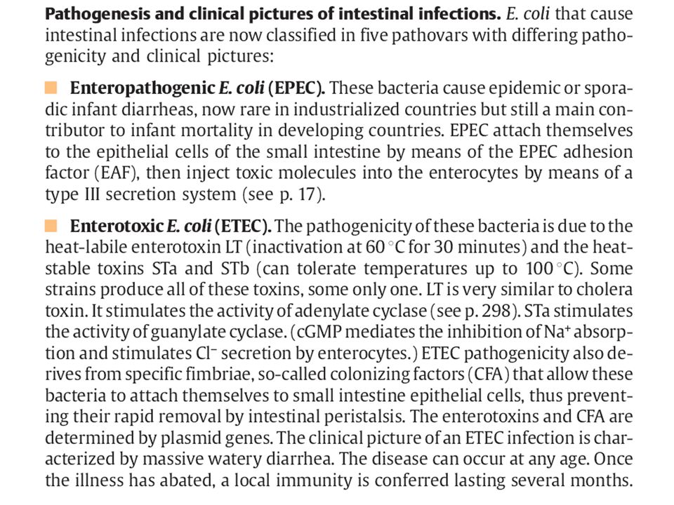
Enteropathogenic E. coli (EPEC) Enterotoxigenic E. coli (ETEC) Enteroinvasive E. coli (EIEC) Enterohaemorrhagic E. Coli (EHEC) Enteroaggressive E. coli (EaggEC) Intestinal infections are caused by the pathovars EPEC, ETEC, EIEC, EHEC, and EAggEC. EPEC and EAggEC frequently cause diarrhea in infants. ETEC produce enterotoxins that cause a choleralike clinical picture. EIEC cause a dysenterylike infection of the large intestine. EHEC produce verocytotoxins and cause a hemorrhagic colitis as well as the rare hemolytic-uremic syndrome. E. coli bacteria infections are diagnosed by means of pathogen identification.
 Enterotoxigenic E. coli (ETEC) Enteroinvasive E. coli (EIEC) Enterohaemorrhagic E. Coli (EHEC) Enteroaggressive E. coli (EaggEC) Intestinal infections are caused by the pathovars EPEC, ETEC, EIEC, EHEC, and EAggEC. EPEC and EAggEC frequently cause diarrhea in infants. ETEC. produce enterotoxins that cause a choleralike clinical picture. EIEC cause a. dysenterylike infection of the large intestine. EHEC produce verocytotoxins. and cause a hemorrhagic colitis as well as the rare hemolytic-uremic syndrome. E. coli bacteria infections are diagnosed by means of pathogen identification.")
16
EPEC Frequently cause diarrhea in infants
Vomiting, fever and prolonged diarrhoea Infants mainly Many serotypes

17
ETEC Enterotoxins that cause watery diarrhoea similar to cholera
Infants and adults Traveler diarrhea Many serotypes Plasmid mediated toxin (HS, HL)
")
18
EIEC Cause a dysentery like infection of the large intestine (similar to shigellosis) Fever and colitis Many serotypes

19
EHEC Produce verocytotoxins and cause a hemorrhagic colitis (damage to vascular endothelia ) Causes life-threatening haemorrhagic diarrhoea All ages

20
EHEC No pus cells and no fever
It can progress to Haemolytic Uremic Syndrome → Renal failure O157:H7 or verocytotoxin-producing E. coli Contaminated meat products, unpasteurized milk and diary products

21
EaggEC Chronic watery diarrhoea Mainly in children

24
Klebsiella species

25
Klebsiella species

26
Klebsiella pneumoniae
Four subspecies: K.p. pneumoniae K.p. aerogenes K.p. ozaenae K.p. rinhoscleromatis

27
Infections caused by Klebsiella species
UTI Wound infections Chest infections

28
Proteus species

29
Proteus species on Blood Agar

30
Medically important Proteus species
P. mirabilis UTI Wound infection Septiceamia Occasionally meningitis and chest infections P. vulgaris UTI and wound infections Alkaline reaction Common cause of male UTI specially those with catheter or cytoscopy

31
Other enterobacteria

32
Other enterobacteria Enterobacter Citrobacter Serratia
Opportunistic pathogens: UTI Wound infections Septiceamia Pulmonary infections

33
Laboratory diagnosis Specimens: Direct examination:
Urine, pus, faeces, CSF, blood, sputum Direct examination: Gram –ve bacilli Few capsulated Culture aerobically at 36-37° C: Blood agar MacConkey agar CLED XLD and DCA

34
UTI CFU/ml Midstream urine Bacterial count
≥105/ml indicate an infection 104/ml doubtful significance ≤103/ml indicate a contamination

35
MacConkey agar showing lactose and non-lactose fermenting colonies

36
Escherichia coli (Gram negative)
")
37
Oxidase test

Similar presentations


 Gram negative bacilli characterized by: - Grow on ordinary media - Aerobic or facultative.>")


>")
 Demonstrated that particular strains were.>")

 Prof. Dr. Ebtisam.F. El Ghazzawi Medical Research Institute (MRI) Alexandria University.>")


>")
