Download presentation
Presentation is loading. Please wait.

1
NERVOUS SYSTEM I & II Chapter 10 &11

2
PNS vs. CNS

4
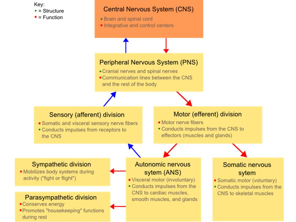
Nervous System Central Nervous System (CNS) – brain and spinal cord
Peripheral Nervous System (PNS) – cranial and spinal nerves
 – cranial and spinal nerves.")
5
PNS The peripheral nervous system is subdivided into:
Sensory division (Afferent division) Motor division (Efferent division) Somatic nervous system– skeletal muscles (voluntary) Autonomic nervous system– smooth and cardiac muscles, glands, etc. (involuntary)
 Motor division (Efferent division) Somatic nervous system– skeletal muscles (voluntary) Autonomic nervous system– smooth and cardiac muscles, glands, etc. (involuntary)")
6
The Sensory-Somatic division of the PNS
The sensory-somatic system consists of two major components: a subsystem for the detection of mechanical stimuli (e.g., light touch, vibration, pressure, and cutaneous tension), and a subsystem for the detection of painful stimuli and temperature. Together, these two subsystems give humans and other animals the ability to identify the shapes and textures of objects, to monitor the internal and external forces acting on the body at any moment, and to detect potentially harmful circumstances. It consists of 12 pairs of cranial nerves and 31 pairs of spinal nerves.
, and a subsystem for the detection of painful stimuli and temperature. Together, these two subsystems give humans and other animals the ability to identify the shapes and textures of objects, to monitor the internal and external forces acting on the body at any moment, and to detect potentially harmful circumstances. It consists of. 12 pairs of cranial nerves and. 31 pairs of spinal nerves.")
7
The autonomic nervous system
The autonomic nervous system consists of sensory neurons and motor neurons that run between the central nervous system (especially the hypothalamus and medulla oblongata) and various internal organs such as the: heart lungs viscera glands (both exocrine and endocrine) It is responsible for monitoring conditions in the internal environment and bringing about appropriate changes in them. The contraction of both smooth muscle and cardiac muscle is controlled by motor neurons of the autonomic system.
 and various internal organs such as the: heart. lungs. viscera. glands (both exocrine and endocrine) It is responsible for monitoring conditions in the internal environment and bringing about appropriate changes in them. The contraction of both smooth muscle and cardiac muscle is controlled by motor neurons of the autonomic system.")
8
Autonomic Nervous System
Is divided into Sympathetic nervous system Parasympathetic Nervous system

9
Sympathetic nervous system
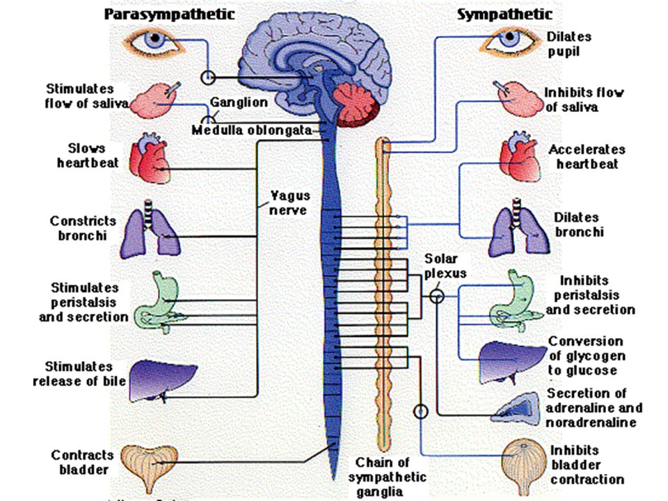
Stimulation of the sympathetic branch of the autonomic nervous system prepares the body for emergencies: for "fight or flight" The neurotransmitter acetylcholine (ACh) stimulates action potentials in the postganglionic neurons. The neurotransmitter released by the postganglionic neurons is noradrenaline (also called norepinephrine). The action of noradrenaline on a particular gland or muscle is excitatory is some cases, inhibitory in others. (At excitatory terminals, ATP may be released along with noradrenaline.) The release of noradrenaline stimulates heartbeat raises blood pressure dilates the pupils dilates the trachea and bronchi stimulates glycogenolysis — the conversion of liver glycogen into glucose shunts blood away from the skin and viscera to the skeletal muscles, brain, and heart inhibits peristalsis in the gastrointestinal (GI) tract inhibits contraction of the bladder and rectum
 stimulates action potentials in the postganglionic neurons. The neurotransmitter released by the postganglionic neurons is noradrenaline (also called norepinephrine). The action of noradrenaline on a particular gland or muscle is excitatory is some cases, inhibitory in others. (At excitatory terminals, ATP may be released along with noradrenaline.) The release of noradrenaline. stimulates heartbeat. raises blood pressure. dilates the pupils. dilates the trachea and bronchi. stimulates glycogenolysis — the conversion of liver glycogen into glucose. shunts blood away from the skin and viscera to the skeletal muscles, brain, and heart. inhibits peristalsis in the gastrointestinal (GI) tract. inhibits contraction of the bladder and rectum.")
10
Parasympathetic Parasympathetic nervous system causes slowing down of the heartbeat (as Loewi demonstrated) lowering of blood pressure constriction of the pupils increased blood flow to the skin and viscera peristalsis of the GI tract

11
Sympathetic vs. Parasympathetic
In short, the parasympathetic system returns the body functions to normal after they have been altered by sympathetic stimulation. In times of danger, the sympathetic system prepares the body for violent activity. The parasympathetic system reverses these changes when the danger is over.

13
Neural Tissue Consists of 2 types of cells Neurons (nerve cells)
Neuroglia (Help, nourish and support neurons) Neurons come from neural stem cells. Neurons do not divide once they mature.
 Neurons come from neural stem cells. Neurons do not divide once they mature.")
14
Neurons React to chemical and physical changes in the environment
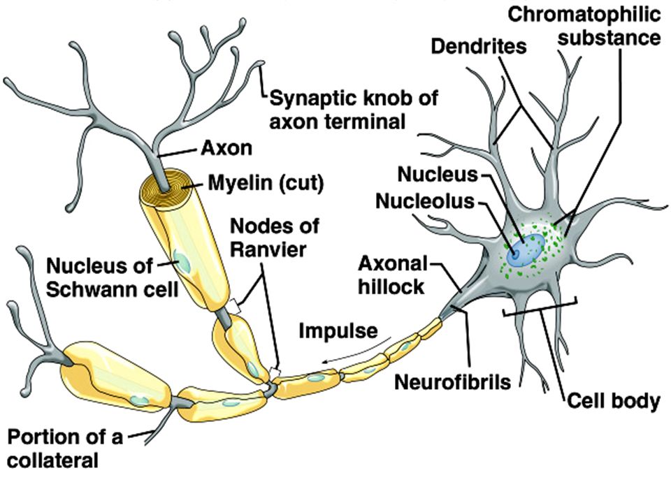
Have highly branched cellular extensions called dendrites that receive impulses Have a soma or “body” Have a long cellular extension called an axon (also called nerve fiber), that carries impulses away from the body. A bundle of axons (nerve fibers) is called a nerve
, that carries impulses away from the body. A bundle of axons (nerve fibers) is called a nerve.")
15
Neurons, cont’d. The soma (cell body) contains Granular cytoplasm
All cellular organelles and cytoskeleton Multiple rough endoplasmic reticula are found in membrane-bound packets called Nissl bodies or chromatophilic substance A network of threads called neurofibrils which extends into the axon to support them Cytoplasm may contain glycogen, lipids and even melanin

17
Dendrites and Axons Highly branched, with thorn-like projections called dendritic spines Axons leave the soma from a slightly elevated region called the axon hillock Axons contain many mitochondria, microfilament and neurofibrils Axons may have branches called collaterals Axon ends have many fine branches called terminals and each one ends with a synaptic knob The synaptic knobs make contact with post synaptic neurons in a synapse. The gap between the synaptic knob of the pre-synaptic neuron and the post-synaptic neuron is called the synaptic cleft.

18
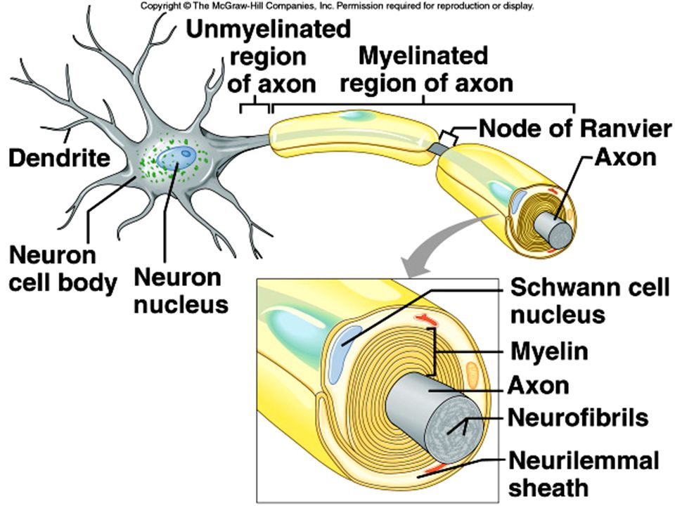
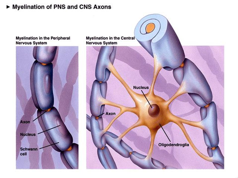
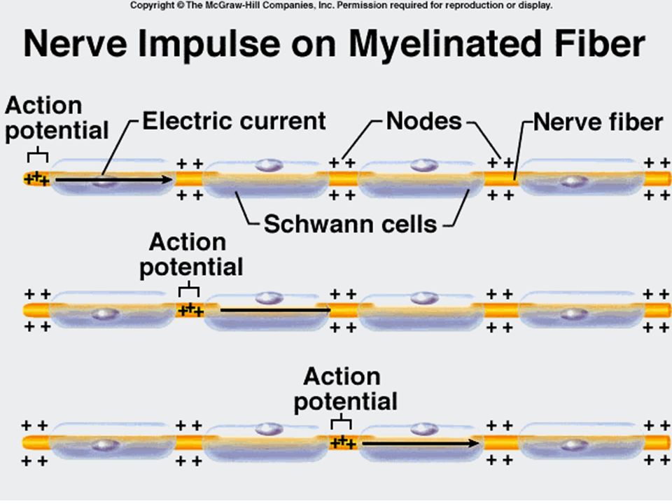
Axons, cont’d. Larger neurons of the peripheral nervous system have layers of lipid-rich coverings around their membranes These are actually neuroglial cells called Schwann cells which wind tightly around the axons The Schwann cell membranes contain myelin, which have more lipid than other cell membranes. So this cell covering around the axon is called a myelin sheath The cytoplasm and organelles of the Schwann cell remains on the outside area and is called the neurolemma or the neurolemmal sheath The gaps between the myelinated segments are called Nodes of Ranvier

20
Unmyelinated Axons These axons lie in the longitudinal groove of the Schwann cells – so the Schwann cells enclose them, but don’t wrap around them

21
Cell membrane, neurilemma & myelin sheath

22
Gray and White Matter Myelinated axons appear white and are found in the brain and spinal cord – a mass of these axons is called white matter Unmyelinated axons appear gray and masses of these are called gray matter – also found in the brain and spinal cord Some CNS (brain and spinal cord) axons have myelin sheaths produced by other neuroglial cells called oligodendrocytes Myelinated axons in the brain and spinal cord (CNS)lack neurolemmas (no cell cytoplasm – just myelin)
 axons have myelin sheaths produced by other neuroglial cells called oligodendrocytes. Myelinated axons in the brain and spinal cord (CNS)lack neurolemmas (no cell cytoplasm – just myelin)")
24
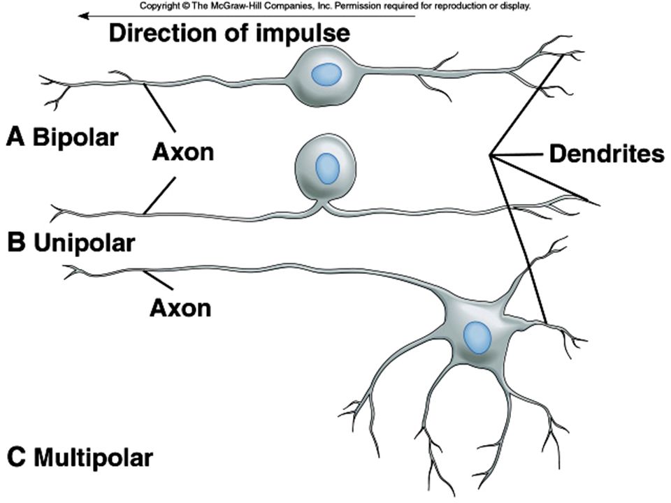
Types of Neurons Neurons can be grouped in two ways: on the basis of
Structural differences (bipolar, unipolar, and multipolar neurons), and by Functional differences (sensory neurons, interneurons, and motor neurons).
, and by. Functional differences (sensory neurons, interneurons, and motor neurons).")
25
Structural Differences
Bipolar neurons are found in the eyes, nose, and ears, and have a single axon and a single dendrite extending from opposite sides of the cell body. Unipolar neurons are found in ganglia outside the CNS and have an axon and a dendrite arising from a single short fiber extending from the cell body. Multipolar neurons have many nerve fibers arising from their cell bodies and are commonly found in the brain and spinal cord.

26
Functional Differences
Sensory neurons (afferent neurons) conduct impulses from peripheral receptors to the CNS and are usually unipolar, although some are bipolar neurons. Interneurons are multipolar neurons lying within the CNS that form links between other neurons. Motor neurons are multipolar neurons that conduct impulses from the CNS to effectors.
 conduct impulses from peripheral receptors to the CNS and are usually unipolar, although some are bipolar neurons. Interneurons are multipolar neurons lying within the CNS that form links between other neurons. Motor neurons are multipolar neurons that conduct impulses from the CNS to effectors.")
28
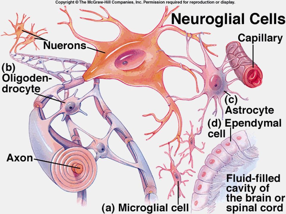
Types of Neuroglial Cells
Astrocytes (CNS) Oligodendrocytes (CNS) Microglia (CNS) Ependyma (CNS) Schwann Cells (PNS only) Neuroglial cells make up more than half of the brain volume
 Oligodendrocytes (CNS) Microglia (CNS) Ependyma (CNS) Schwann Cells (PNS only) Neuroglial cells make up more than half of the brain volume.")
29
Astrocytes Most abundant glial cell in CNS
Star-shaped, found between neurons and blood vessels Help in blood-brain barrier Metabolize glucose and maintain concentrations of important ions Form scar tissue upon brain injury, to fill in gaps left by dead neurons Gap junctions between astrocytes Produce nerve growth factors

30
Oligodendrocytes Myelinate neurons of the CNS by sending out processes that wrap around the axons Each oligodendrocyte can myelinate several axons These axons do not have a neurilemma, since the cell body of the oligodendrocytes is not wrapped around the axon

31
Microglia Small, with fewer cellular extensions than other glial cells
Phagocytic - help neurons by devouring bacteria and cellular debris Numbers increase during brain infections

32
Ependyma Cuboidal or columnar shaped, with cilia
Line the inner tube of the spinal cord Line the internal spaces of the brain called ventricles and also cover the capillaries or Choroid Plexuses of the brain – where they regulate the composition of CSF (Cerebrospinal Fluid) Form a one-cell thick membrane that allow diffusion of ions and molecules from brain ventricles and interstitial fluid of the brain tissues
 Form a one-cell thick membrane that allow diffusion of ions and molecules from brain ventricles and interstitial fluid of the brain tissues.")
34
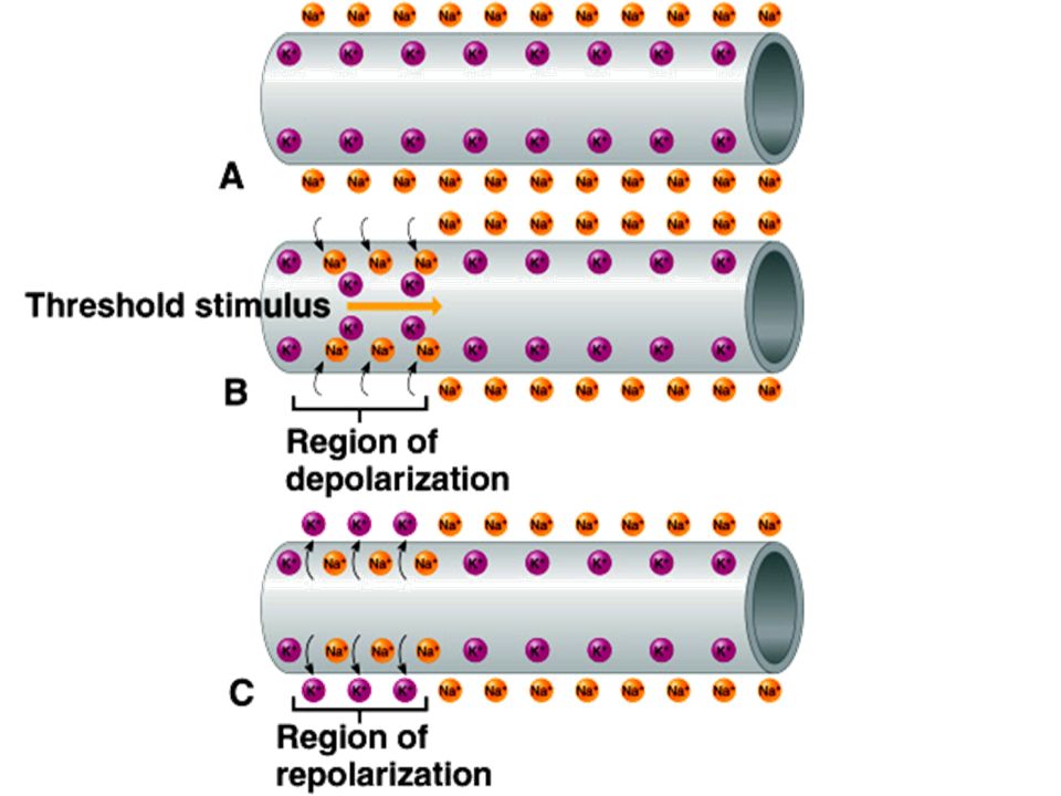
Nerve Impulses A cell membrane is usually polarized, with an excess of negative charges on the inside of the membrane; polarization is important to the conduction of nerve impulses. Potassium ions pass through the membrane more readily than do sodium ions, making potassium ions a major contributor to membrane polarization.

35
Resting Potential Due to active transport, the cell maintains a greater concentration of sodium ions outside and a greater concentration of potassium ions inside the membrane. The inside of the membrane also has excess negative charges, while the outside has more positive charges. This separation of charge, or potential difference, is called the resting potential. (-70mV)
")
36
Resting Potential

37
Action Potential At threshold potential, membrane permeability to sodium suddenly changes in the region of stimulation. As sodium channels open, sodium ions rush in, and the membrane potential changes and becomes depolarized. At the same time, potassium channels open to allow potassium ions to leave the cell, the membrane becomes repolarized, and resting potential is reestablished. This rapid sequence of events is the action potential. The active transport mechanism then works to maintain the original concentrations of sodium and potassium ions.

39
Impulse Conduction Unmyelinated fibers conduct impulses over their entire membrane surface. Myelinated fibers conduct impulses from node of Ranvier to node of Ranvier, a phenomenon called saltatory conduction. Saltatory conduction is many times faster than conduction on unmyelinated neurons.

41
Direction of impulse travel
Synaptic Knob of axon

42
Excitatory and Inhibitory Actions
Neurotransmitters that increase postsynaptic membrane permeability to sodium ions may trigger impulses and are thus excitatory. Other neurotransmitters may decrease membrane permeability to sodium ions, reducing the chance that it will reach threshold, and are thus inhibitory. The effect on the postsynaptic neuron depends on which presynaptic knobs are activated.

43
The Synapse Neurons communicate with adjacent neurons via a synapse – a small space between them is the synaptic cleft

44
Axon terminals (Synaptic knobs) have neurotransmitters in synaptic vesicles and dendrites have receptors for the neurotransmitters. Neurotransmitters are released into the synaptic cleft

45
Neurotransmitters Neurotransmitters are chemicals that are used to relay, amplify and modulate electrical signals between a neuron and another cell. It is synthesized within the presynaptic neuron; It is available in sufficient quantity in the presynaptic neuron to exert an effect on the postsynaptic neuron; A biochemical mechanism for its inactivation is always present.

47
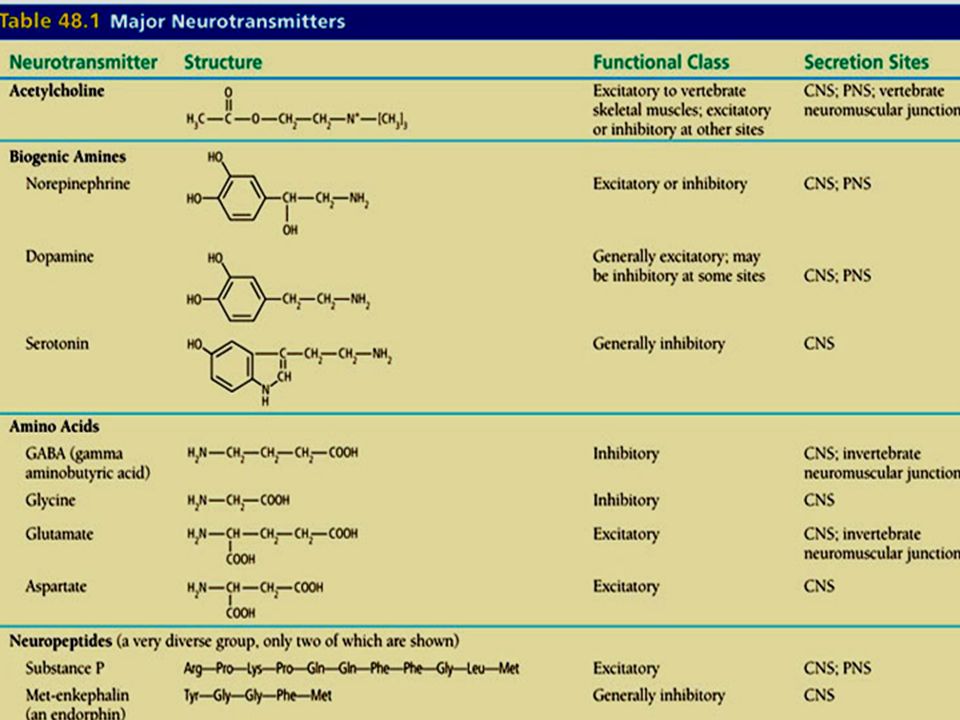
Neurotransmitters At least 50 kinds of neurotransmitters are produced by the nervous system, most of which are synthesized in the cytoplasm of the synaptic knobs and stored in synaptic vesicles. When an action potential reaches the synaptic knob, calcium ions rush inward and, in response, some synaptic vesicles fuse with the membrane and release their contents to the synaptic cleft. Enzymes in synaptic clefts and on postsynaptic membranes rapidly decompose the neurotransmitters after their release. Destruction or removal of neurotransmitter prevents continuous stimulation of the postsynaptic neuron.

48
Some examples of neurotransmitter action
Acetylcholine - voluntary movement of the muscles Epinephrine (aka Adrenalin) – “Flight or Fight” preparation Norepinephrine - wakefulness or arousal Dopamine - voluntary movement and emotional arousal Serotonin - memory, emotions, wakefulness, sleep and temperature regulation GABA (gamma aminobutyric acid) - motor behavior Glutamate is an excitatory relative of GABA. Glycine - spinal reflexes and motor behaviour Neuromodulators - sensory transmission-especially pain
 – Flight or Fight preparation. Norepinephrine - wakefulness or arousal. Dopamine - voluntary movement and emotional arousal. Serotonin - memory, emotions, wakefulness, sleep and temperature regulation. GABA (gamma aminobutyric acid) - motor behavior. Glutamate is an excitatory relative of GABA. Glycine - spinal reflexes and motor behaviour. Neuromodulators - sensory transmission-especially pain.")
49
Impulse Processing How impulses are processed is dependent upon how neurons are organized in the brain and spinal cord. Neuronal Pools Neurons within the CNS are organized into neuronal pools with varying numbers of cells. Each pool receives input from afferent nerves (Sensory neurons) and processes the information according to the special characteristics of the pool.
 and processes the information according to the special characteristics of the pool.")
50
Facilitation A particular neuron of a pool may receive excitatory or inhibitory stimulation; if the net effect is excitatory but subthreshold, the neuron becomes more excitable to incoming stimulation (a condition called facilitation).
.")
51
Convergence A single neuron within a pool may receive impulses from two or more fibers (convergence), which makes it possible for the neuron to summate impulses from different sources.
, which makes it possible for the neuron to summate impulses from different sources.")
52
Divergence Impulses leaving a neuron in a pool may be passed into several output fibers (divergence), a pattern that serves to amplify an impulse.
, a pattern that serves to amplify an impulse.")
53
Types of Nerves A nerve is a bundle of nerve fibers held together by layers of connective tissue. Nerves can be sensory, motor, or mixed, carrying both sensory and motor fibers.

54
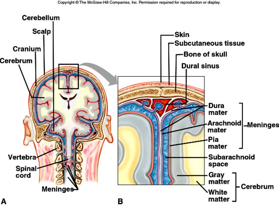
Meninges A. The brain and spinal cord are surrounded by membranes called meninges that lie between the bone and the soft tissues. B. The outermost meninx is made up of tough, white dense connective tissue, contains many blood vessels, and is called the dura mater. 1. It forms the inner periosteum of the skull bones. 2. In some areas, the dura mater forms partitions between lobes of the brain, and in others, it forms dural sinuses. 3. The sheath around the spinal cord is separated from the vertebrae by an epidural space. C. The middle meninx, the arachnoid mater, is thin and lacks blood vessels. 1. It does not follow the convolutions of the brain. 2. Between the arachnoid and pia maters is a subarachnoid space containing cerebrospinal fluid. D. The innermost pia mater is thin and contains many blood vessels and nerves. 1. There are cauliflower-like masses of capillaries called choroid plexuses in the pia mater. Ependymal cells line these choroid plexuses which secrete CSF.

57
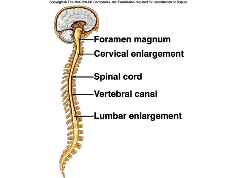
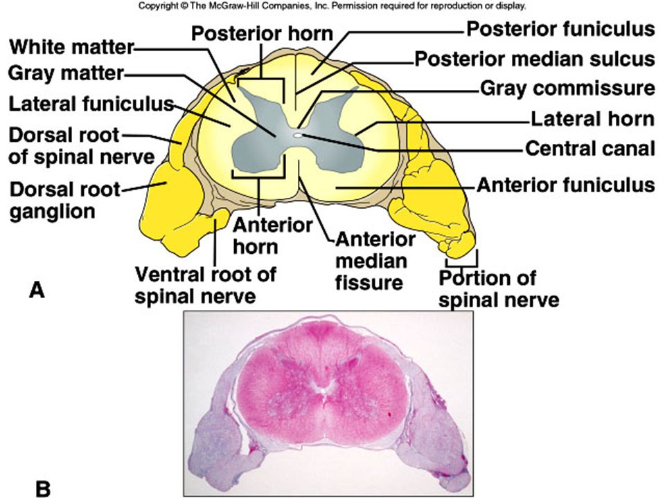
Spinal Cord A. The spinal cord begins at the base of the brain and extends as a slender cord to the level of the intervertebral disk between the first and second lumbar vertebrae. B. Structure of the Spinal Cord 1. The spinal cord consists of 31segments, each of which gives rise to a pair of spinal nerves. 2. A cervical enlargement gives rise to nerves leading to the upper limbs, and a lumbar enlargement gives rise to those innervating the lower limbs. 3. Two deep longitudinal grooves (anterior median fissure and posterior median sulcus) divide the cord into right and left halves. 4. White matter, made up of bundles of myelinated nerve fibers (nerve tracts), surrounds a butterfly-shaped core of gray matter housing interneurons. 5. A central canal contains cerebrospinal fluid. C. Functions of the Spinal Cord 1. The spinal cord has two major functions: to transmit impulses to and from the brain, and to house spinal reflexes. 2. Tracts carrying sensory information to the brain are called ascending tracts; descending tracts carry motor information from the brain. 3. The names that identify nerve tracts identify the origin and termination of the fibers in the tract. 4. Many spinal reflexes also pass through the spinal cord.
 divide the cord into right and left halves. 4. White matter, made up of bundles of myelinated nerve fibers (nerve tracts), surrounds a butterfly-shaped core of gray matter housing interneurons. 5. A central canal contains cerebrospinal fluid. C. Functions of the Spinal Cord. 1. The spinal cord has two major functions: to transmit impulses to and from the brain, and to house spinal reflexes. 2. Tracts carrying sensory information to the brain are called ascending tracts; descending tracts carry motor information from the brain. 3. The names that identify nerve tracts identify the origin and termination of the fibers in the tract. 4. Many spinal reflexes also pass through the spinal cord.")
59
There are: 12 pairs of cranial nerves 31 pairs of spinal nerves
Cranial nerves are nerves that emerge directly from the brain. Only the first and the second pair emerge from the cerebrum , the remaining ten pairs emerge from the brainstem 31 pairs of spinal nerves spinal nerves emerge from segments of the spinal cord

63
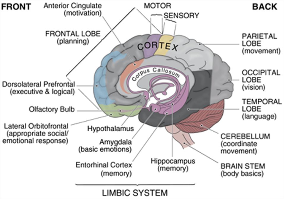
Lobes of the Brain

64
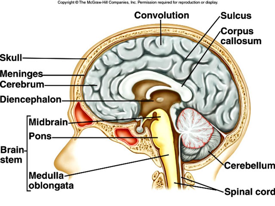
The Brain A. The brain is the largest, most complex portion of the nervous system, containing 100 billion multipolar neurons. B. The brain can be divided into the 1)cerebrum (largest portion and associated with higher mental functions) 2) Diencephalon (contains thalamus, hypothalamus, parts of pituitary – controls autonomic nervous system) 3)cerebellum (coordinates muscular activity), 4)brain stem (coordinates and regulates visceral activities).
cerebrum (largest portion and associated with higher mental functions) 2) Diencephalon (contains thalamus, hypothalamus, parts of pituitary – controls autonomic nervous system) 3)cerebellum (coordinates muscular activity), 4)brain stem (coordinates and regulates visceral activities).")
65
The Cerebrum 1. The cerebrum is the largest portion of the mature brain, consisting of two cerebral hemispheres. 2. A deep ridge of nerve fibers called the corpus callosum connects the hemispheres. 3. The surface of the brain is marked by convolutions, sulci, and fissures. 4. The lobes of the brain are named according to the bones they underlie and include the frontal lobe, parietal lobe, temporal lobe, occipital lobe, and insula. 5. A thin layer of gray matter, the cerebral cortex, lies on the outside of the cerebrum and contains 75% of the cell bodies in the nervous system. 6. Beneath the cortex lies a mass of white matter made up of myelinated nerve fibers connecting the cell bodies of the cortex with the rest of the nervous system.

66
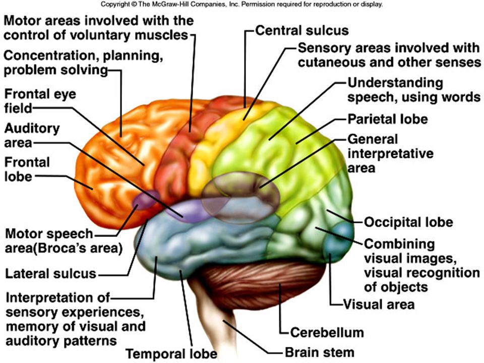
Functions of the Cerebrum
The cerebrum provides higher brain functions, such as interpretation of sensory input, initiating voluntary muscular movements, memory, and integrating information for reasoning. Functional Regions of the Cerebral Cortex a. The functional areas of the brain overlap, but the cortex can generally be divided into motor, sensory, and association areas. b. The primary motor areas lie in the frontal lobes, anterior to the central sulcus and in its anterior wall. c. Broca's area, anterior to the primary motor cortex, coordinates muscular activity to make speech possible. d. Above Broca's area is the frontal eye field that controls the voluntary movements of the eyes and eyelids. e. The sensory areas are located in several areas of the cerebrum and interpret sensory input, producing feelings or sensations. f. Sensory areas for sight lie within the occipital lobe. g. The various association areas of the brain analyze and interpret sensory impulses and function in reasoning, judgment, emotions, verbalizing ideas, and storing memory. h. Association areas of the frontal lobe control a number of higher intellectual processes. i. A general interpretive area is found at the junction of the parietal, temporal, and occipital lobes, and plays the primary role in complex thought processing.

67
understanding of written and spoken language
Speech Production

69
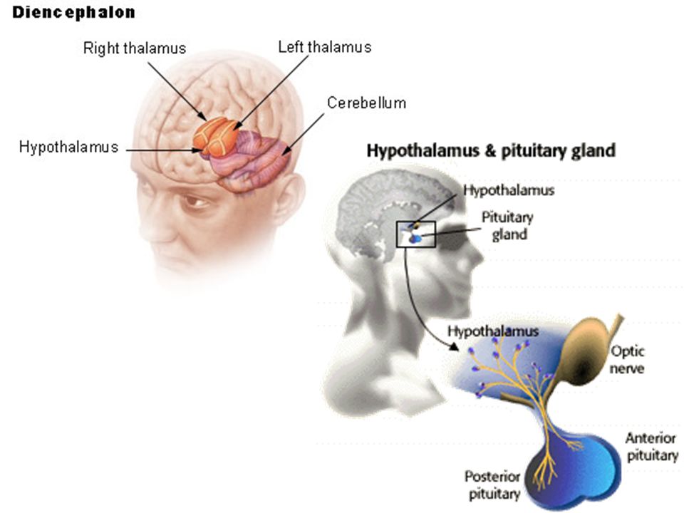
The Diencephalon The diencephalon lies above the brain stem and contains the thalamus and hypothalamus, the optic tracts and optic chiasma, the infundibulum (attachment for the pituitary), the posterior pituitary, mammillary bodies, and the pineal gland. a. The thalamus functions in sorting and directing sensory information arriving from other parts of the nervous system, performing the services of both messenger and editor. b. The hypothalamus maintains homeostasis by regulating a wide variety of visceral activities and by linking the endocrine system with the nervous system. The hypothalamus regulates heart rate and arterial blood pressure, body temperature, water and electrolyte balance, hunger and body weight, movements and secretions of the digestive tract, growth and reproduction, and sleep and wakefulness. The limbic system, in the area of the diencephalon, controls emotional experience and expression. a. By generating pleasant or unpleasant feelings about experiences, the limbic system guides behavior that may enhance the chance of survival.
, the posterior pituitary, mammillary bodies, and the pineal gland. a. The thalamus functions in sorting and directing sensory information arriving from other parts of the nervous system, performing the services of both messenger and editor. b. The hypothalamus maintains homeostasis by regulating a wide variety of visceral activities and by linking the endocrine system with the nervous system. The hypothalamus regulates heart rate and arterial blood pressure, body temperature, water and electrolyte balance, hunger and body weight, movements and secretions of the digestive tract, growth and reproduction, and sleep and wakefulness. The limbic system, in the area of the diencephalon, controls emotional experience and expression. a. By generating pleasant or unpleasant feelings about experiences, the limbic system guides behavior that may enhance the chance of survival.")
71
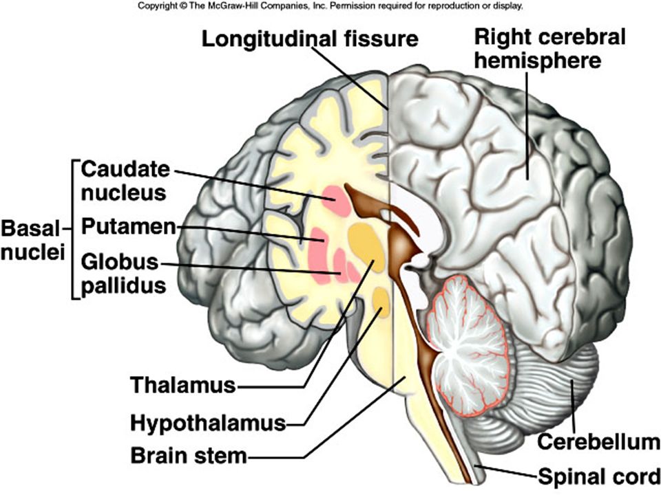
Basal ganglia (in the region of the diencephalon)
1 These are masses of gray matter located deep within the cerebral hemispheres that relay motor impulses from the cerebrum and help to control motor activities by producing inhibitory dopamine. Basal ganglia include the caudate nucleus, the putamen, and the globus pallidus. 2 2 3

72
Limbic System shares the same area as the diencephalon
Include the hippocampus, amygdala, fronix, septum functions include: Emotion and emotional memory (amygdala) behavior Formation of long term memory (hippocampus) Olfaction (olfactory bulb)
 behavior. Formation of long term memory (hippocampus) Olfaction (olfactory bulb)")
73
Brain Stem The brain stem, consisting of the midbrain, pons, and medulla oblongata, lies at the base of the cerebrum, and connects the brain to the spinal cord. 1. Midbrain a. The midbrain, located between the diencephalon and pons, contains bundles of myelinated nerve fibers that convey impulses to and from higher parts of the brain, and masses of gray matter that serve as reflex centers. b. The midbrain contains centers for auditory and visual reflexes. 2. Pons a. The pons, lying between the midbrain and medulla oblongata, transmits impulses between the brain and spinal cord, and contains centers that regulate the rate and depth of breathing.

74
The Brain Stem, Cont’d. 3. Medulla Oblongata
a. The medulla oblongata transmits all ascending and descending impulses between the brain and spinal cord. b. The medulla oblongata also houses nuclei that control visceral functions, including the cardiac center that controls heart rate, the vasomotor center for blood pressure control, and the respiratory center that works, along with the pons, to control the rate and depth of breathing. c. Other nuclei in the medulla oblongata are associated with coughing, sneezing, swallowing, and vomiting. 4. Reticular Formation a. Throughout the brain stem, hypothalamus, cerebrum, cerebellum, and basal ganglia, is a complex network of nerve fibers connecting tiny islands of gray matter; this network is the reticular formation. b. Decreased activity in the reticular formation results in sleep; increased activity results in wakefulness. c. The reticular formation filters incoming sensory impulses.

75
Cerebellum 1. The cerebellum is made up of two hemispheres connected by a vermis. 2. A thin layer of gray matter called the cerebellar cortex lies outside a core of white matter. 3. The cerebellum communicates with other parts of the central nervous system through cerebellar peduncles. 4. The cerebellum functions to integrate sensory information about the position of body parts and coordinates skeletal muscle activity and maintains posture.

76
Reticular Formation Decreased activity in the reticular formation results in sleep; increased activity results in wakefulness

77
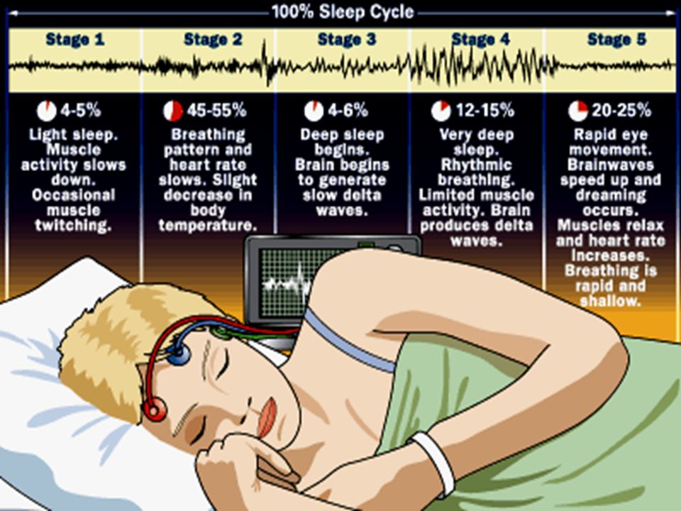
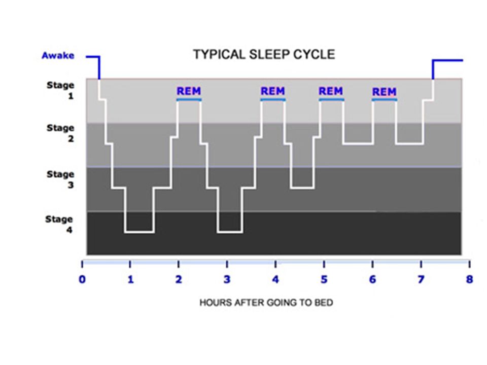
Slow wave sleep and REM alternate
Two types of sleep NREM (No Rapid Eye Movement) - Slow wave – results from tiredness, is dreamless and can be light or heavy - Lasts 70 – 90 minutes Low BP restful low respiratory rate REM – Rapid Eye Movement – or paradoxical sleep Some areas of brain are active Lasts 5 – 15 minutes Dream sleep – very important and needed – if you do not have REM sleep for even one night, you make up for it the next night Irregular heart rate and breathing, twitching of muscles, eyes move rapidly under lids Slow wave sleep and REM alternate
 - Slow wave – results from tiredness, is dreamless and can be light or heavy. - Lasts 70 – 90 minutes. Low BP. restful. low respiratory rate. REM – Rapid Eye Movement – or paradoxical sleep. Some areas of brain are active. Lasts 5 – 15 minutes. Dream sleep – very important and needed – if you do not have REM sleep for even one night, you make up for it the next night. Irregular heart rate and breathing, twitching of muscles, eyes move rapidly under lids. Slow wave sleep and REM alternate.")
80
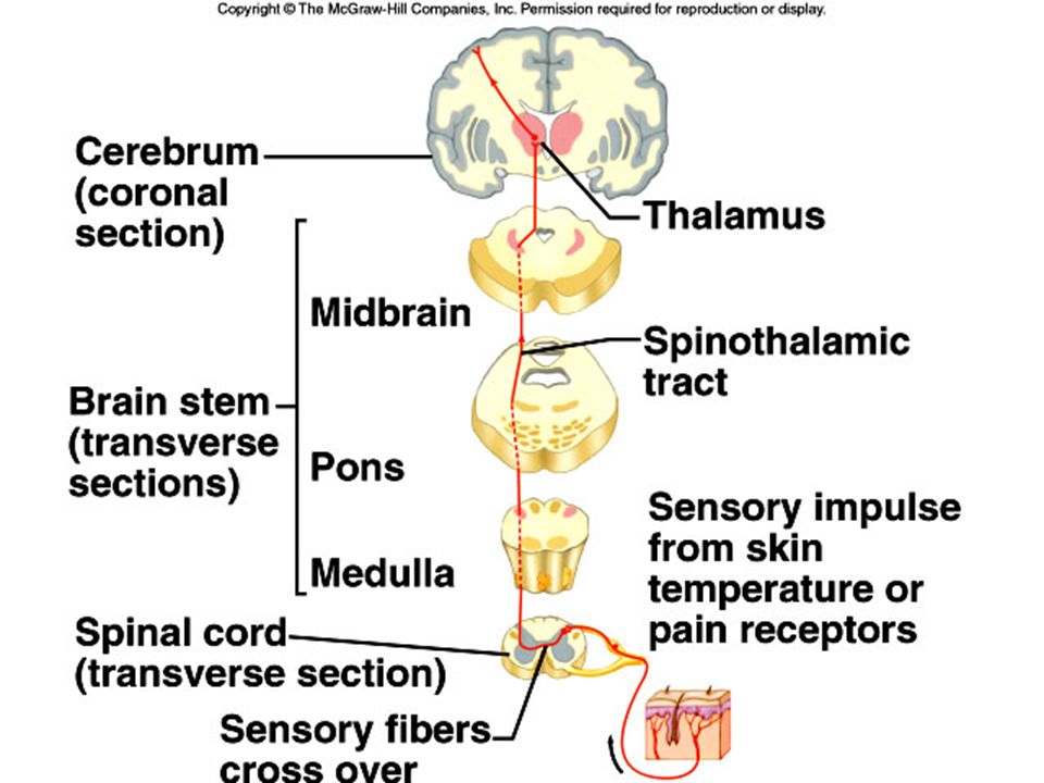
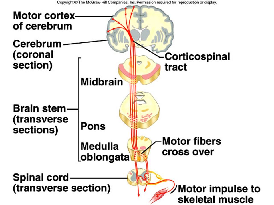
Hemisphere Dominance Sensory and motor fibers alike cross over in the spinal cord or brain stem so centers in the right hemisphere are interpreting or controlling the left side of the body, and vice versa. a. Both cerebral hemispheres function in receiving and analyzing sensory input and sending motor impulses to the opposite side of the body. b. Most people exhibit some type of hemisphere dominance for the language-related activities of speech, writing, and reading. c. The left hemisphere is dominant in 90% of the population, although some individuals have the right hemisphere as dominant, and others show equal dominance in both hemispheres. d. The non-dominant hemisphere specializes in nonverbal functions and controls emotions and intuitive thinking.

81
The left and right hemispheres are connected with a bundle of nerve fibers called the Corpus Callosum

82
Long term memory is limitless!
Learning is the acquisition of knowledge – memory is the persistance of the new knowledge. Short term – Series of electrical impulses in neurons and when the stimulation is gone, so is the memory – also called “working memory” Long term - The neurons actually change – make more synapses (trillions) and better synaptic transmission – once these synapses are forged, they do not change for years Long term memory is limitless!
 and better synaptic transmission – once these synapses are forged, they do not change for years. Long term memory is limitless!")
83
The Amygdala There has been a growing debate on the function of the amygdala, an almond-shaped sub-cortical structure in the limbic system (temporal lobe). It receives electrical signals carrying auditory information. New research published in Journal of Neuroscience suggests that the amygdala plays a pivotal role in the initial process of storing memory elsewhere in the brain. The amygdala appears to decide which experiences are important enough to store a decision based on the emotional significance of the events in a decoding process that affects both learning and memory.
. It receives electrical signals carrying auditory information. New research published in Journal of Neuroscience suggests that the amygdala plays a pivotal role in the initial process of storing memory elsewhere in the brain. The amygdala appears to decide which experiences are important enough to store a decision based on the emotional significance of the events in a decoding process that affects both learning and memory.")
84
Memory Storage = Cerebral Cortex
Scientific studies have not shown the existence of a central memory manager. This means that each of the highly subdivided segments of the cerebral cortex is responsible not only for its function but also for its own memory storage. Findings indicate that recognition comes from groups of neurons working together to identify items. These groups may be very specific and respond only to a single item or may respond to a broad range of similar objects or activity. These individual qualities such as shape, color, location, motion, etc. are divided up by the cortical regions.

85
Homunculus = Little Man

86
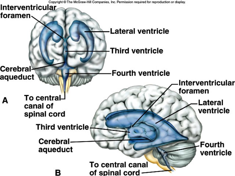
Ventricles and Cerebrospinal Fluid
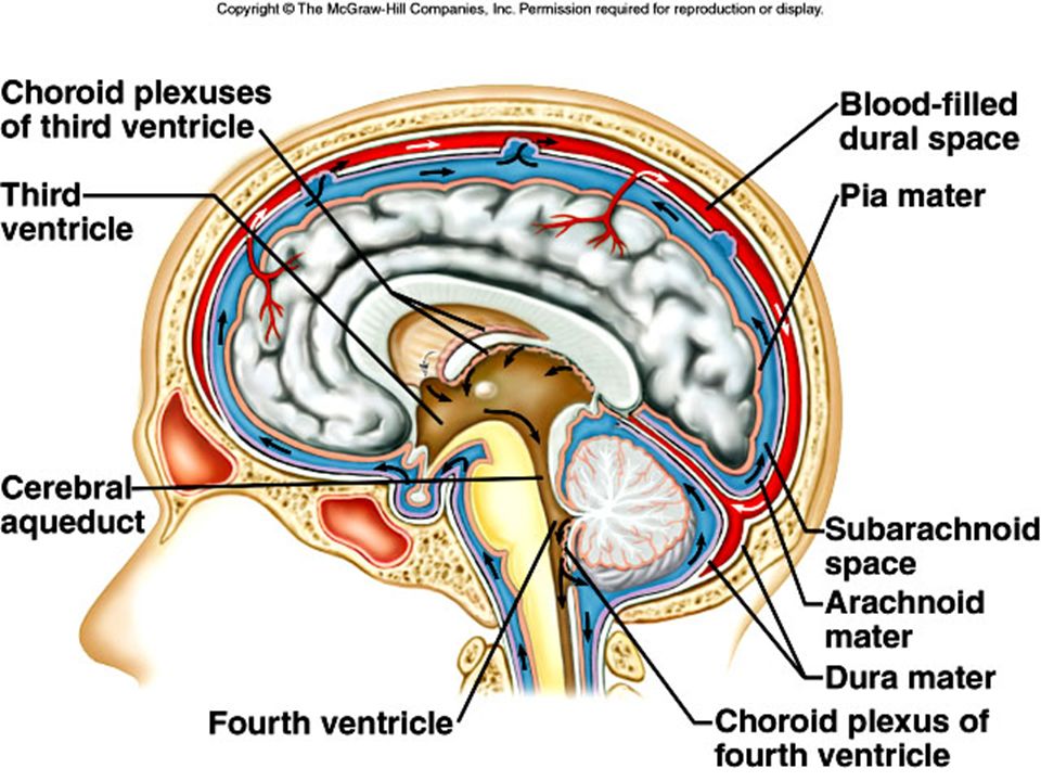
1. The ventricles are a series of connected cavities within the cerebral hemispheres and brain stem. 2. The ventricles are continuous with the central canal of the spinal cord, and are filled with cerebrospinal fluid. 3. Choroid plexuses, specialized capillaries from the pia mater, secrete cerebrospinal fluid (ependymal cells of the choroid plexuses). a. Most cerebrospinal fluid arises in the lateral ventricles. 4. Cerebrospinal fluid has nutritive as well as protective (cushioning) functions.
. a. Most cerebrospinal fluid arises in the lateral ventricles. 4. Cerebrospinal fluid has nutritive as well as protective (cushioning) functions.")
88
Pituitary gland The pituitary gland has two sections: the posterior and the anterior pituitary. These sections have different origins and produce different hormones. The posterior pituitary derives from nervous system tissue and produces neurohormones. The anterior pituitary derives from epithelial tissue and produces hormones with endocrine functions.

89
Neurohormones Neurohormonal activity is distinguished from that of classical neurotransmitters as it can have effects on cells distant from the source of the hormone. Neurohormones include: GnRH Gonadotropin releasing hormone CRH Corticotropin releasing hormone TRH Thyrotropin-releasing hormone Dopamine Prolactin inhibiting hormone Orexin(aka hypocretin) Stimulates appetite
 Stimulates appetite.")
93
Peripheral Nervous System
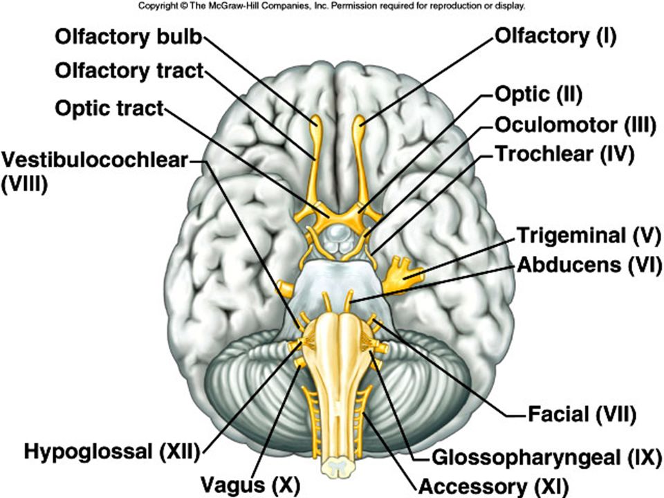
The peripheral nervous system (PNS) consists of the cranial and spinal nerves that arise from the central nervous system and travel to the remainder of the body. The PNS is made up of the somatic nervous system that oversees voluntary activities, and the autonomic nervous system that controls involuntary activities. Cranial Nerves . 1. Twelve pairs of cranial nerves arise from the underside of the brain, most of which are mixed nerves. 2. The 12 pairs are designated by number and name and include the olfactory, optic, oculomotor, trochlear, trigeminal, abducens, facial, vestibulocochlear, glossopharyngeal, vagus, accessory, and hypoglossal nerves. 3. Refer to Figure 9.31 and Table 9.6 for cranial nerve number, name, type, and function.
 consists of the cranial and spinal nerves that arise from the central nervous system and travel to the remainder of the body. The PNS is made up of the somatic nervous system that oversees voluntary activities, and the autonomic nervous system that controls involuntary activities. Cranial Nerves . 1. Twelve pairs of cranial nerves arise from the underside of the brain, most of which are mixed nerves. 2. The 12 pairs are designated by number and name and include the olfactory, optic, oculomotor, trochlear, trigeminal, abducens, facial, vestibulocochlear, glossopharyngeal, vagus, accessory, and hypoglossal nerves. 3. Refer to Figure 9.31 and Table 9.6 for cranial nerve number, name, type, and function.")
95
Peripheral Nervous System, Cont’d.
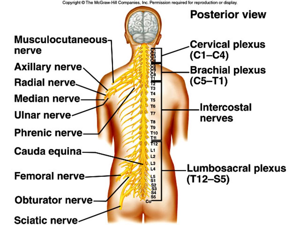
2. Spinal Nerves 1. Thirty-one pairs of mixed nerves make up the spinal nerves. 2. Spinal nerves are grouped according to the level from which they arise and are numbered in sequence, beginning with those in the cervical region. 3. Each spinal nerve arises from two roots: a dorsal or sensory root, and a ventral or motor root. 4. The main branches of some spinal nerves form plexuses. 5. Cervical Plexuses a. The cervical plexuses lie on either side of the neck and supply muscles and skin of the neck. 6. Brachial Plexuses a. The brachial plexuses arise from lower cervical and upper thoracic nerves and lead to the upper limbs. 7. Lumbrosacral Plexuses a. The lumbrosacral plexuses arise from the lower spinal cord and lead to the lower abdomen, external genitalia, buttocks, and legs.

97
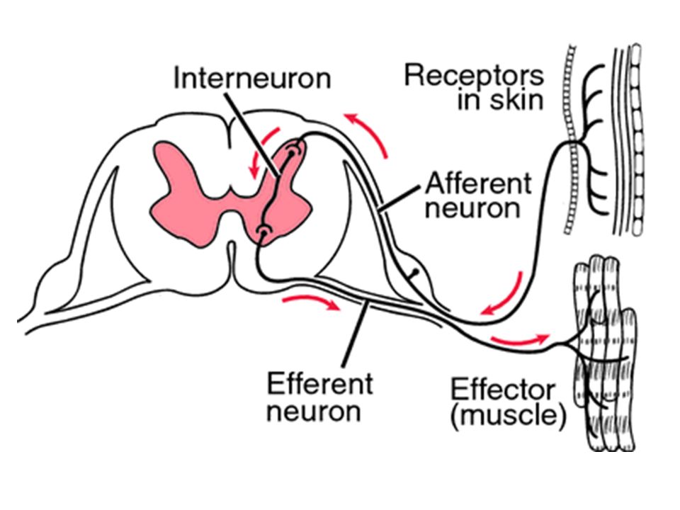
The Reflex Arc A reflex arc is a neural pathway that controls an action reflex. In higher animals, most sensory neurons do not pass directly into the brain, but synapse in the spinal cord. This characteristic allows reflex actions to occur relatively quickly by activating spinal motor neurons without the delay of routing signals through the brain, although the brain will receive sensory input while the reflex action occurs.

99
Autonomic Nervous System
The autonomic nervous system has the task of maintaining homeostasis of visceral activities without conscious effort. General Characteristics 1. The autonomic nervous system includes two divisions: the sympathetic and parasympathetic divisions, which exert opposing effects on target organs. a. The parasympathetic division operates under normal conditions. b. The sympathetic division operates under conditions of stress or emergency.

100
Parasympathetic Division of the Autonomic Nervous System
Works under normal conditions

101
Sympathetic Division of the Autonomic Nervous System
Works under conditions of stress or emergency

102
Autonomic Nerve Fibers
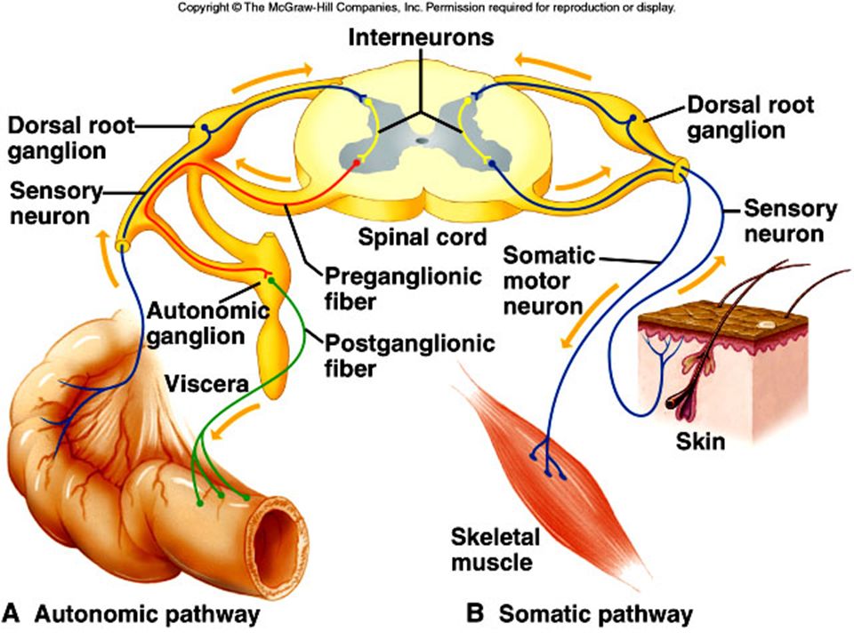
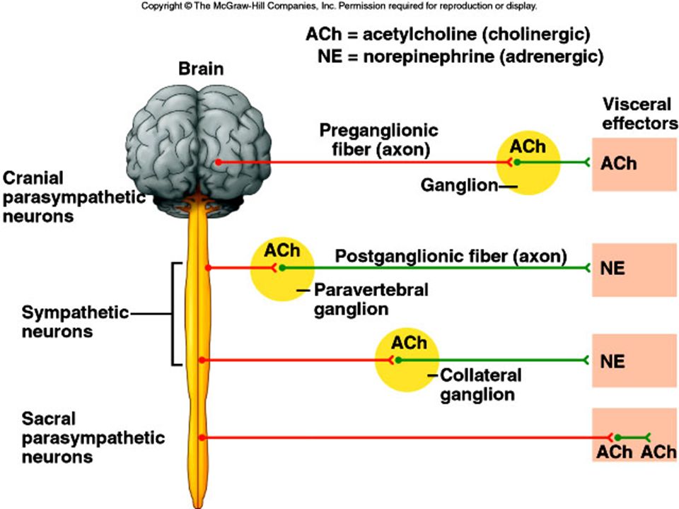
In the autonomic motor system, motor pathways include two fibers: a preganglionic fiber that leaves the CNS, and a postganglionic fiber that innervates the effector. 1. Sympathetic Division a. Fibers in the sympathetic division arise from the thoracic and lumbar regions of the spinal cord, and synapse in paravertebral ganglia (like the dorsal root ganglia) close to the vertebral column. b. Postganglionic axons lead to an effector organ. 2. Parasympathetic Division a. Fibers in the parasympathetic division arise from the brainstem and sacral region of the spinal cord, and synapse in ganglia close to the effector organ.
 close to the vertebral column. b. Postganglionic axons lead to an effector organ. 2. Parasympathetic Division. a. Fibers in the parasympathetic division arise from the brainstem and sacral region of the spinal cord, and synapse in ganglia close to the effector organ.")
104
Autonomic Neurotransmitters
Preganglionic fibers of both sympathetic and parasympathetic divisions release acetylcholine. Parasympathetic postganglionic fibers are cholinergic fibers and release acetylcholine. (Normal muscle function) Sympathetic postganglionic fibers are adrenergic and release norepinephrine. (Adrenaline for stress – flight or fight) The effects of these two divisions, based on the effects of releasing different neurotransmitters to the effector, are generally antagonistic.
 Sympathetic postganglionic fibers are adrenergic and release norepinephrine. (Adrenaline for stress – flight or fight) The effects of these two divisions, based on the effects of releasing different neurotransmitters to the effector, are generally antagonistic.")
106
Control of Autonomic Activity
a. The autonomic nervous system is largely controlled by reflex centers in the brain and spinal cord. b. The limbic system - a combination of the cerebral cortex, hypothalamus, thalamus, etc. - alter the reactions of the autonomic nervous system through emotional influence.

Similar presentations
>")


 Pia mater -inner membrane, contains.>")


![The Nervous System. Divisions of the Nervous System Central Nervous System [CNS] = Spinal Cord Brain Peripheral Nervous System [PNS]= Spinal Nerves.](/16/5239306/big_thumb.jpg "The Nervous System. Divisions of the Nervous System Central Nervous System [CNS] = Spinal Cord Brain Peripheral Nervous System [PNS]= Spinal Nerves.>")


 Bone – Cranium, Vertebrae 2) Meninges – Three connective tissue membranes covering the brain and spinal cord a) Dura Mater – outermost,>")

 Brain and spinal cord Peripheral Nervous System (PNS) ◦ nerves.>")


