Download presentation
Presentation is loading. Please wait.

1
EEG & Sleep

2
EEG: definition It is record of variations in brain potential It is record of electrical activity of brain/neurons in different phases e.g. during sleep, wakefulness and epilepsy.

3
E.E.G Carried out by placing electrodes on surface of scalp. Sometimes placed directly on surface of cerebral cortex, e.g., during neurosurgical operations or in experimental animals. Such a record is called Electro- Corticogram (ECOG)
.")
4
E.E.G E.E.G was 1 st recorded by a German Psychiatrist Hans Berger. There are 2 methods for EEG recording Recording of EEG can be unipolar or bipolar. In unipolar EEG, active electrode is placed on surface of scalp, while inactive or indifferent is placed at a distant point, like tip of 7 th cervical vertebra. In bipolar EEG, both electrodes are active & placed on surface of scalp.

5
E.E.G In routine E.E.G, 20 electrodes are placed on scalp at different points to record EEG. In normal EEG, 4 types of waves can be seen: alpha, beta, theta & delta, in different phases. Character of each wave is described as 1- its intensity/voltage 2- frequency

6
Alpha waves: Waves of quiet wakefulness / waves of inattentiveness: Frequency: 8-13 / sec Voltage: 50 micro-volts Relaxed awareness These are recorded when a person is awake but mentally relaxed & inattentive, e.g., lying comfortably in a quiet room, eyes are closed & person is mentally relaxed & inattentive.

7
Alpha waves: Waves of quiet wakefulness / waves of inattentiveness: When a person opens eyes or the brain becomes active by thinking process or solving a problem, alpha waves disappear. Frequency of alpha waves decreases by decreased body temperature, decreased glucocorticoid secretion, hypoglycemia & increase in pCO2. (due to cold temp. / empty stomach / during suffocation, one cant relax)
.")
9
Alpha waves: Waves of quiet wakefulness / waves of inattentiveness: Best recorded from parietal & occipital regions. Thalamo-cortical connections are important for it.

10
Beta waves: waves of alertness / wakefulness / desynchronized waves Frequency: 14-80 cycles/sec Amplitude / voltage: 20 microvolts. Awareness with concentrated attention Recorded when brain is highly active. Best recorded from parietal & frontal regions. Recorded during REM sleep. Appear on eye opening

11
Theta waves: Frequency: 4-7 / sec Voltage: 10 microvolts Best recorded from parietal & temporal regions. Recorded during light sleep. Recorded in adults during states of frustration & disappointment. In children normally recorded in awake E.E.G. Also recorded in brain disorders like Grand Mal Epilepsy. In degenerative brain disorders.

12
Delta Waves: Very slow waves. Frequency: 0.5 - 3 / sec Voltage: 100 microvolts Recorded in deep & restful sleep. Also recorded in coma, general anesthesia & in epilepsy. In organic brain disorders.

13
Brain waves in normal E.E.G

14
Physiological basis of E.E.G: Electrical activity recorded in EEG, is mainly from superficial layers of cerebral cortex which have number of dendrites on which many nerve terminals synapse. Some terminals are excitatory (EPSP is produced), some are inhibitory (IPSP is produced). Electrical activity recorded in EEG, is summation of EPSPs & IPSPs
, some are inhibitory (IPSP is produced). Electrical activity recorded in EEG, is summation of EPSPs & IPSPs.")
16
Physiological basis of E.E.G: Amplitude of waves in EEG depends on how much the waves are synchronized / coordinated. If waves are synchronized, there is increased amplitude. If desynchronized, there are deflections in different directions & these neutralize each other, resulting into a small amplitude like in beta waves. During each wave there is synchronous discharge/ activation of neurons.

17
Physiological basis of E.E.G: Thalamo-cortical connections are important, mainly in recording of alpha waves. If these fibers are cut, alpha waves disappear & delta waves appear.

18
Clinical use of E.E.G: EEG reflects functional state of brain. Recorded as an investigation in patients. Also recorded for research purpose. Recorded for many hours in epilepsy cases. EEG machines are now computerized & there is automatic analysis of EEG.

19
Clinical use of E.E.G: Helps to diagnose SOL (Space occupying lesion) in skull, which may be infective, neoplastic, traumatic or vascular. It helps in diagnosis of epilepsy & its types. It helps in diagnosis of psychopathic disorders.

20
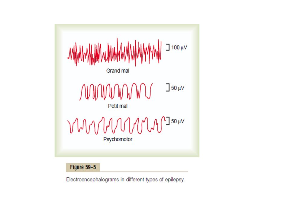
Clinical use of E.E.G: In Grand Mal epilepsy, there may be theta or delta waves (high voltage waves) in EEG. In Petit Mal epilepsy, there is spike & dome pattern. In Psychomotor epilepsy, mainly delta waves are seen.

21
Clinical use of E.E.G: E.E.G silence is sure indication of brain death.

22
Epilepsy Epilepsy (also called “seizures”) is characterized by uncontrolled excessive activity of either part or all of CNS. Attack occurs when basal level of excitability of neurons crosses threshold. Epilepsy can be classified into three major types: grand mal epilepsy, petit mal epilepsy, and focal epilepsy.

26
Grand Mal Epilepsy/Generalized epilepsy Grand mal epilepsy is characterized by extreme neuronal discharges in all areas of the brain cerebral cortex, deep parts of cerebrum, and brain stem. Discharges transmitted into the spinal cord sometimes cause generalized tonic seizures of the entire body, followed by alternating tonic and spasmodic muscle contractions called tonic-clonic seizures

27
person bites his tongue difficulty in breathing & cyanosis urination and defecation can occur grand mal seizure lasts from a few sec to 3 to 4 min. It is also characterized by post seizure depression of the entire nervous system.

28
Factors which produce epilepsy Strong emotional stimuli Hyperventilation or alkalosis Effects of some drugs e.g. phenylenetetrazole High fever Loud noises Bright light Traumatic lesion in any part of brain

30
Petit mal/Partial epilepsy Person suddenly becomes unconscious. Convulsions do not occur Muscles of face show twitching & blinking of eyes Afterwards person become normal It occurs in late childhood Absence syndrome/ absence epilepsy Excitatory thalamocortical neurons

31
Focal Epilepsy It involves only localized area of brain (cerebral cortex or deep parts of cerebrum) The abnormality starts from a particular area and spreads to the adjacent area.
 The abnormality starts from a particular area and spreads to the adjacent area.")
32
Focal Epilepsy Two types 1- Jacksonian epilepsy 2- Psychomotor epilepsy Causes 1- scar tissue in brain, 2- Tumor, 3- some destroyed part of brain tissue In Jacks, as the wave of excitation spreads over motor cortex, it causes progressive march

33
Of muscle contrations throughout the opposite side of body. Beginning in mouth region and marching progressively downwards to legs.

34
Psychomotor epilepsy It is characterized by emotional outburst such as abnormal rage,anxiety,fear or discomfort. There is amnesia or confused mental state for some period. The cause, are the abnormalities in temporal lobe & tumor in hypothalamus and limbic system.

35
SLEEP “Period of inactivity during which there is unconsciousness from which person can be aroused by sensory & other stimuli”. Unconsciousness during surgical anesthesia, epilepsy & coma should not be considered as sleep.

36
Lack of SLEEP Sleep is essential for normal life. It restores a balance between different parts of nervous system. If a person is not allowed to sleep for 2-3 days, certain effects are seen: Loss of concentration Slow thought making Loss of memory irritability

37
Lack of SLEEP If insomnia is further prolonged, person may develop: Dysarthria (defect of speech) Tremors Abnormal gait
 Tremors Abnormal gait")
38
Requirement of SLEEP Varies with age: Infants: 20-24 hrs Young children: 12 hrs Young adults: 7-9 hrs Old age: 5-7 hrs

39
Relationship of SLEEP with ANS: During sleep, generally there is Sympathetic inhibition & Parasympathetic stimulation

40
Types of SLEEP SLOW WAVE / Non- REM sleep / Delta wave sleep REM sleep / paradoxical sleep

41
SLOW WAVE / Non-REM sleep / Delta wave sleep Deep & restful sleep. If a person is tired, he passes into deep sleep in 1 hr. On average it constitutes 75% of total sleep during a night. Dreams can be seen but are not remembered, so cannot be recalled. Muscle tone decreases.

42
SLOW WAVE / Non-REM sleep / Delta wave sleep Slowing of heart rate & respiratory rate BMR decrease There is GH secretion Pupillary constriction. Sleep walking (somnambulism) may be seen during slow wave sleep
 may be seen during slow wave sleep.")
43
REM sleep / paradoxical sleep Occurs in periods lasting for 5-30 min. Each period is repeated at every 90 min. There are 4-6 periods of REM sleep during a night. It constitutes 25% of total night sleep.

44
REM sleep / paradoxical sleep Its duration is different in different age groups. There is more REM sleep as age advances. If a person is tired, less REM sleep at night. If a person has taken rest during day time, more REM sleep at night.

45
REM sleep / paradoxical sleep There is active dreaming & dreams can be recalled. It is difficult to arouse the person from REM sleep as compared to non REM sleep but Usually in the morning, person wakes up from REM sleep.

46
REM sleep / paradoxical sleep During REM sleep, brain is highly active, so beta waves are recorded from EEG. Muscle tone increases. Rapid movement of eye. Twitching in different parts of body. Mild convulsions.

47
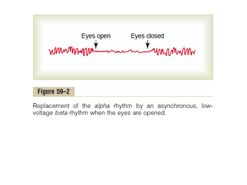
REM sleep / paradoxical sleep Respiratory & heart rate becomes irregular during REM sleep. Increased secretion of corticosteroid hormones. In males, may be erection (parasympathetic stimulation) Teeth grinding (BRUXISM) occurs. Replacement of alpha rhythm by asynchronous rhythm on opening eye.
 Teeth grinding (BRUXISM) occurs. Replacement of alpha rhythm by asynchronous rhythm on opening eye..")
48
Replacement of alpha rhythm by asynchronous rhythm on opening eye:

49
Types of SLEEP SLOW WAVE / Non-REM sleep / Delta wave sleep 75% sleep Dreaming without memory Increased parasympathetic stimulation Decreased muscle tone Bed-wetting in children REM sleep / paradoxical sleep 25% sleep Active dreaming with memory Increased sympathetic stimulation Increased muscle tone, muscle twitching & convulsions.

50
Types of SLEEP SLOW WAVE / Non-REM sleep / Delta wave sleep Decreased heart rate Decreased respiratory rate Pupil constriction Easy to arouse from sleep Brain is not active REM sleep / paradoxical sleep Irregular heart rate Irregular respiratory rate No constriction of pupil Difficult to arouse from sleep, but gets up spontaneously in the morning Brain is active

51
Types of SLEEP SLOW WAVE / Non- REM sleep / Delta wave sleep Increased GH No erection No bruxism Sleep walking REM sleep / paradoxical sleep Increased corticosteroid Erection Bruxism No sleep walking

52
Changes in EEG when a person goes to sleep:

53
Mechanism of sleep There is a cycle of wakefulness & sleep. When a person is awake, gradually neurons in reticular activating system become less & less active & there is also activation of certain sleep centers. This results into sleep.

54
Mechanism of sleep During hours of sleep, neurons in reticular activating system become progressively more & more active, leading to wakefulness.

55
Sleep centers: 1) LOCUS CERULEUS: Location: At junction of midbrain & pons. Neurons in this locus secrete nor epinephrine at nerve endings of nerve fibers. Nerve fibers from these neurons pass to reticular formation.

56
Sleep centers: This center (Locus Ceruleus) is involved in REM sleep, when brain is highly active. So perhaps nor-epinephrine secreting neurons are involved (sympathetic stimulation in REM sleep) Ach secreting neurons in reticular formation of upper brain-stem are also involved.
 Ach secreting neurons in reticular formation of upper brain-stem are also involved..")
57
PGO spikes In REM sleep there are PGO spikes (large phasic potentials in groups of 3-5). These spikes are due to Acetylcholine secreting neurons in this pathway of producing REM sleep. Only the tone of neck muscles is dec., other muscles keep their tone. But at the same time there is locus ceruleus dependent inhibition of voluntary act.

58
Sleep centers: 2) RAPHE MAGNUS NUCLEUS: Midline linear nuclei in upper pons & lower medulla. Fibers from here pass to reticular formation, hypothalamus, limbic system & also spinal cord. These fibers synapse with pain-inhibitory neurons in dorsal horn of spinal cord (analgesia system).
..")
59
Sleep centers: There is release of serotonin at nerve endings of these fibers. Raphe Magnus Nucleus is involved in Deep Slow Wave sleep (NREM sleep). Serotonin inhibitors wakefulness. Stimulation of SCN of Anterior hypothalamus, certain thalamic nuclei & portion of nucleus of tractus solitarius NREM sleep.
. Serotonin inhibitors wakefulness. Stimulation of SCN of Anterior hypothalamus, certain thalamic nuclei & portion of nucleus of tractus solitarius NREM sleep..")
60
Muramyl dipeptide induced sleep: Experimental observation: In animals kept awake for 2-3 days, muramyl dipeptide & other sleep promoting factors are produced in CSF of brain stem which can be later detected in blood & urine. When muramyl dipeptide is injected to some other animal, it immediately passes into sleep.

61
DISORDERS OF SLEEP: 1) Somnambulism / sleep walking 2) Bedwetting in children 3) Bruxism 4) Insomnia 5) Narcolepsy 6) Sleep apnea
 Somnambulism / sleep walking 2) Bedwetting in children 3) Bruxism 4) Insomnia 5) Narcolepsy 6) Sleep apnea")
62
1) Somnambulism / sleep walking Occurs during slow wave sleep / NREM. More common in male children. Episode of sleep walking may remain for many minutes. Person walks with open eyes, obstacles are avoided during walking. Person wakes up unaware of sleep walking.

63
2) Bedwetting in children Also called Nocturnal enuresis. May be due to parasympathetic dominance, as it occurs in slow wave sleep.

64
3) Bruxism Teeth grinding Occurs during active sleep (REM sleep)
 Bruxism Teeth grinding Occurs during active sleep (REM sleep)")
65
4) Insomnia Inability to sleep, although sufficient facilities & time is available for sleep. Reason: Psychological or medical.

66
5) Narcolepsy: There are attacks of intense desire to sleep during day time Person cannot resist to sleep in the day Attack may last for seconds to minutes
 Narcolepsy: There are attacks of intense desire to sleep during day time Person cannot resist to sleep in the day Attack may last for seconds to minutes")
67
Cause of Narcolepsy: Etiology: Considered to be hypothalamic disorder Evidence: Other features of hypothalamic disorders are present, e.g., obesity, polyuria, sexual retardation.

68
6) Sleep apnea During sleep, breathing stops suddenly. May be repeated 100’s of times in severe cases. When breathing stops, person wakes up, takes a few breaths & then tries to go to sleep.

69
6) Sleep apnea In the morning, person is fatigued & drowsy. There may be features of respiratory failure without respiratory disease.

70
6) Sleep apnea ETIOLOGY: Exact cause ?? POSSIBLE CAUSES: Obesity Airway obstruction Disease of CNS
 Sleep apnea ETIOLOGY: Exact cause POSSIBLE CAUSES: Obesity Airway obstruction Disease of CNS")
71
Story of sleep disorders: Somoo (somnambulism / sleep walking) knocks mom’s bedroom door, while sleeping. Complains of wetting his bed (bed-wetting) Mom reacts by Bruxism / teeth grinding Mom shouts: You disturbed my sleep, I cannot sleep at night because of you!! (insomnia) Mom adds: I will now go to sleep at my work place in the day! (narcolepsy) Mom continues: I am so tired, that I can hardly breathe (sleep apnea)
 Mom reacts by Bruxism / teeth grinding Mom shouts: You disturbed my sleep, I cannot sleep at night because of you!. (insomnia) Mom adds: I will now go to sleep at my work place in the day. (narcolepsy) Mom continues: I am so tired, that I can hardly breathe (sleep apnea).")
Similar presentations

>")


>")
, Wakefulness and Sleep.>")

>")

 Preconscious:>")