Download presentation
Presentation is loading. Please wait.

1
PowerPoint Lecture Outlines to accompany
Hole’s Human Anatomy and Physiology Eleventh Edition Shier w Butler w Lewis Chapter 16 Copyright © The McGraw-Hill Companies, Inc. Permission required for reproduction or display.

2
Chapter 16 Lymphatic System and Immunity
network of vessels that assist in circulating fluids closely associated with the cardiovascular system transports excess fluid away from interstitial spaces transports fluid to the bloodstream transports fats to bloodstream help defend the body against diseases

3
Lymphatic Pathways

4
Lymphatic Capillaries
microscopic closed-ended tubes in interstitial spaces of most tissues

5
Lymphatic Vessels walls are similar but thinner than those of veins
composed of three layers endothelial lining (inner) smooth muscle (middle) connective tissue (outer) larger vessels lead to lymph nodes and then to larger lymphatic trunks
 smooth muscle (middle) connective tissue (outer) larger vessels lead to lymph nodes and then to larger lymphatic trunks.")
6
Collecting Ducts Right lymphatic duct Thoracic duct
drains lymph from the upper right side of the body Thoracic duct drains lymph from the rest of the body

7
Lymph Trunks & Ducts Vessels unite to form trunks & thoracic ducts
Right side head, arm & chest empty into right lymphatic duct and rest of body empties into thoracic duct Lymph is dumped directly into left & right subclavian veins

8
Summary of Lymphatic Pathway
Lymphatic vessel to afferent lymphatic to lymph node to efferent lymphatic

9
Tissue Fluid and Lymph Lymph
tissue fluid that has entered a lymphatic capillary Lymph formation dependent on tissue fluid formation

10
Tissue Fluid Formation
originates from plasma contains water and dissolved substances contains smaller proteins which create colloid osmotic pressure

11
Lymph Formation increasing hydrostatic pressure within interstitial spaces forces tissue fluid into lymphatic capillaries resultant fluid is lymph this process prevents accumulation of excess tissue fluid or edema

12
Lymph Function absorption of dietary fats delivers fats to bloodstream
collection of excess interstitial fluids delivers excess fluids to bloodstream delivers foreign particles to lymph nodes

13
Lymphatic Capillaries
Found throughout the body except in avascular tissue (cartilage, epidermis & cornea) Structure is designed to let tissue fluid in but not out anchoring filaments keep tube from collapsing under outside pressure overlapping endothelial cells open when tissue pressure is high (one-way valve)
 Structure is designed to let tissue fluid in but not out. anchoring filaments keep tube from collapsing under outside pressure. overlapping endothelial cells open when tissue pressure is high (one-way valve)")
14
Lymph Movement action of skeletal muscles respiratory movements
smooth muscle in larger lymphatic vessels valves in lymphatic vessels

15
Major Organs of Lymphatic System

16
Lymph Nodes - Overview Lymph nodes are encapsulated oval structures located along lymphatic vessels T cells, macrophages, follicular dendritic cells, and B cells. Lymph enters nodes through afferent lymphatic vessels, is filtered to remove damaged cells and microorganisms, and exits through efferent lymphatic vessels. Foreign substances filtered by the lymph nodes are trapped by nodal reticular fibers. Macrophages then destroy some foreign substances by phagocytosis and lymphocytes bring about the destruction of others by immune responses. Lymph nodes are the site of proliferation of plasma cells and T cells. Location of the lymph nodes and the direction of lymph flow is important in the diagnosis and prognosis of the spread of cancer by metastasis

17
Lymph Nodes

18
Locations of Lymph Nodes
cervical region axillary region supratrochlear region inguinal region pelvic cavity abdominal cavity thoracic cavity

19
Functions of Lymph Nodes
filter potentially harmful particles from lymph immune surveillance by macrophages and lymphocytes areas of lymphocyte production

20
Lymphatic Nodules Concentrations of lymphatic tissue not surrounded by a capsule scattered throughout connective tissue of mucous membranes mucosa-associated lymphoid tissue (MALT) Peyer’s patches in the ileum of the small intestine Appendix Tonsils form ring at top of throat adenoids (pharyngeal tonsil) palatine tonsils (on each side wall) lingual tonsil in the back of the tongue
 Peyer’s patches in the ileum of the small intestine. Appendix. Tonsils form ring at top of throat. adenoids (pharyngeal tonsil) palatine tonsils (on each side wall) lingual tonsil in the back of the tongue.")
21
Thymus small in an adult site of T lymphocyte production
secretes thymosins

22
Thymus Gland Large organ in infants (70 g) but atrophied as adult (3 g) 2 lobed organ located in mediastinum Capsule & trabeculae divide it into lobules Each lobule has cortex & medulla Cortex tightly packed lymphocytes & macrophages Medulla reticular epithelial cells produces thymic hormones Hassall’s corpuscles

23
Spleen largest lymphatic organ
located in upper left abdominal quadrant sinuses filled with blood contains two tissue types white pulp lymphocytes red pulp red blood cells macrophages platelets

24
Spleen The red pulp consists of venous sinuses filled with blood and splenic cords consisting of RBCs, macrophages, lymphocytes, plasma cells, and granulocytes. Macrophages remove worn-out or defective RBCs, WBCs, and platelets. The spleen stores blood platelets in the red pulp. The red pulp is involved in the production of blood cells during the second trimester of pregnancy.

25
Body Defenses Against Infection
pathogen disease causing agent bacteria, viruses, complex microorganisms, spores of multicellular organisms innate defenses general defenses protects against many pathogens adaptive defenses immunity more specific memory carried out by lymphocytes

26
Innate (Nonspecific) Defenses
 Defenses")
27
Inflammation Response
KNOW in order!!!

28
Adaptive (Specific) Defenses or Immunity
Two key features Specificity Memory

29
Adaptive (Specific) Defenses or Immunity
resistance to particular pathogens or to their toxins or metabolic by-products based on the ability to distinguish “self” from “non-self” antigens elicit immune responses

30
Antigens proteins polysaccharides glycoproteins glycolipids
most effective are large and complex haptens are small molecules that are not antigenic by themselves

31
Lymphocyte Origins Insert figure 16.16

32
Lymphocyte Functions T cells secrete lymphokines (cytokines)
help activate T cells cause T cell proliferation activate cytotoxic T cells stimulate leukocyte production stimulate B cells to mature activate macrophages secrete toxins that kill cells secrete growth-inhibiting factors secrete interferon cellular immune response

34
T Cells and the Cellular Immune Response
requires antigen-presenting cell requires MHC antigens types of T cells helper T cell (T4) cytotoxic T cell (T8) memory T cell
 cytotoxic T cell (T8) memory T cell.")
35
Lymphocyte Functions B cells differentiate into plasma cells
produce antibodies humoral immune response

36
Comparison of T and B Cells
Know!!!

37
Maturation of T and B Cells
T cells mature in thymus cell-mediated response killer cells attack antigens helper cells costimulate T and B cells effective against fungi, viruses, parasites, cancer, and tissue transplants B cells in bone marrow antibody-mediated response plasma cells form antibodies effective against bacteria

38
Types of Immune Response
Cell-mediated immunity (CMI) refers to destruction of antigens by T cells. particularly effective against intracellular pathogens, such as fungi, parasites, and viruses; some cancer cells; and foreign tissue transplants. CMI always involves cells attacking cells. Antibody-mediated (humoral) immunity (AMI) refers to destruction of antigens by antibodies. works mainly against antigens dissolved in body fluids and extracellular pathogens, primarily bacteria, that multiply in body fluids but rarely enter body cells. Often a pathogen provokes both types of immune response.
 refers to destruction of antigens by T cells. particularly effective against intracellular pathogens, such as fungi, parasites, and viruses; some cancer cells; and foreign tissue transplants. CMI always involves cells attacking cells. Antibody-mediated (humoral) immunity (AMI) refers to destruction of antigens by antibodies. works mainly against antigens dissolved in body fluids and extracellular pathogens, primarily bacteria, that multiply in body fluids but rarely enter body cells. Often a pathogen provokes both types of immune response.")
39
Antigens Molecules or bits of foreign material (see slide 30 above)
entire microbes, parts of microbes, bacterial toxins, pollen, transplanted organs, incompatible blood cells Required characteristics to be considered an antigen immunogenicity = ability to provoke immune response reactivity = ability to react to cells or antibodies it caused to be formed Get past the bodies nonspecific defenses enter the bloodstream to be deposited in spleen penetrate the skin & end up in lymph nodes penetrate mucous membrane & lodge in associated lymphoid tissue

40
Major Histocompatibility Complex Antigens
Almost all cells have unique surface markers (1000s molecules) integral membrane proteins called HLA antigens MHC-I molecules are built into cell membrane of all cells except red blood cells Function if cell is infected with virus, MHC-I contain bits of virus marking cell so T cells recognize the problem Some cells also display MHC class II antigens. MHC-II markers seen only on membrane of antigen presenting cells (macrophages, B cells, thymus cells) if antigen presenting cells (macrophages or B cells) ingest foreign proteins, they will display as part of their MHC-II
 integral membrane proteins called HLA antigens. MHC-I molecules are built into cell membrane of all cells except red blood cells. Function. if cell is infected with virus, MHC-I contain bits of virus marking cell so T cells recognize the problem. Some cells also display MHC class II antigens. MHC-II markers seen only on membrane of antigen presenting cells (macrophages, B cells, thymus cells) if antigen presenting cells (macrophages or B cells) ingest foreign proteins, they will display as part of their MHC-II.")
41
Pathways of Antigen Processing
B and T cells must recognize a foreign antigen before beginning their immune response B cells can bind to antigen in extracellular fluid T cells can only recognize fragments of antigens that have been processed and presented to them as part of a MHC molecule Helper T cells “see” antigens if part of MHC-II molecules on surface of antigen presenting cell Cytotoxic T cells “see” antigens if part of MHC-I molecules on surface of body cells

42
Processing of Exogenous Antigens
Cells called antigen-presenting cells (APCs) process exogenous antigens (antigens formed outside the body) and present them together with MHC class II molecules to T cells. APCs include macrophages, B cells, and dendritic cells. The presentation of exogenous antigens together with MHC II molecules on antigen presenting cells alerts T cells that “intruders are present”.
 process exogenous antigens (antigens formed outside the body) and present them together with MHC class II molecules to T cells. APCs include macrophages, B cells, and dendritic cells. The presentation of exogenous antigens together with MHC II molecules on antigen presenting cells alerts T cells that intruders are present .")
43
Processing of Exogenous Antigens
Foreign antigen in body fluid is phagocytized by APC macrophage, B cell, dendritic cell (Langerhans cell in skin) Antigen is digested and fragments are bound to MHC-II molecules stuck into antigen presenting cell membrane APC migrates to lymphatic tissue to find T cells
 Antigen is digested and fragments are bound to MHC-II molecules stuck into antigen presenting cell membrane. APC migrates to lymphatic tissue to find T cells.")
44
Processing of Endogenous Antigens
Endogenous antigens are synthesized within the body and include viral proteins or proteins produced by cancer cells Most of the cells of the body can process endogenous antigens Fragments of endogenous antigen are associated with MHC I molecules inside the cell. The antigen MHC I complex moves to the cell’s surface where it alerts T cells.

45
T Cell and B Cell Activation

47
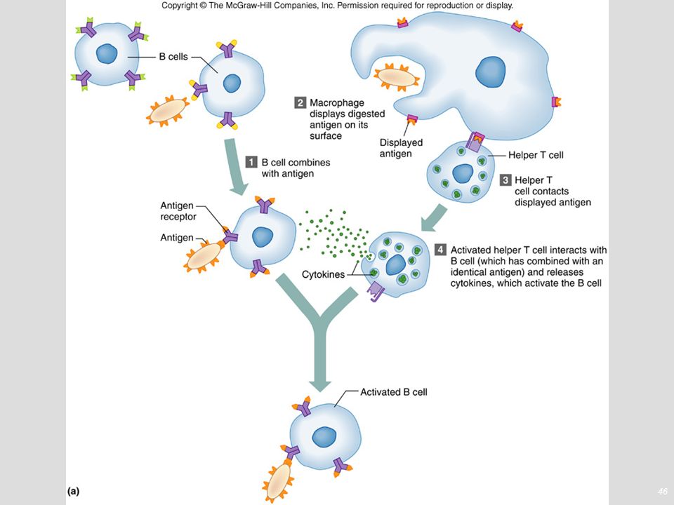
B Cell Activation, Stimulation and Proliferation

48
B Cell Proliferation and Differentiation

49
Activation, Proliferation, and Differentiation of T Cells
T cell receptors recognize antigen fragments associated with MHC molecules on the surface of a body cell. Proliferation of T cells requires costimulation, by cytokines such as interleukin-1 (IL-1) and interleukin-2 (IL-2), or by pairs of plasma membrane molecules, one on the surface of the T cell and a second on the surface of an APC.
 and interleukin-2 (IL-2), or by pairs of plasma membrane molecules, one on the surface of the T cell and a second on the surface of an APC.")
50
Activation, Proliferation & Differentiation of Cytotoxic T Cells
Receptor on CD8 cell binds to foreign antigen fragment part of MHC-I Costimulation from helper T cell prevents accidental immune response Proliferates & differentiates into population (clone) of Tc cells and memory Tc cells Occurs in secondary lymphatic organs such as lymph node
 of Tc cells and memory Tc cells. Occurs in secondary lymphatic organs such as lymph node.")
51
Activation, Proliferation & Differentiation of Helper T Cells
Receptor on CD4 cell binds to foreign antigen fragment associated with MHC-II Costimulation with interleukin Proliferates & differentiates into population (clone) of TH cells and long-lived memory TH cells
 of TH cells and long-lived memory TH cells.")
52
Overview of Mature T Cells
Helper T (TH) cells, or T4 cells, display CD4 protein, recognize antigen fragments associated with MHC-II molecules, and secrete several cytokines, most important, interleukin-2, which acts as a costimulator for other helper T cells, cytotoxic T cells, and B cells Cytotoxic T (TC) cells, or T8 cells, develop from T cells that display CD8 protein and recognize antigen fragments associated with MHC-I molecules. Memory T cells are programmed to recognize the original invading antigen, allowing initiation of a much swifter reaction should the pathogen invade the body at a later date.
 cells, or T4 cells, display CD4 protein, recognize antigen fragments associated with MHC-II molecules, and secrete several cytokines, most important, interleukin-2, which acts as a costimulator for other helper T cells, cytotoxic T cells, and B cells. Cytotoxic T (TC) cells, or T8 cells, develop from T cells that display CD8 protein and recognize antigen fragments associated with MHC-I molecules. Memory T cells are programmed to recognize the original invading antigen, allowing initiation of a much swifter reaction should the pathogen invade the body at a later date.")
53
Steps in Antibody Production

54
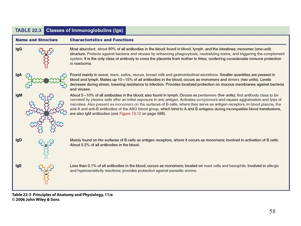
Antibody Structure Antibodies consist of heavy and light chains and variable and constant portions Based on chemistry and structure, antibodies are grouped into five principal classes each with specific biological roles (IgG, IgA, IgM, IgD, and IgE).
.")
55
Antibody Molecules

56
Types of Immunoglobulins

57
Antibody Actions

59
Antibody Actions Neutralization of antigen by blocking effects of toxins or preventing its attachment to body cells Immobilize bacteria by attacking cilia/flagella Agglutinate & precipitate antigens by cross-linking them causing clumping & precipitation Complement activation Enhancing phagocytosis through precipitation, complement activation or opsonization (coating with special substance)
")
60
Immune Responses

61
Classifications of Immunity

62
Allergic Reactions Immune attacks against nonharmful substances that can damage tissues

63
Allergic Reactions Type I immediate-reaction allergy
occurs minutes after contact with allergen hives hay fever asthma eczema gastric disturbances anaphylactic shock

64
Allergic Reactions Type II antibody-dependent cytotoxic reaction
takes 1-3 hours to develop transfusion reaction Type III immune-complex reaction takes 1-3 hours to develop antibody complexes cannot be cleared from body damage of body tissues

65
Allergic Reactions Type IV delayed-reaction allergy
results from repeated exposure to allergen eruptions and inflammation of the skin takes about 48 hours to occur

66
Transplantation and Tissue Rejection
Tissue rejection reaction resembles cellular immune response against antigens important to match MHC antigens immunosuppressive drugs used to prevent rejection Transplanted tissues and organs cornea kidney liver pancreas heart bone marrow skin

67
Rejection mechanisms Rejection is an adaptive immune response and is mediated through both T cell mediated and humoral immune (antibodies) mechanisms. The number of mismatched alleles determines the speed and magnitude of the rejection response. Different grafts usually have a proclivity to a certain mechanism of rejection.
 mechanisms. The number of mismatched alleles determines the speed and magnitude of the rejection response. Different grafts usually have a proclivity to a certain mechanism of rejection.")
68
Transplant Rejection Organ/tissue Mechanism
Blood Antibodies, (iso-hemagglutinins) IgM Kidney Antibodies, CMI Heart Antibodies, CMI Skin, CMI Bone marrow, CMI Cornea Usually accepted unless vascularized, CMI
 IgM. Kidney Antibodies, CMI. Heart Antibodies, CMI. Skin, CMI. Bone marrow, CMI. Cornea Usually accepted unless vascularized, CMI.")
69
Autoimmunity inability to distinguish “self” from “non-self”

Similar presentations