Download presentation
Presentation is loading. Please wait.

1
Hip, Pelvis and Thigh : Anatomy, Evaluation

2
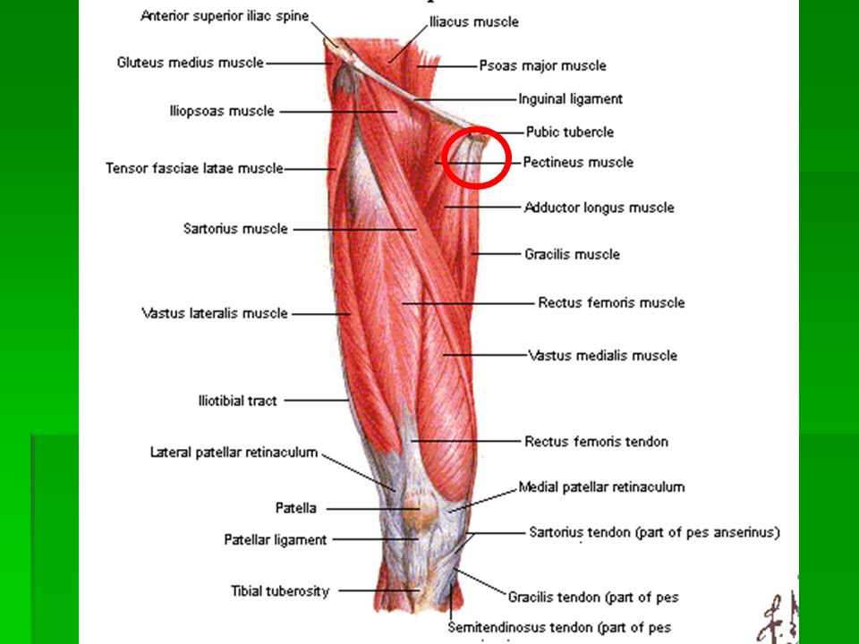
BONY ANATOMY

6
Hip Capsule Ligaments Iliopsoas bursa

10
Hip - Anatomy Multiaxial ball & socket joint Acetabulum 1/2 sphere Femoral head 2/3 sphere Strong ligaments & capsule Maximally stable

14
Observation Gait Posture Balance Limb position shortened, adducted, medially rotated abducted, laterally rotated shortened, laterally rotated Leg shortening

15
Inspection Pelvic unleveling (iliac crest levels) Pelvic rotation (PSIS levels) If asymmetric, measure leg lengths
 Pelvic rotation (PSIS levels) If asymmetric, measure leg lengths")
16
Leg Length Measurements Eyeball method Measurement method

18
Range of Motion Flexion: 110 to 120 degrees Extension: 10 to 15 degrees

19
Abduction: 30 to 50 degrees Adduction: 30 degrees

20
External rotation: 40 to 60 degrees Internal rotation: 30 to 40 degrees

21
Examination Strength testing isometric eccentric knee extension knee flexion

22
Hip Flexion Strength Iliopsoas, rectus femoris, sartorius, tensor fascia lata, pectineus

23
Hip Extension Strength Hamstrings, gluteus maximus

24
Hip Adduction Strength Adductor longus, adductor brevis, adductor magnus, gracilis, pectineus, oburator externus

25
Hip Abduction Testing Gluteus medius, gluteus minimus, tensor fascia lata

26
Internal Rotation Strength Gluteus medius, gluteus minimus, tensor fascia lata

27
External Rotation Strength Piriformis, Obturator internus & externus, Superior/inferior Gemelli, Quadratus femoris, Gluteus maximus

28
Special Tests Patrick’s Test (FAbER) hip joint SI joint
 hip joint SI joint")
29
Gaenslen’s Sign Pain at ipsilateral SIJ is positive test

30
Special Tests modified Thomas Test hip flexor and quad flexibility

31
Special Tests Ober Test iliotibial band flexibility

32
Special Tests Piriformis Test Piriformis flexibility or pain

33
Special Tests Popliteal Angle Hamstring flexibilty

35
Special Tests Labral Injury FAdAxL: flexion, Adduction, Axial Load + some IR/ER pain +/- click

36
Weber-Barstow Maneuver *Can measure true vs. apparent

37
Gross Leg Length Discrepancy Magee 4 th Edition – pg. 628

38
Prone Knee Flexion Test for Tibial Shortening Magee 4 th Edition - pg. 630

39
Thomas Test 3 Muscle Kendall test As above….but also look at…. IP = hip flexor and hip ER RF = hip flexor and knee extensor TFL/ITB = hip flexor and hip abductor Magee - 4 th Edition

40
Ely’s Test Prone, passive knee flexion Positive for RF tightness if pelvic anterior tilting / hip flexion accompanies knee flexion before end range and if asymmetrical in bilateral comparison Magee 4 th Edition

41
FAIR Test Cleland, J. – Orthopedic Clinical Examination Fishman et. al (2002) Archives of Physical Medicine – 10 yr. Piriformis study Sen..88 Spec..83 +LR= 5.2 -LR=.14 (+) = pain at intersection of sciatic nerve and piriformis
 Archives of Physical Medicine – 10 yr. Piriformis study Sen..88 Spec..83 +LR= 5.2 -LR=.14 (+) = pain at intersection of sciatic nerve and piriformis.")
42
Ober Test Magee 4 th Edition – pg. 633 Reese and Bandy (2003) JOSPT Ober Test Modified Ober Test (4-5 0 > Ober test) Ober ICC=.90 Modified Ober ICC=.91
 JOSPT Ober Test Modified Ober Test (4-5 0 > Ober test) Ober ICC=.90 Modified Ober ICC=.91.")
43
Leg Length Tests True Leg Length Discrepancy Measure ASIS to medial malleolus Positive = 1-1.5 cm Apparent (Functional) Leg Length Umbilicus to Medial malleolus
 Leg Length Umbilicus to Medial malleolus")
44
Trendelenberg Test Pt Position = standing on one leg with WB leg being the involved limb Positive = pelvis on opposite side drops Indications = weak Gluteua medius

45
Kendall Test Pt Position = supine with knees bent over the table Evaluation One hand under lordotic curve Passively flex hip to chest Allow opposite leg to rest on table Positive = knee move into extension or leg rises off table

46
Thomas Test Pt Position = supine with both leg on table Evaluation One hand under lumbar region Passively flex one leg to chest Positive = straight leg raises off table Increased lordotic curve

47
Measurements True leg length Measure from A.S.I.S to inferior border of medial malleolus

48
Measurements True Shortening In true shortening the affected limb is physically shorter than the other and this may be caused by pathology proximal or distal to the trochanters. True shortening from causes distal to the trochanters most frequently results from previous fractures of the femur or tibia or growth disturbance (e.g. from polio or epiphyseal trauma). Proximal to the trochanters causes include femoral neck fractures, OA and hip dislocation.
. Proximal to the trochanters causes include femoral neck fractures, OA and hip dislocation..")
49
Measurements Apparent leg length Measure from tip of xiphoid process to inferior border of medial malleolus Apparent Shortening In apparent shortening the limb is not altered in length, but appears shortened. This may be as a result of an adduction contracture of the hip joint, which has to be compensated for by tilting of the pelvis, or SIJ pathology causing pelvic rotation.

50
Movement Expected Range of Movement Flexion:0-130 Degrees Abduction:0-45 Degrees Adduction:0-30 Degrees MR:0-45 Degrees LR:0-60 Degrees Extension:0-20 Degrees

51
Movements Thomas’ test: Place your left hand in hollow of lumbar spine Flex hip and knee of unaffected side Look to see if hip of the affected side lifts from bed Flexion: Flex hip and knee of affected side and note ROM (130°)
")
52
Movements Abduction: Stabilise pelvis and hold ankle with other hand Abduct and note ROM (45°) Adduction: As above and note ROM (30°)
 Adduction: As above and note ROM (30°)")
53
Movements Rotation: Flex hip and knee to 90 degrees, externally and internally rotate Note ROM (45°) Abnormal Movement (telescoping): Alternately push and pull leg along its long axis – demonstrates marked instability
 Abnormal Movement (telescoping): Alternately push and pull leg along its long axis – demonstrates marked instability")
54
Trendelenberg Test Used to assess the ability of the hip abductors to stabilise the pelvis on the femur. A positive test demonstrates that the hip abductors are not functioning. Causes: Disturbance in pivotal mechanism – dislocation or subluxation of hip, shortening of femoral neck Weakness of the hip abductors e.g. myopathy, neuropathy

55
Trendelenberg Test The test is performed with the patients back to the examiner. The model stands on the normal leg and flexes the knee of the other leg to a right angle. The pelvis should remain level or tilt slightly upwards on the unsupported side. The model then stands on the affected leg and flexes the knee of the other leg. If the pelvis tilts downwards on the unsupported side, then this confirms a positive Trendelenberg sign.

56
Trendelenberg Test

Similar presentations


>")


