Download presentation
Presentation is loading. Please wait.

1
PTA 106 Unit 2 Lecture 1

2
Position of the heart and Associated Structures Coronary trivia Pumps blood through 60,000 miles of blood vessels Pumps about 3,600 gal per day 2.6 million gal per year

3
Approximate Location of the heart projected to the surface Landmarks Superior R point: Is at the superior border of the R 3 rd costal cartilage Superior L point: Is located at the inferior border of the L 2 nd costal cartilage Inferior L point: (the apex) is located at of the heart in the L 5 th intercostal space Inferior R point: Is located at the superior border of the sixth R costal catilage
 is located at of the heart in the L 5 th intercostal space Inferior R point: Is located at the superior border of the sixth R costal catilage")
4
Layers of the heart wall and it’s associated membranes

5
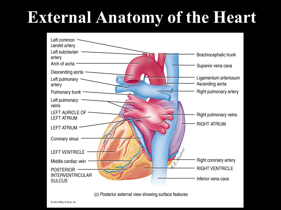
External Anatomy of the Heart

7
Internal Anatomy of the Heart

8
Position and Function of the Cardiac valves

9
Circulations Patterns of the Heart

10
Coronary Vessels and circulation

11
Cardiac Conduction Systems: the Heart pacemaker

12
Physiology of Cardiac Muscle Contraction 1.Action potential initiated by the SA node 2.Action potential conducted to the purkinje fibers 3.Depolarization of sarcolemma opens voltage-gated fast Na+ channels causing rapid depolarization 4.Prolonged depolarization called the “plateau” involves opening of voltage-gated slow Ca2+ channels

13
Physiology of Cardiac Muscle Contraction 5.Repolarization is caused by opening of voltage-gated K+ channels 6.The prolonged depolarization causes an absolute refractory period where the cardiac muscle can not respond to additional stimulus.

14
The parts of an Electrocardiogram (EKG) during a cardiac cycle P wave = atrial depolarization (Large P = atrial enlargement) QRS complex = ventricular depolarization (Large Q = myocardial infarction) T Wave = ventricular repolarization (Flat T = coronary artery disease) P-Q interval = Time required for conduction from SA node to purkinje fibers
 during a cardiac cycle P wave = atrial depolarization (Large P = atrial enlargement) QRS complex = ventricular depolarization (Large Q = myocardial infarction) T Wave = ventricular repolarization (Flat T = coronary artery disease) P-Q interval = Time required for conduction from SA node to purkinje fibers")
15
The parts of an Electrocardiogram (EKG) during a cardiac cycle S-T segment = Time when ventricular myocardia is depolarized (elevated S-T indicates acute myocardial infraction} Q-T interval= time form start of ventricular depolarization to ventricular repolarization. (Lengthened by myocardial damage)
.")
16
The Cardiac Cycle: Atrial Systole Atrial Diastole Ventricular fillling Ventricular Ejection Ventricular Systole Ventricular Diastole Isovolumetric Contraction Isovolumetric Relaxation

17
The Cardiac Cycle: End-diastolic volume End-systolic volume

18
Cardiac Output (CO) CO = volume of blood ejected from the left ventricle into the Aorta each minute. CO = SV x HR SV = stroke volume, volume of blood ejected from ventricle (70 ml) HR = Heart rate, heartbeats per minute
 HR = Heart rate, heartbeats per minute.")
19
Cardiac Output (CO) Factors the effect SV 1. Preload: degree of stretch of the myocardium before contraction 2. Contractility: force of contraction of the ventricular myocardium 3. Afterload: Force or pressure that the ventricular myocardium must exceeded to open the semilunar valves.

20
Nervous Control of Cardiac Activity

Similar presentations


 –Contracts.>")


