Download presentation
Presentation is loading. Please wait.

1
Chronic inflammation - If damaging stimulus is of short duration & does not persist the : - Tissue damage - Acute inflammation - Exudate - Organization of exudate - Granulation tissue - Fibrous scar

2
- If damage stimulus persists the process of continuing tissues necrosis , organization & repair all occur concurrently = chronic inflammation - Acute inflammation , immune system activated around the area of damage & tissues Infiltrated by activated lymphoid cells ; persists until damaging stimulus removed or neutralized -Histology : necrotic cell debris , acute inflammatory exudate , vascular & fibrous granulation tissue , lymphoid cells , macrophages & collagenous scar.

3
- The result of a balance between continuing tissue damage on the one hand & eradication of the damaging stimulus followed by healing & scar formation on the other - If the damaging stimulus eradicated or neutralized then further tissue necrosis does not occur & the repair response progresses to complete scarring - If the damaging stimulus cannot be eradicated or neutralized the balance between tissue damage & tissue repair is maintained in a stalemate & thus chronic inflammation will persist , often for years.

4
Example- chronic peptic ulcer
- Protective mechanism of upper alimentary track breaks down - HCL & proteolytic enzymes destroy epithelium & supporting stroma ulceration of the wall stomach or duodenum persistent damage - Acute inflammatory reaction occurs : formation of exudate close to the acid exposed surface while in the depths of the ulcer( farthest from the acid) attempts are made to organize the exudate & granulation tissue forms collagenous scar - Establishment ulcer has all these processes occuring simultaneously - Treatment aim = remove or greatly reduce acid & enzymes secreted Perforate , heal or persist
 attempts are made to organize the exudate & granulation tissue forms collagenous scar. - Establishment ulcer has all these processes occuring simultaneously. - Treatment aim = remove or greatly reduce acid & enzymes secreted Perforate , heal or persist.")
5
In chronic inflammation : - Key effector cells are lymphoid cells & macrophages ( tissue – based immune response to the damaging agent - Macrophages – phagocytic & activated to fulfill other immunological & secretory functions - Chronic inflammatory cells = lymphocytes , plasma cells , & macrophages

8
Macrophages : - Converted from inactive monocytes by trophic signals ( e.g gamma interferon) - Morphology changes with a subcellular apparatus to synthesize protein - voluminous cytoplasm = epitheloid cell - Fusion of activated macrophages multinucleate histiocytic giant cells - Now have phagocytic & secretory functions against injurious agents & impt. In cell-mediated immunity ( e.g antigen presentation)
 .")
9
Macrophages secrete : - Mediators of acute inflammation
- Platelet activating factor & arachidonic acid metabolites - Highly reactive oxygen metabolites - Bacterial & cell killing - Proteases & hydrolytic enzymes - Dissolution of extracellular matrix - Cytokines Il-1 & TNF alpha - Fibroblast proliferation & collagen synthesis –repair - Growth factors ( PDGF,EGF,FGF) - Stimulate growth of B.Vs & division /migration of fibroblasts
 - Stimulate growth of B.Vs & division /migration of fibroblasts.")
11
Chronic inflammation – Key facts : - Predisposed by factors that prevent elimination of a damaging stimulus - Tissue damaging , acute inflammation , granulation tissue , repair , & immune response all take place concurrently - Infiltration by lymphocytes demonstrates immune response in tissues - May develop after acute inflammation or may be a primary response to certain stimuli - Predisposing factors include persistent damaging stimulus , inadequate host response to infection , & persistent autoimmune disease

12
Granulomatous inflammatory reactions - In certain disease , acute inflammatory response dominated by neutrophils is transient & quickly replaced by immune-based cellular reaction w/aggregation of lymphocytes & macrophages - Granulomas = discrete clusters of macrophages & a pattern of this is granulomatous inflammation

13
- examples of damaging stimuli that provoke granulomatous inflammatory reaction :
- Microorganisms of low inherent pathogenicity but excite an immune response : Ex. Mycobacteria intracellular pathogens with resistant lipoprotein coating to the cell membrane ( TB , leprosy) . - Non-living foreign material deposited in tissues ( e.g. Keratin , urate crystals , inhaled inorganic dust ) , cannot be destroyed by neutrophil enzymes. - Certain fungi ; cannot be death with neutrophils - Unknown factors ( e.g. Sarcoidosis)
 . - Non-living foreign material deposited in tissues ( e.g. Keratin , urate crystals , inhaled inorganic dust ) , cannot be destroyed by neutrophil enzymes. - Certain fungi ; cannot be death with neutrophils. - Unknown factors ( e.g. Sarcoidosis)")
14
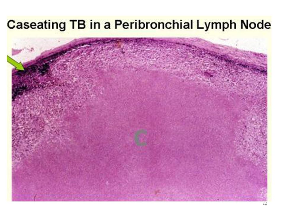
1) Tuberculosis – type IV hypersensitivity reaction
- Granulomatous inflammation example - M. tuberculosis inhaled into alveolar spaces of lung ; other tissues affected transient marked immune – mediated response - T-cell produce cytokines - Neutrophils cannot degrade cell wall so acute inflammation becomes chronic inflammation Cytokines aggregate macrophages ( granulomas called tubercles) central caseous necrosis ( ricotta cheese appearance)
 central caseous necrosis ( ricotta cheese appearance)")
15
Tuberculous granuloma :
- Central necrosis surrounded by a collection of large , activated macrophages that have a minimal resemblance to epithelial cells & thus termed epithelioid cells. - Langhans giant cells = activated macrophages that fuse to form large multinucleated cells ; many nuclei around the periphery & a large central cytoplasmic mass - Outside & around the macrophages is a collar border of lymphocytes - Over time some fibroblasts appear in the lymphocytic collar & outside it ( macrophage cytokine recruitment)
")
17
Tuberculous granuloma ; - Outcome of tubercles depends on balance of between conflict of extension of infection Vs. Containment /healing/eradication - Factors predisposing to extension : - ingestion of large numbers of highly virulent organisms - Poor immune response ( malnutrition , extreme youth or old age , intercurrent disease , immunosuppressive therapy) - Factors to containment / eradication : - Ingestion of small numbers of poorly virulent organisms - Good immune response ( health , immunization ) - Antibiotic administration
 - Factors to containment / eradication : - Ingestion of small numbers of poorly virulent organisms - Good immune response ( health , immunization ) - Antibiotic administration .")
18
Tuberculosis : - Most severe in patients with poor natural immunity ( e.g. , malnourished , poor ) or immunosuppressed via AIDS or iatrogenically ( e.g. , transplant patients) - New miliary tuberculosis strains are resistant to formerly successful medications. - Three pulmonary tissue patterns ( host immunity dependent ) - Primary – no previous exposure - Secondary – previously exposed , sensitized to organism - Exposure , but immunosuppression primary
 or immunosuppressed via AIDS or iatrogenically ( e.g. , transplant patients) - New miliary tuberculosis strains are resistant to formerly successful medications. - Three pulmonary tissue patterns ( host immunity dependent ) - Primary – no previous exposure - Secondary – previously exposed , sensitized to organism - Exposure , but immunosuppression primary .")
19
Primary tuberculosis :
- Organism inhaled , proliferate in the lung alveoli at periphery of lung , just below pleura Ghon focus - Organism causes cell death in adjacent lung - Ineffective acute inflammatory response with bacterial organism not destroyed Bacteria then conveyed to local nodes at lung hilum proliferate weeks , immune response develops macrophages recruited by T-cells to lung & nodes granulomatous inflammation & caseation : - Often no clinical symptoms - Outcome depends on balance of host response & virulence of organisms .

20
-In primary tuberculosis vast majority of Ghon focus & caseating granulomas in lymph nodes ( i.e. primary complex) heal by deposition of collagen around the tubercles centrally yellowish caseation surrounded by wall of dense collagen with calcium salts deposited , in both , at times. - In primary T.B ; once immune system exposed to organism then patient has been sensitized & the disease does not progress as it is walled off by the dense collagen but viable organisms may remain walled off within the healed primary complex ( latent T.B.)
")
21
- in primary T.B. ; if patient is not able to mount a vigorous immune & reparative response then further spread of organism occurs with continuing enlargement of caseating granulomas in the lymph nodes ( progressive primary TB) - Enlarging nodes erode through bronchus wall or into thin-walled blood vessel - Ghon focus usually remains small although rarely it may rupture through the visceral pleura discharge organisms into pleural cavity tuberculosis pleurisy
 - Enlarging nodes erode through bronchus wall or into thin-walled blood vessel - Ghon focus usually remains small although rarely it may rupture through the visceral pleura discharge organisms into pleural cavity tuberculosis pleurisy .")
23
Primary T.B : - Tuberculous caseous material with live tubercle bacilli pass down bronchi & bronchioles via gravity furthest reaches of the lung confluent caseating granulomatous lesions ( galloping consumption) rapidly fatal. -Tuberculous caseous material with live tubercle bacilli erode blood vessel wall blood stream …….many parts of the body ( miliary T.B – kidney , liver , spleen) , if into pulmonary artery remainder of lung.
 , if into pulmonary artery remainder of lung.")
25
Secondary T.B.: - Organisms acquired exogenously or from heald primary tuberculosis - Assmann focus : an apical lung lesion that begins as a small caseating tuberculous granuloma. - Histopathology is similar to Ghon focus with central caseous necrosis surrounded by granulomatous inflammatory response - Usually destruction of lung cavitation - Little involvement of lymph nodes due to vigorous tissue-based hypersensitivity response - Therefore , outcome of infection depends entirely on what happens to Assmann focus

26
- If vigorous immune response then healing of the apical lesion via exact process as for Ghon focus so containment of the infection with no further spread of the organism - But , if fibrous wall breaks down then latent TB can lead to spreading infection at a much later stage ( reactivated fibrocaseous TB) - if poor immune response then progressive enlargement of apical lesion with caseous necrosis destroying lung tissue ……large caseous mass surrounded by thin cellular reaction with little collagen wall ( progressive pulmonary TB) with increase risk of erosion into blood vessels or airways. - Release of tubercle bacilli into main bronchi allow them to be coughed into the atmosphere as droplets…..transmission to other people( open TB) & producing TB bronchopneumonia ( passage down to lower lobes)
 & producing TB bronchopneumonia ( passage down to lower lobes) .")
28
Metastatic ( isolated organ) TB : - Only small number of tubercle bacilli escape into blood & if host defense effective then most die but some can settle in specific organ & remain dormant there for years : - Especially in adrenal glands , kidney , fallopian tubes , epididymis , brain & meninges , & bones/joints - Can proliferate & produce overt disease at later date
 TB : - Only small number of tubercle bacilli escape into blood & if host defense effective then most die but some can settle in specific organ & remain dormant there for years : - Especially in adrenal glands , kidney , fallopian tubes , epididymis , brain & meninges , & bones/joints - Can proliferate & produce overt disease at later date")
29
Reactivated pulmonary TB ; - After years of primary or secondary walled off focus with immune defense waning the organisms escape into adjacent lung …………….proliferate rapidly ………..tissue necrosis ……new caseation & granulomatous inflammation ……rapid spread ……miliary TB or bronchopneumonia TB & commonly fatal

30
TB facts : - Caused by Mycobacterium tuberculosis
- Induces type IV hypersensitivity response - Histopathology hallmarks is caseating granulomatous inflammation -Main site of infection is in lungs - Lung infection in childhood comprises Ghon focus & nodal disease ( primary complex) - Infection in adult life causes Assman focus - Blood-borne spread lead to miliary TB - Bronchial spread leads to tuberculous bronchopneumonia - Reactivation of disease may take place in later life if host response is weakened.
 - Infection in adult life causes Assman focus. - Blood-borne spread lead to miliary TB. - Bronchial spread leads to tuberculous bronchopneumonia. - Reactivation of disease may take place in later life if host response is weakened.")
31
Ghon Complex

32
Cells in Granuloma

33
Tuberculous Granuloma

34
Epitheloid cells in Granuloma

35
Caseation Necrosis

36
Typical cavitating granuloma

37
Primary or Ghon’s Complex
Primary tuberculosis is the pattern seen with initial infection with tuberculosis in children. • Reactivation, or secondary tuberculosis, is more typically seen in adults

38
Cavitary Tuberculosis
When necrotic tissue is coughed up cavity. • Cavitation is typical for large granulomas. • Cavitation is more common in the secondary reactivation tuberculosis - upper lobes.

39
Cavitary Secondary TB

40
Lung TB -Cavitation

41
Systemic Miliary TB

42
Adrenal TB -Addison Disease

43
Testes TB Orchitis.

44
TB Peritonitis + liver Miliary TB

45
TB Brain – Caudate n.

46
TB Intestine

47
Prostate TB

48
Spinal TB -Potts Disease

49
Chronic inflammation : other mycobacterium diseases
- M. avium -intracellular : macrophage laden with organism in many organs or caseating granuloma of lung ; AIDS patients. - Chronic inflammatory granulomatous response with little induction of acute inflammatory response : - Leprosy (M . leprae) – chronic , indolent mainly skin ; granulomas may form( Tuberculous form) or if immunity low then intracellular proliferation in phagocytic cells ( lepromatous form) - Bovine TB – cervical neck nodes infected due to drinking infected cow,s milk - M. marinum – chronic skin lesions usually hands - M. scrofulaceum – enlarged lymph nodes of the neck
 – chronic , indolent mainly skin ; granulomas may form( Tuberculous form) or if immunity low then intracellular proliferation in phagocytic cells ( lepromatous form) - Bovine TB – cervical neck nodes infected due to drinking infected cow,s milk. - M. marinum – chronic skin lesions usually hands. - M. scrofulaceum – enlarged lymph nodes of the neck.")
50
Other causes of granulomatous inflammation :
2- foreign material ( organic or inorganic ) introduced into tissue commonly excites a predominantly macrophagic reaction ; neutrophils are unable to phagocytize & destroy the material …….. - Granulomatous aggregates with giant cells form around foreign material or irregular , ill-defined : - Endogenous : Keratin , urate crystals ,degenerated altered collagen , degenerated altered elastin ( artery walls) - Exogenous : sutures , talcum powder , vegetable matter
 introduced into tissue commonly excites a predominantly macrophagic reaction ; neutrophils are unable to phagocytize & destroy the material …….. - Granulomatous aggregates with giant cells form around foreign material or irregular , ill-defined : - Endogenous : Keratin , urate crystals ,degenerated. altered collagen , degenerated altered elastin ( artery walls) - Exogenous : sutures , talcum powder , vegetable matter.")
52
3 - Sarcoidosis : Other causes of granulomatous inflammation :
- Etiology unknown - Clinical : - Lungs & lymph node enlargement – can result in pulmonary fibrosis - Slowly progressive but often bums out - Discrete granulomas with histiocytic giant cells mainly in lymph nodes , lungs ,liver , spleen & skin ( rarely brain , bone) - Granulomas are multiple , slowly increase in size often becoming confluent - Histopathology resembles TB although no true caseation ; giant cells may have laminated calcific spherical concretions ( Schaumann bodies ) , or stellate shapes ( asteroid bodies)
 - Granulomas are multiple , slowly increase in size often becoming confluent. - Histopathology resembles TB although no true caseation ; giant cells may have laminated calcific spherical concretions ( Schaumann bodies ) , or stellate shapes ( asteroid bodies)")
53
4-Rhinoscleroma - A chronic granulamotus disease caused by Klebsiella rhinoscleromatous. - It affect the mucus membrane of nose, tonsils, pharynx, and larynx. Microscopic picture: - Squamous metaplasia, - Vascular CT core with chronic inflammatory cells and fibroblasts. - Mickulicz cells : degenerated Histocytes - Russell bodies: defective Plasma cells

54
Macroscopic picture: Effects:-
The lesion starts as diffuse thickening or small nodule, Gradually increased in size and infiltrate the surroundings. Effects:- - Ulceration - Obstruction - Pre-cancerous

55
5 - Actinomycosis It is a chronic suppurative granulomatous disease caused by a fungus called actinomyces bovis (or gram positive anaerobic bacilli). Tissue reaction: - Chronic inflammatory reaction - Multiple abscesses filled with pus ( yellowish pus called sulpher granules ) , and colonies of organisms. Sites : Cervico – facial – Ileocaecal - Pulmonary
 , and colonies of organisms. Sites : Cervico – facial – Ileocaecal - Pulmonary.")
56
Infection

57
Infection means tissue invasion by pathogenic organism .
The result of infection depends on : - Dose & virulence of organism - Body resistance ( immunity ) If the defense mechanisms failed to localized infection , organisms or their toxins reach the circulation & produces their specific manifestations.
 If the defense mechanisms failed to localized infection , organisms or their toxins reach the circulation & produces their specific manifestations.")
58
Pathogenesis Immunity Infection

59
Toxaemias - Toxaemia means circulation of bacterial toxins in the blood with production of clinical and pathological manifestations. - It could be acute if a large doses of toxins reaches the blood within a short time ; e.g Pneumonia , diphtheria & cholera. - Or could be chronic if a small doses of toxins reaches the blood within a long time ; e.g .TB

60
Clinical manifestations of toxaemia
Affect the brain: fever, headache, body aches, rigors, tachycardia. -Affect the kidney: tubular necrosis, acute renal failure, adrenal necrosis. Affect the heart: toxic myocarditis. Affect the liver: fatty liver Peripheral neuritis Toxic shock - Amyloidosis especially in chronic toxaemia.

61
Bacteraemia - Its effect depends on:-
- It means circulation of bacteria from aseptic focus ( sinusitis , otitis media , tonsillitis , .. ) , and in early stages of some diseases e.g. typhoid fever. - Its effect depends on:- - Body resistance : activity of RES - Dose and virulence of the organisms : large doses or highly virulence organisms could causes serious diseases such as ; subacute bacterial endocarditis , pyelonephritis , osteomyelitis.
 , and in early stages of some diseases e.g. typhoid fever. - Its effect depends on:- - Body resistance : activity of RES. - Dose and virulence of the organisms : large doses or highly virulence organisms could causes serious diseases such as ; subacute bacterial endocarditis , pyelonephritis , osteomyelitis.")
62
Septicemia - The causes include:- - Lowering of the body resistance
- It means multiplication of bacteria and their toxins with production of sever clinical manifestations: - The causes include:- - Lowering of the body resistance - Virulent pyogenic organisms e.g. - Gonococci in meningitis - Staph aureus in osteomyelitis - Pneumococci in labor pneumonia - Hemolytic streptococci in puerperal sepsis

63
Manifestations of septicemia
- Fever, headache, body aches, malaise,.. - Petechial hemorrhage in skin and mucus membranes due to toxic capillaries. - Fatty changes of the liver - Hemolysis - Acute bacterial endocarditis - Toxic myocarditis

64
Pyaemia - It means production of multiple small abscesses in different organs due to circulation and impaction of septic emboli started from aseptic focus. - According to the site of the original septic focus ; pyaemia can be classified into : Systemic or Portal

65
- The abscesses are formed primary in the lungs.
A -Systemic pyaemia - The abscesses are formed primary in the lungs. - Causes: any purulent inflammation in areas drained by systemic venous circulation; e.g. : - Acute bacterial endocarditis - Carbuncles and abscesses - Osteomyelitis - Face suppuration

66
B - Portal pyaemia -The abscesses are primary formed in the liver - Causes : any purulent inflammation in areas drained by portal venous circulation ; e.g .: - Acute suppurative appendicitis - Septic ulcer in GIT - Suppurative cholecystitis - Septic thrombosed piles

Similar presentations


 INFLAMMATION>")

 Environmental.>")


, calor (heat), tumor (swelling), dolor (pain), and loss of function.>")


 System. Lymph Fluid in the tissue spaces that carries protein molecules and other substances back into the blood.>")
