Download presentation
Presentation is loading. Please wait.

1
SUPPURATIVE LUNG DISEASES
Bronchiectasis

2
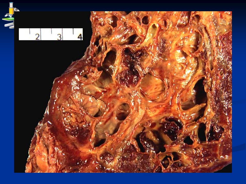
* Def: persistent dilatation of medium sized bronchi accompanied by suppurative inflammation of their walls. * Etio-pathogenesis: I. Weakening of the bronchial wall by; a. Chronic suppurative inflammation: due to recurrent septic bronchopneumonia. b. Congenital weakness: leads to congenital bronchiectasis. II. Bronchial obstruction: by foreign body, bronchial secretion or tumor.

3
- The surrounding alveoli are: fibrotic and collapsed.
* N/E: - Dilated bronchi: Cylindrical, fusiform or saccular Bilateral. Basal. Has patchy distribution. The bronchial lumen contains pus. The bronchial mucosa: ulceratd. - The surrounding alveoli are: fibrotic and collapsed. - The pleura shows: pleurisy - Draining hilar L. nodes: enlarged

5
* Complications: Spread of infection: direct, lymphatic and blood.
Hemoptysis. Lung abscess (post-bronchiectatic lung abscess). 2ry amyloidosis. Lung fibrosis. Bronchogenic carcinoma (squamous cell carcinoma).
. 2ry amyloidosis. Lung fibrosis. Bronchogenic carcinoma (squamous cell carcinoma).")
6
PNEUMONITIS

7
* Classification: 1. Bacterial pneumonia: lobar pneumonia & bronchopneumonia. 2. Viral (interstitial) pneumonia: influenza, measles, chicken pox. 3. Loeffler’s (parasitic) pneumonia: Bilharziasis, ascaris & ankylostomiasis. 4. Granulomatous pneumonia: T.B, sarcoidosis, leprosy, syphilis, actinomycosis . 5. Lipoid pneumonia: due to aspiration of oily nasal drops. 6. Irradiation pneumonia.
 pneumonia: Bilharziasis, ascaris & ankylostomiasis. 4. Granulomatous pneumonia: T.B, sarcoidosis, leprosy, syphilis, actinomycosis . 5. Lipoid pneumonia: due to aspiration of oily nasal drops. 6. Irradiation pneumonia.")
8
LOBAR PNEUMONIA * Def: acute diffuse fibrinous inflammation of one or more lung lobes. * Etiology: Age: middle age. Predisposing factors: low resistance. Causative organism: pneumococci. Route of infection: droplet infection.

9
* Pathogenesis: Pneumococci are inhaled to reach alveoli. They cause acute inflammaion with excess fluid exudate. This fluid exudate pass from one alveolus to another rapidly through the inter- alveolar pores of cohn to involve the whole lung lobe. The fluid exudate expel air away from the alveoli producing a firm airless lobe leading to consolidation (hepatisation) of the affected lobe.
 of the affected lobe.")
10
1. Duration 2. Gross findings: the affected lobe is:
4. Stage of resolution 3. Stage of gray hepatization 2. Stage of red hepatization 1. Stage of congestion 1. Duration 9th – day 21 5th – 8th day 2nd – 4th day 1st day 2. Gross findings: the affected lobe is: - Since there is no necrosis, healing by resolution occurs. - Enlarghed. - Gray. - Consistency: consolidated (hepatized). - Cut section: dry. - Pleurisy. - Enlarged hilar LNs. - Enlarged. - Red. - Size: Enlarged. - Color: Red. - Consistency: like wet sponge. - Cut section: exudes frothy fluid. - The covering pleura: normal - The hilar L.Ns.: normal
. - Cut section: dry. - Pleurisy. - Enlarged hilar LNs. - Enlarged. - Red. - Size: Enlarged. - Color: Red. - Consistency: like wet sponge. - Cut section: exudes frothy fluid. - The covering pleura: normal. - The hilar L.Ns.: normal.")
11
3. Stage of gray hepatization 2. Stage of red hepatization
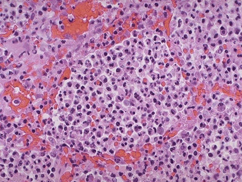
4. Stage of resolution 3. Stage of gray hepatization 2. Stage of red hepatization 1. Stage of congestion 3. M/P a. Alveolar capillariers: Less congested b. Alveolar walls: thin. c. Alveolar spaces: Show dead bacteria, shrinked fibrin, hemolysed RBCs, excess polymorphs and macrophages. Congested. b. Alveolar walls: thickened. Show bacteria, fibrin, RBCs and polymorphs. Show bacteria & fluid exudate.

12
3. Stage of gray hepatization 2. Stage of red hepatization
4. Stage of resolution 3. Stage of gray hepatization 2. Stage of red hepatization 1. Stage of congestion 4. Clinical course - Fever, cough, dyspnea and chest pain. - At about 9th day the disease ends by crisis (sudden improvement), however death may occur due to severe toxaemia.
, however death may occur due to severe toxaemia.")
13
3. Stage of gray hepatization 2. Stage of red hepatization
4. Stage of resolution 3. Stage of gray hepatization 2. Stage of red hepatization 1. Stage of congestion 5. Complications Spread of infection: direct, lymphatic and blood (toxaemia, septicaemia). Lung fibrosis due to failure of resolution. Post-pneumonic lung abscess.
. Lung fibrosis due to failure of resolution. Post-pneumonic lung abscess.")
16
BRONCHOPNEUMONIA * Def: Acute suppurative inflammation of bronchioles and adjacent alveoli characterized by patchy lung consolidation. * Etilogy: Age: extremes of age (young & elderely). Predisposing factors: low resistance and bronchitis. Causative bacteria: staphylococci, streptococci & H. influenza. Route of infection: endogenous invaders and exogenous invaders (droplet infection).
. Predisposing factors: low resistance and bronchitis. Causative bacteria: staphylococci, streptococci & H. influenza. Route of infection: endogenous invaders and exogenous invaders (droplet infection).")
17
* N/E: Bilateral. Basal. Multiple consolidated yellowish patches exuding pus on pressure. Several patches may coalesce to produce confluent bronchopneuomonia. Enlarged hilar L. nodes.

19
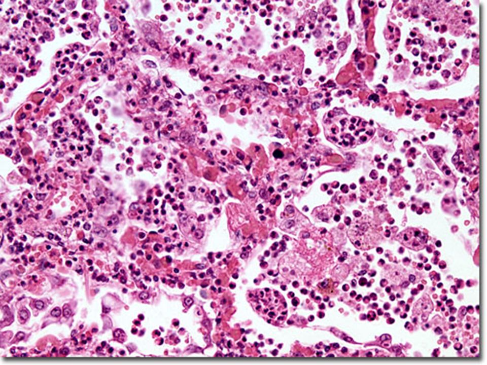
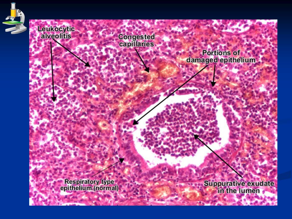
* M/P: I. The bronchioles show:
Their lumen shows: necrotic epithelial cells, polymorphs & pus cells. Their lining: ulceration. Their walls: congested capillaries, neutrophils and pus cells & exudate. II. The adjacent alveoli show: 3 successive zones: zone of alveolitis then zone of alveolar collapse and a zone of alveolar dilatation (compensatory emphysema).
.")
21
* Complications: Spread of infection: direct, lymphatic and blood (toxaemia, septicaemia). Lung fibrosis due to failure of resolution. Post-pneumonic lung abscess. Bronchiectasis.

Similar presentations

>")
 Environmental.>")


