Download presentation
Presentation is loading. Please wait.

1
HEART DISEASES Lecture I
Associate Professor Dr. Alexey Podcheko Spring 2015

2
INTENDED LEARNING OUTCOMES
CONGENITAL (CHD): 1. To know Cardiac Development 2. To know causes of CHD 3. Pathophysiology and clinical presentations of: ASD, VSD, patent ductus arteriosus (PDA) Tetralogy of Fallot Transposition of the great arteries Persistent Truncus arteriosus Tricuspid atresia Total anomalous pulmonary venous connection COARCTATION of aorta pulmonary stenosis/atresia Aortic stenosis/atresia 2
: 1. To know Cardiac Development. 2. To know causes of CHD. 3. Pathophysiology and clinical presentations of: ASD, VSD, patent ductus arteriosus (PDA) Tetralogy of Fallot. Transposition of the great arteries. Persistent Truncus arteriosus. Tricuspid atresia. Total anomalous pulmonary venous connection. COARCTATION of aorta. pulmonary stenosis/atresia. Aortic stenosis/atresia. 2.")
3
THE HEART Normal Pathology Heart Failure: L, R Heart Disease
Congenital: LR shunts, RL shunts, Obstrustive Ischemic: Angina, Infarction, Chronic Ischemia, Sudden Death Hypertensive: Left sided, Right sided Valvular: AS, MVP, Rheumatic, Infective, Non-Infective, Carcinoid, Artificial Valves Cardiomyopathy: Dilated, Hypertrophic, Restrictive, Myocarditis, Other Pericardium: Effusions, Pericarditis Tumors: Primary, Effects of Other Primaries Transplants

4
NORMAL Features 6000 L/day 250 to 300 gm in females and 300 to 350 gm in males, or roughly 0.4% to 0.5% of body weight 40% of all deaths (2x cancer) Wall thickness ~ pressure (i.e., a wall is only as thick as it has to be) -Right ventricle to 0.5 cm -Left ventricle 1.3 to 1.5 cm -Atria =.2 cm Heart weight varies with body height and weight; it normally averages approximately. The usual thickness of the free wall of the right ventricle is 0.3 to 0.5 cm, and that of the left ventricle 1.3 to 1.5 cm.
 Wall thickness ~ pressure. (i.e., a wall is only as thick as it has to be) -Right ventricle to 0.5 cm. -Left ventricle 1.3 to 1.5 cm. -Atria =.2 cm. Heart weight varies with body height and weight; it normally averages approximately. The usual thickness of the free wall of the right ventricle is 0.3 to 0.5 cm, and that of the left ventricle 1.3 to 1.5 cm.")
5
Normal Features, Starling's Law of the Heart
ability of the heart to change its force of contraction and therefore stroke volume in response to changes in venous return is called the Frank- Starling mechanism (or Starling's Law of the heart).
.")
6
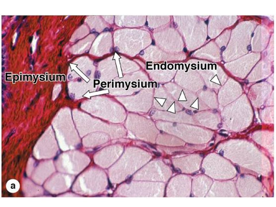
NORMAL Features The efficient pumping of blood by the heart to the entire body requires the normal function of each of its key components: 1. the myocardium 2. valves 3. conduction system 4. coronary arterial circulation

7
A B Which one is cardiac tissue?
Cardiac muscle cell has one nucleus (sometimes two). Each cardiac muscle cell is branched, forming a tissue that is appropriately described as a functional syncytium Intercalated disks junctions types : Fascia adherens Desmosomes Gap junctions (exchange of ions)
. Each cardiac muscle cell is branched, forming a tissue that is appropriately described as a functional syncytium. Intercalated disks junctions types : Fascia adherens. Desmosomes. Gap junctions (exchange of ions)")
9
CONDUCTION SYSTEM of the HEART
Needed for coordinated contraction of the cardiac muscle Presented by specialized excitatory and conducting myocytes Key components : (1) the sinoatrial (SA) pacemaker of the heart, (near the junction of the right atrial appendage and the superior vena cava) (2) the AV node (in the right atrium along the atrial septum) (3) the bundle of His (courses from the right atrium to the summit of the ventricular septum) (4) the right and left bundle branches, Left bundle brunch has anterior and posterior fascicles!!! (5) Purkinje network
 the sinoatrial (SA) pacemaker of the heart, (near the junction of the right atrial appendage and the superior vena cava) (2) the AV node (in the right atrium along the atrial septum) (3) the bundle of His (courses from the right atrium to the summit of the ventricular septum) (4) the right and left bundle branches, Left bundle brunch has anterior and posterior fascicles!!! (5) Purkinje network.")
10
S.A. NodeAV NodeBundle of HIS L. Bundle, R. Bundle
The specialized myocytes of the heart’s conduction system, running sub-endocardially, have this unique appearance. S.A. NodeAV NodeBundle of HIS L. Bundle, R. Bundle 10

11
Coronary arterial circulation
To meet their energy needs, cardiac myocytes rely almost exclusively on oxidative phosphorylation, which is manifest by the abundant mitochondria that are found in these cells cardiac myocytes extremely vulnerable to ischemia Coronary arteries: -epicardial coronary arteries: Left Coronary artery (LCA) left anterior descending (LAD) left circumflex (LCX) arteries, both arising from branches of the left (main) coronary artery 2. Right Coronary artery (RCA) Marginal Posterior descending artery (in 85% of cases) and nodal artery Most coronary arterial blood flow to the myocardium occurs during ventricular diastole
 left anterior descending (LAD) left circumflex (LCX) arteries, both arising from branches of the left (main) coronary artery. 2. Right Coronary artery (RCA) Marginal. Posterior descending artery (in 85% of cases) and nodal artery. Most coronary arterial blood flow to the myocardium occurs during ventricular diastole.")
12
Whichever artery winds up supplying the posterior interventricular septum is said to be “DOMINANT”
12

13
VALVES AV: TRICUSPID MITRAL SEMILUNAR: PULMONIC AORTIC 13

14
Valves Their function depends on the mobility, pliability, and structural integrity of their delicate flaps, called leaflets (in the tricuspid and mitral valves) or cusps (in the aortic and pulmonary valves, also known as the semilunar valves). layered architecture : 1. lamina ventricularis (tight network of reticular fibres ) 2. lamina radialis (radial orientated collagenous and elastic fibres) 3. lamina spongiosa (loosely arranged reticular fibres with bundles of collagenous and some elastic fibres) 4. lamina fibrosa (Circular arranged collagen fibres ) 5. lamina arterialis
 or cusps (in the aortic and pulmonary valves, also known as the semilunar valves). layered architecture : 1. lamina ventricularis (tight network of reticular fibres ) 2. lamina radialis (radial orientated collagenous and elastic fibres) 3. lamina spongiosa (loosely arranged reticular fibres with bundles of collagenous and some elastic fibres) 4. lamina fibrosa (Circular arranged collagen fibres ) 5. lamina arterialis.")
15
Schematic drawing of aortic root structures after longitudinal opening of the root.

16
VALVES The function of the semilunar valves depends on the integrity and coordinated movements of the cuspal attachments. Dilation of the aortic root can hinder cooptation of the aortic valve cusps during closure, yielding regurgitation. The competence of the atrioventricular valves depends on not only the leaflets and their attachments, but also on tendinous connections to the papillary muscles of the ventricular wall.

17
Heart Pathology In the United States, heart disease accounts for nearly 40% of all postnatal deaths, totaling about 750,000 individuals annually; this is nearly 1.5 times the number of deaths caused by all forms of cancer combined one third of Americans have one or more types of cardiovascular disease

18
HEART DISEASE CONGENITAL (CHD) ISCHEMIC (IHD) HYPERTENSIVE (HHD)
VALVULAR (VHD) MYOPATHIC (MHD) Does this look like it covers all bases? Ans: YES You can always logically remember heart diseases as being in one of these 5 categories. 18
 MYOPATHIC (MHD) Does this look like it covers all bases Ans: YES. You can always logically remember heart diseases as being in one of these 5 categories. 18.")
19
INTENDED LEARNING OUTCOMES
CONGENITAL (CHD): 1. To know Cardiac Development 2. To know causes of CHD 3. Pathophysiology and clinical presentations of: ASD, VSD, patent ductus arteriosus (PDA) Tetralogy of Fallot Transposition of the great arteries Persistent Truncus arteriosus Tricuspid atresia Total anomalous pulmonary venous connection COARCTATION of aorta pulmonary stenosis/atresia Aortic stenosis/atresia Does this look like it covers all bases? Ans: YES You can always logically remember heart diseases as being in one of these 5 categories. 19
: 1. To know Cardiac Development. 2. To know causes of CHD. 3. Pathophysiology and clinical presentations of: ASD, VSD, patent ductus arteriosus (PDA) Tetralogy of Fallot. Transposition of the great arteries. Persistent Truncus arteriosus. Tricuspid atresia. Total anomalous pulmonary venous connection. COARCTATION of aorta. pulmonary stenosis/atresia. Aortic stenosis/atresia. Does this look like it covers all bases Ans: YES. You can always logically remember heart diseases as being in one of these 5 categories. 19.")
20
CONGENITAL HEART DEFECTS
Faulty embryogenesis (week 3-8) Usually MONO-morphic (i.e., SINGLE lesion) Approximately half of congenital cardiovascular malformations are diagnosed in the first year of life May not be evident until adult life (Coarctation, ASD) Overall incidence 1% of USA births Most defects are sporadic, but there are some genetic syndromes associated with congenital heart disease 20
 Usually MONO-morphic (i.e., SINGLE lesion) Approximately half of congenital cardiovascular malformations are diagnosed in the first year of life. May not be evident until adult life (Coarctation, ASD) Overall incidence 1% of USA births. Most defects are sporadic, but there are some genetic syndromes associated with congenital heart disease. 20.")
21
Incidence per Million Live Births
Most Common CHD in US Malformation Incidence per Million Live Births % 1.Ventricular septal defect 4482 42 2. Atrial septal defect 1043 10 3. Pulmonary stenosis 836 8 Patent ductus arteriosus 781 7 Tetralogy of Fallot 577 5 Coarctation of aorta 492 5 Atrioventricular septal defect 396 4 Aortic stenosis Do the NAMES of these congenital heart conditions adequately describe the pathology? Ans: YES Why have I highlighted the “D”s and the “T”s? Ans: D = L shunt, T= RL shunt (cyanosis, or “blue” babies). 388 4 Transposition of great arteries 388 4 Truncus arteriosus 136 1 Total anomalous pulmonary venous connection 120 1 <1 Tricuspid atresia 21
 Transposition of great arteries Truncus arteriosus Total anomalous pulmonary venous connection <1 Tricuspid atresia. 21.")
22
Cardiac Development Day 15: heart cell precursors of mesoderm moving to the mid-line in two migratory waves to create a crescent of cells consisting of the first and second heart fields Day 20 (Week3), the initial cell crescent develops into a beating tube
, the initial cell crescent develops into a beating tube.")
23
Cardiac Development 3. Day 28 (WEEK4) beginning formation of heart chambers, rightward looping of the heart tube, cardiac neural crest cells (blue) migrate (arrow) into the outflow tract and pattern the bilaterally symmetric aortic arch arteries 4. Day 50 (WEEK 7) Septation of the ventricles, atria, and atrioventricular valves (AVV) results in the appropriately configured four-chambered heart
 beginning formation of heart chambers, rightward looping of the heart tube, cardiac neural crest cells (blue) migrate (arrow) into the outflow tract and pattern the bilaterally symmetric aortic arch arteries. 4. Day 50 (WEEK 7) Septation of the ventricles, atria, and atrioventricular valves (AVV) results in the appropriately configured four-chambered heart.")
24
GENETICS of CHD Most common genetic cause of congenital heart disease is trisomy 21 (Down syndrome) Trisomy 13, 15, 18 or Turner (XO) – Coarctation of Aorta! 22q11.2 deletion (DiGeorge syndrome aka CATCH22)Tetralogy of Fallot, aortic arch abnormalities Marfan Syndrome – cystic medial degeneration of aorta – aortic aneurism Tuberous Sclerosis (mutation of tumor suppressor genes TSC) – valvular obstructions due to cardiac rhabdomyomas in newborns Friderich Ataxia – Hypertrophic cardiomyopathy 24
 – Coarctation of Aorta! 22q11.2 deletion (DiGeorge syndrome aka CATCH22)Tetralogy of Fallot, aortic arch abnormalities. Marfan Syndrome – cystic medial degeneration of aorta – aortic aneurism. Tuberous Sclerosis (mutation of tumor suppressor genes TSC) – valvular obstructions due to cardiac rhabdomyomas in newborns. Friderich Ataxia – Hypertrophic cardiomyopathy. 24.")
25
Father has VSD – for child risk is 2%
Mother has VSD – for child risk 6-10%

26
ENVIRONMENT RUBELLA TERATOGENS gestational diabetes
Folic acid deficiency 26

27
CHD CLASSIFICATION I. A shunt is an abnormal communication between chambers or blood vessels LR SHUNTS: all “D’s” in their names (ASD, VSD, ASVD, and patent ductus arteriosus- PDA ) NO cyanosis Pulmonary hypertension present Prolonged pulmonary hypertension is IRREVERSIBLE RL SHUNTS: all “T’s” in their names (Tetralogy of Fallot, Transposition of the great arteries, persistent Truncus arteriosus, Tricuspid atresia, Total anomalous pulmonary venous connection) CYANOSIS (i,.e., “blue” babies) VENOUS EMBOLI become SYSTEMIC II. OBSTRUCTIONS (coarctation of the aorta, aortic valvular stenosis, pulmonary valvular stenosis ) 27
 NO cyanosis. Pulmonary hypertension present. Prolonged pulmonary hypertension is IRREVERSIBLE. RL SHUNTS: all T’s in their names (Tetralogy of Fallot, Transposition of the great arteries, persistent Truncus arteriosus, Tricuspid atresia, Total anomalous pulmonary venous connection) CYANOSIS (i,.e., blue babies) VENOUS EMBOLI become SYSTEMIC. II. OBSTRUCTIONS (coarctation of the aorta, aortic valvular stenosis, pulmonary valvular stenosis ) 27.")
28
LEFT to RIGHT SHUNTS, NON-cyanotic
28

29
Septum Primum Septum Secundum Ventricular Septum
Septum primum migrates down Septum Primum Septum Secundum migrates up Septum Secundum Ventricular Septum migrates up Ventricular Septum Formation of Atrial and Ventricular defects

31
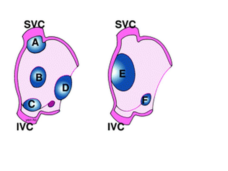
ASD Abnormal, fixed opening in the atrial septum caused by incomplete tissue formation that allows communication of blood between the left and right atria Usually asymptomatic until adulthood 3 Main Types: SECUNDUM (90%): Defective fossa ovalis (B, E) PRIMUM (5%): Next to AV valves, mitral cleft defect –it associated with Down Syndrome!!! (D) SINUS VENOSUS (5%): Next to SVC with anomalous pulmonary veins draining to SVC or IVC (A and C) 31
: Defective fossa ovalis (B, E) PRIMUM (5%): Next to AV valves, mitral cleft defect –it associated with Down Syndrome!!! (D) SINUS VENOSUS (5%): Next to SVC with anomalous pulmonary veins draining to SVC or IVC (A and C) 31.")
32
Patent foramen ovale A small hole created by an open flap of tissue in the atrial septum at the oval fossa. Normally, foramen ovale is an important functional right-to-left shunt that allows oxygen-rich blood from the placenta to bypass the not yet inflated lungs The hole is forced shut at birth in 80% of cases Sustained pulmonary hypertension or even transient increases in right-sided pressures, such as occurs during a bowel movement, coughing, or sneezing, can produce brief periods of right-to-left shunting, with the possibility of paradoxical embolism

33
ASD Clinical Features Splitting of S1 and S2 sounds, crescendo- decrescendo systolic murmur in the second intercostal space at the upper left sternal border. Paradoxal embolism (emboli from veins in systemic arteries) ASDs do not become symptomatic before age 30 (why?) Irreversible pulmonary hypertension is unusual, but LARGE defects may progress to pulmonary hypertension and reverse of the shunt which will lead to cyanosis (aka Eisenmenger syndrome)
 ASDs do not become symptomatic before age 30 (why ) Irreversible pulmonary hypertension is unusual, but LARGE defects may progress to pulmonary hypertension and reverse of the shunt which will lead to cyanosis (aka Eisenmenger syndrome)")
34
ASD treatment and prognosis
Surgical or catheter-based closure of an ASD reverses the hemodynamic abnormalities and prevents complications, including heart failure, paradoxical embolization, and irreversible pulmonary vascular disease. Long-term survival is comparable to that of a normal population

35
VSD Most common CHD defect
Incomplete closure of the ventricular septum Only 30% are isolated (Often associated with TETRALOGY of FALLOT) MC Associated with fetal alcohol syndrome 35
 MC Associated with fetal alcohol syndrome. 35.")
36
VSD Morphology 90% involve the membranous septum
Infundibular VSD - below the pulmonary valve Muscular septum VSD, likely to have multiple holes (“Swiss- cheese” septum ) SMALL ones often close spontaneously LARGE ones progress to pulmonary hypertension and reverse of the shunt which will lead to cyanosis (aka Eisenmenger syndrome)
 SMALL ones often close spontaneously. LARGE ones progress to pulmonary hypertension and reverse of the shunt which will lead to cyanosis (aka Eisenmenger syndrome)")
37
Infundibular VSD - below the pulmonary valve
37

38
Clinical presentation of VSD
Low Pitched HoloSystolic murmur, accentuation with handgrip exercise Splitting of S2 Polycythemia Clubbing of digits Paradoxal embolization Rx: Surgical closure

39
A 7-year old boy is brought to the pediatrician by his mother for a routine check-up. He has no complains. Cardiac auscultation findings at the left sternal border are given below. The findings accentuate with the handgrip exercise Which of The following is the most likely diagnosis? A Atrial septal defect B. Hypertrophic cardiomyopathy C. Patent ductus arteriousus D. Ventricular septal defect E. Aortic regurgitation

40
Physiologic ways to increase intensivity of murmurs

41
PDA (Persistent ductus arteriosus)
DA should be closed within hrs after birth 90% of PDAs isolated 10% associated with VSD, or coarctation of the aorta, or pulmonary or aortic valve stenosis Associated with congenital rubella PGE kEEps PDA open At the beginning is left to right shunt but later is switched to right to left (Eisenmenger syndrome) 41
 41.")
42
PDA Clinic HOLOSYSTOLIC HARSH, machinery- like murmur
LR, possibly RL as pulmonary hypertension approaches systemic pressure with development of cyanosis Closing the defect may be life saving Indomethacin causes closure of PDA

43
A 10-year-old immigrant from Eastern Europe is brought to the physician because of exertional dyspnea and easy fatigability. According to his parents, he was diagnosed with a congenital heart disease in infancy for which they refused treatment. They cannot recall the details of his diagnosis. Physical examination reveals toe cyanosis and clubbing but no finger abnormalities. This patient most likely suffers from which of the following? A. Primum-type atrial septal defect B. Secundum-type atrial septal defect C. Ventricular septal defect D. Patent ductus arteriosus E. Coarctation of the aorta F. Tetralogy of Fallot

44
AVSD Associated with defective, inadequate AV valves
The two most common forms are partial AVSD (consisting of a primum ASD and a cleft anterior mitral leaflet, causing mitral insufficiency) and complete AVSD (a hole in the center of the heart). More that 1/3 of all patients with a complete AVSD have Down syndrome. Rx: surgery 44
 and complete AVSD (a hole in the center of the heart). More that 1/3 of all patients with a complete AVSD have Down syndrome. Rx: surgery. 44.")
45
RL SHUNT – 5Ts Tetralogy of Fallot Transposition of great arteries
Truncus arteriosus Total anomalous pulmonary venous connection Tricuspid atresia All the RL congenital shunts are CYANOTIC, and have T’s in their names. 45

46
RL SHUNTS TETRALOGY of FALLOT most COMMON
1) LARGE Ventricular Septal Defect 2) OBSTRUCTION to blood flow from RV into Pulmonary artery 3) Aorta OVERRIDES the VSD 4) Right Ventricular Hypertrophy SURVIVAL DEPENDS on SEVERITY of SUBPULMONIC STENOSIS Can be a “PINK” tetrology if pulmonic obstruction is small, but the greater the obstruction, the greater is the RL shunt 46
 LARGE Ventricular Septal Defect. 2) OBSTRUCTION to blood flow from RV into Pulmonary artery. 3) Aorta OVERRIDES the VSD. 4) Right Ventricular Hypertrophy. SURVIVAL DEPENDS on SEVERITY of SUBPULMONIC STENOSIS. Can be a PINK tetrology if pulmonic obstruction is small, but the greater the obstruction, the greater is the RL shunt. 46.")
47
CLASSICAL “TETROLOGY” of FALLOT
1) VSD, large 2) OBSTRUCTION to RV flow 3) Aorta OVERRIDES the VSD 4) RVH (not shown on this picture) CLASSICAL “TETROLOGY” of FALLOT 10% alive at 20 years and 3% at 40 years Clinical Features: CLASSICAL “TETROLOGY” of FALLOT: 1) VSD, large 2) OBSTRUCTION to RV flow 3) Aorta OVERRIDES the VSD 4) RVH 47
 VSD, large. 2) OBSTRUCTION to RV flow. 3) Aorta OVERRIDES the VSD. 4) RVH (not shown on this picture) CLASSICAL TETROLOGY of FALLOT. 10% alive at 20 years and 3% at 40 years. Clinical Features: CLASSICAL TETROLOGY of FALLOT: 1) VSD, large. 2) OBSTRUCTION to RV flow. 3) Aorta OVERRIDES the VSD. 4) RVH. 47.")
48
Clinical features Physical exercises cause cyanosis in this patients (cyanotic spells) Squatting alleviate cyanosis !!! Degree of pulmonic stenosis determines extent of shunting and cyanosis CXR – boot shaped heart Auscultation: diamond shaped systolic murmur

49
A 2-week-old girl is found to have a harsh murmur along the left sternal border. The parents report that the baby gets “blu-ish” when she cries or drinks from her bottle. Echocardiogram reveals a congenital heart defect associated with pulmonary stenosis, ventricular septal defect, dextroposition of the aorta, and right ventricular hypertrophy. What is the appropriate diagnosis? (A) Atrial septal defect (B) Coarctation of aorta, postductal (C) Coarctation of aorta, preductal (D) Tetralogy of Fallot (E) Truncus arteriosus
 Atrial septal defect. (B) Coarctation of aorta, postductal. (C) Coarctation of aorta, preductal. (D) Tetralogy of Fallot. (E) Truncus arteriosus.")
50
TGA (TRANSPOSITION of GREAT ARTERIES)
Creates two not linked circuits: PA arises from LV Aorta arises from RV NEEDS a SHUNT for survival PDA or PFO (65%), “unstable” shunt VSD (35%), “stable” shunt Q:What will happen if we close PDA in patient with TGA? A: 50
, unstable shunt. VSD (35%), stable shunt. Q:What will happen if we close PDA in patient with TGA A: 50.")
51
TGA Clincal Features Associated with maternal diabetes
RV>LV in thickness Need to administer PgE to keep PDA open until surgery Fatal in first few months if not corrected Rx: Prostaglandin E Surgical “switching” Balloon Septostomy

52
Possible USMLE Scenario
A 27-year-old woman gives birth to a term infant after an uncomplicated pregnancy and delivery. The infant is cyanotic at birth. Two months later, physical examination shows the infant to be at the 37th percentile for height and weight. The representative gross appearance of the infant's heart is shown in figure. What is the most likely diagnosis? (A) Tetralogy of Fallot (B) Pulmonic stenosis (C) Truncus arteriosus (D) Transposition of the great vessels (E) Aortic stenosis
 Tetralogy of Fallot. (B) Pulmonic stenosis. (C) Truncus arteriosus. (D) Transposition of the great vessels. (E) Aortic stenosis.")
53
PERSISTENT TRUNCUS ARTERIOSUS
Single large vessel arising from both ventricles!!! Developmental failure of separation of the embryologic truncus arteriosus into the aorta and pulmonary artery Cyanosis Danger of irreversible pulmonary hypertension Early diastolic decrescendo murmur 53

54
A 2-week-old boy is irritable and feeding poorly
A 2-week-old boy is irritable and feeding poorly. On physical examination, the infant is irritable, diaphoretic, tachypneic, and tachycardic. There is circumoral cyanosis, which is not alleviated by nasal oxygen. A systolic thrill and holosystolic murmur are heard along the left sternal border. An echocardiogram reveals a heart defect in which the aorta and pulmonary artery form a single vessel that overrides a ventricular septal defect. What is the appropriate diagnosis? (A) Atrial septal defect (B) Coarctation of aorta, preductal (C) Patent ductus arteriosus (D) Tetralogy of Fallot (E) Truncus arteriosus
 Atrial septal defect. (B) Coarctation of aorta, preductal. (C) Patent ductus arteriosus. (D) Tetralogy of Fallot. (E) Truncus arteriosus.")
55
TRICUSPID ATRESIA Tricuspid valve orifice fails to form Hypoplastic RV
Needs a shunt, ASD, VSD, or PDA Signs: Soft S1 sound Low PCWP (wedge pressure) RA hypertrophy – dilation of P wave on ECG Early Cyanosis and high mortality in first week 55
 RA hypertrophy – dilation of P wave on ECG. Early Cyanosis and high mortality in first week. 55.")
56
Main message: pulmonary atresia, tricuspid atresia, Tetralogy of Fallot, Transposition of the great vessels – administer prostaglandin to keep DA open or child will die!

57
Total Anomalous Pulmonary Venous Connection (TAPVC)
PULMONARY VEINS do NOT go into LA, but into L. innominate v. or coronary sinus Needs a PFO or a VSD HYPOPLASTIC LA Do not administer Prostaglandin E1 57

58
OBSTRUCTIVE CHD COARCTATION of aorta Pulmonary stenosis/atresia
Aortic stenosis/atresia 58

59
COARCTATION of AORTA Coarctation=narrowing of aorta
Two major forms: INFANTILE and ADULT (pre- and postductal) “Infantile” form: hypoplasia of the aortic arch proximal to a patent ductus arteriosus symptomatic in early childhood 59
 Infantile form: hypoplasia of the aortic arch proximal to a patent ductus arteriosus. symptomatic in early childhood. 59.")
60
Infantile form (preductal) COARCTATION of AORTA
Associated with presence of patent ductus arteriosus Presents with lower part of the body cyanosis, upper extremities are ok XO’s (Turner syndrome) frequently have it with shortening of 4th metacarpal bone and horse-shoe kidney!!!
 frequently have it with shortening of 4th metacarpal bone and horse-shoe kidney!!!")
61
Adult form of COARCTATION of AORTA
Discrete ridgelike infolding of the aorta, just opposite the closed ductus arteriosus (ligamentum arteriosum) distal to the arch vessels Bicuspid aortic valve 50% of the time (prone to aortic stenosis) Associated with congenital aortic stenosis, ASD, VSD, mitral regurgitation, or berry aneurysms of the circle of Willis in the brain
 distal to the arch vessels. Bicuspid aortic valve 50% of the time (prone to aortic stenosis) Associated with congenital aortic stenosis, ASD, VSD, mitral regurgitation, or berry aneurysms of the circle of Willis in the brain.")
62
Clinical manifestations Adult form of COARCTATION of AORTA
Hypertension in the upper extremities Development of collateral circulation between the precoarctation arterial branches and the postcoarctation arteries through enlarged intercostal and internal mammary arteries, which produce radiographically visible erosions (“notching”) of the undersurfaces of the ribs Murmurs are present throughout systole, ; sometimes a thrill Cardiomegaly due to left ventricular pressure-overload hypertrophy
 of the undersurfaces of the ribs. Murmurs are present throughout systole, ; sometimes a thrill. Cardiomegaly due to left ventricular pressure-overload hypertrophy.")
63
Prognosis for Adult form of COARCTATION of AORTA
Patients with adult-type coarctation of the aorta commonly die of hypertension- associated complications: left ventricular failure ruptured dissecting aortic aneurysm intracranial hemorrhage (due to the increased incidence of congenital berry aneurysms of the Circle of Willis)
")
64
A 22-year-old Caucasian male presents to the emergency room complaining of severe headaches and vomiting. Soon after, he slips into a coma and dies. Autopsy shows a ruptured cerebral aneurysm with extensive intracranial hemorrhage. This patients condition is most likely associated with: A. Primum-type atrial septal defect B. Secundum-type atrial septal defect C. Ventricular septal defect D. Patent ductus artenosus E. Coarctation of the aorta F. Tetralogy of Fallot

65
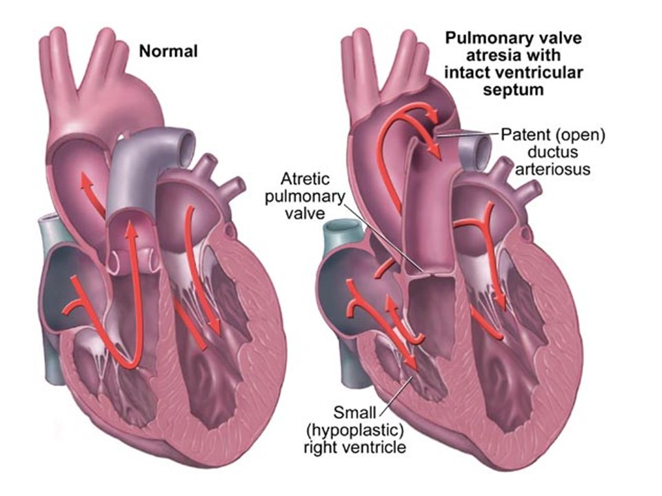
Pulmonary Valve Stenosis and Atresia
PS- an obstruction at the pulmonary valve Lesion can be isolated or part of a more complex anomaly—either tetralogy of Fallot or transposition of the great arteries Right ventricular hypertrophy is often Poststenotic dilation of the pulmonary artery due to injury of the wall by “jetting” blood. When the valve is entirely atretic, there is an ASD and blood reaches the lungs through a patent ductus arteriosus. Clinical severity ~ stenosis severity 65

67
AORTIC VALVE STENOSIS/ATRESIA
Stenosis - Three locations: valvular, subvalvular, supravalvular Atresia leads to underdevelopment (hypoplasia) of the left ventricle and ascending aorta, sometimes accompanied by dense, porcelain-like left ventricular endocardial fibroelastosis The ductus arteriosus is open to allow blood flow to the aorta and coronary arteries when the ductus will be closed it will lead to the child death Less severe degrees of congenital aortic stenosis may be compatible with long survival Congenital aortic stenosis is an isolated lesion in 80% of cases. 67
 of the left ventricle and ascending aorta, sometimes accompanied by dense, porcelain-like left ventricular endocardial fibroelastosis. The ductus arteriosus is open to allow blood flow to the aorta and coronary arteries. when the ductus will be closed it will lead to the child death. Less severe degrees of congenital aortic stenosis may be compatible with long survival. Congenital aortic stenosis is an isolated lesion in 80% of cases. 67.")
68
MITRAL VALVE PROLAPSE: CLINICAL FEATURES
displacement of an abnormally thickened mitral valve leaflet into the left atrium during systole Usually asymptomatic Mid-systolic “click” Holosystolic murmur if regurg. present Occasional chest pain, dyspnea 97% NO untoward effects 3% Infective endocarditis, mitral insufficiency, arrythmias, sudden death 68

69
Ebstein anomaly Congenital heart defect in which the septal leaflet of the tricuspid valve is displaced towards the apex of the right ventricle of the heart Etiology: Fetal exposure to Lithium !!!! Systolic murmur of tricuspid regurgitation Mid-diastolic murmur along the lower left sternal border Right atrial hypertrophy WPW syndrome on ECG

Similar presentations


 tricuspid valve 2. Hypoplastic right ventricle 3. Ventricular septal defect 4. Atrial.>")


