Download presentation
Presentation is loading. Please wait.

1
Human Anatomy, Larry M. Frolich, Ph.D. Cartilage--function, types, location Bone Tissue--structure, types Long Bone Structure and Development Most common bone problems Fractures Osteoporosis Cartilage and Bone

2
Human Anatomy, Larry M. Frolich, Ph.D. What is cartilage? Skeletal tissue--maintains certain shape and form Very resilient (bouncy or rubbery), mostly water Grows fast--forms embryonic skeleton
, mostly water Grows fast--forms embryonic skeleton.")
3
Human Anatomy, Larry M. Frolich, Ph.D. Kinds of cartilage Hyaline cartilage--most common, found in joints Elastic cartilage--epiglottis, ear Fibrocartilage--annular fibrosis of intervertebral disk, menisci of knee

4
Human Anatomy, Larry M. Frolich, Ph.D. M & M Figure 6.1

5
Human Anatomy, Larry M. Frolich, Ph.D. Bones provide: Support and movement (limbs, axial skeleton) Protection (skull bones) Mineral storage Blood cell development (long bone marrow) Bone is made up of: 35% collagen, ground substance and cells 65% inorganic calcium (hydroxyapetite)
 Protection (skull bones) Mineral storage Blood cell development (long bone marrow) Bone is made up of: 35% collagen, ground substance and cells 65% inorganic calcium (hydroxyapetite).")
6
Human Anatomy, Larry M. Frolich, Ph.D. Bone is alive!! Bone cell types: Osteoblasts: Make and deposit components of bone extracellular matrix Osteoclasts: Degrade and resorb bone for remodeling Osteocytes: “watcher cells” Sit in bone and monitor its current status

7
Human Anatomy, Larry M. Frolich, Ph.D. Types of bony tissue Compact Bone Dense tissue at surface of bones Haversian canals Osteocytes in lacunae Highly vascularized Fig. 6.6, p. 138

8
Human Anatomy, Larry M. Frolich, Ph.D.

9
Types of bony tissue Spongy bone Trabeculae (oriented to give mechanical strength) Interior of long bones, skull bones Epiphyses of long bones Intramembranous ossification (osteoblasts lay down bone around blood vessels in connective tissues of dermis (after 8 weeks of development)
 Interior of long bones, skull bones Epiphyses of long bones Intramembranous ossification (osteoblasts lay down bone around blood vessels in connective tissues of dermis (after 8 weeks of development)")
10
Human Anatomy, Larry M. Frolich, Ph.D. Structure of a long bone Diaphysis (shaft) Epiphysis Proximal Distal Compact bone Spongy bone Periosteum Medullary cavity Articular/hyaline cartilage Nutrient V/A/N Epyphyseal (growth) plates Fig. 6.3, p. 135
 Epiphysis Proximal Distal Compact bone Spongy bone Periosteum Medullary cavity Articular/hyaline cartilage Nutrient V/A/N Epyphyseal (growth) plates Fig. 6.3, p")
11
Human Anatomy, Larry M. Frolich, Ph.D.

12
Bone Tissue within a Bone

13
Human Anatomy, Larry M. Frolich, Ph.D. Why do bones need to “remodel?”

14
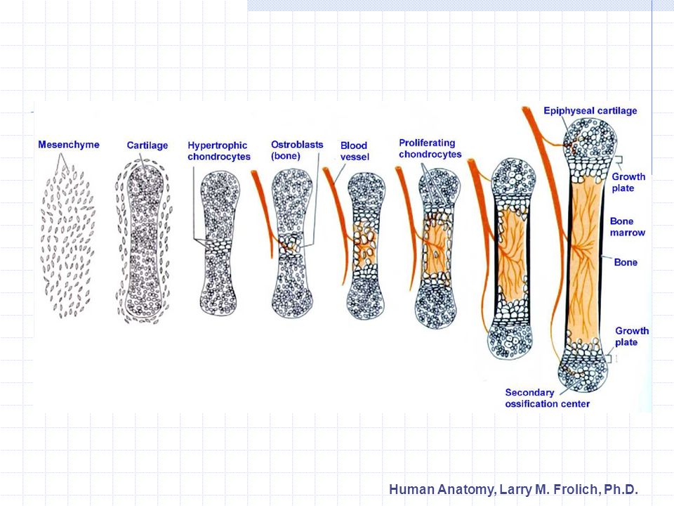
Human Anatomy, Larry M. Frolich, Ph.D. Endochondral Ossification 1. Cartilage model 2. Bone collar forms in diaphysis (dense bone) Cartilage chondrocytes in center of diaphysis die and cartilage disintegrates 3. Periosteal bud enters diaphysis Makes spongy bone at ends of diaphysis (primary ossification center) 4. Epiphysis begins to ossify (secondary ossification center) 5. Hyaline cartilage remains only at Epiphyseal surfaces (articular surfaces of joints) Epiphyseal growth plates between diaphysis and epiphysis (primary and secondary ossification centers on either side) Fig. 6.9, p. 141
 Cartilage chondrocytes in center of diaphysis die and cartilage disintegrates 3. Periosteal bud enters diaphysis Makes spongy bone at ends of diaphysis (primary ossification center) 4. Epiphysis begins to ossify (secondary ossification center) 5. Hyaline cartilage remains only at Epiphyseal surfaces (articular surfaces of joints) Epiphyseal growth plates between diaphysis and epiphysis (primary and secondary ossification centers on either side) Fig. 6.9, p")
15
Human Anatomy, Larry M. Frolich, Ph.D.

17
Endochondral ossification centers—newly formed bone within cartilage shown is stained red

18
Human Anatomy, Larry M. Frolich, Ph.D. Osteoclasts Osteoblasts “Dig holes” with hydrochloric acid Degrades calcium Phagocytize collagen fibers and dead osteocytes Line tubes (Haversian canals) left by osteoclasts Lay down new bone in circular concentric lamellae Unique to warm- blooded animals-- dinosaurs???
 left by osteoclasts Lay down new bone in circular concentric lamellae Unique to warm- blooded animals-- dinosaurs .")
19
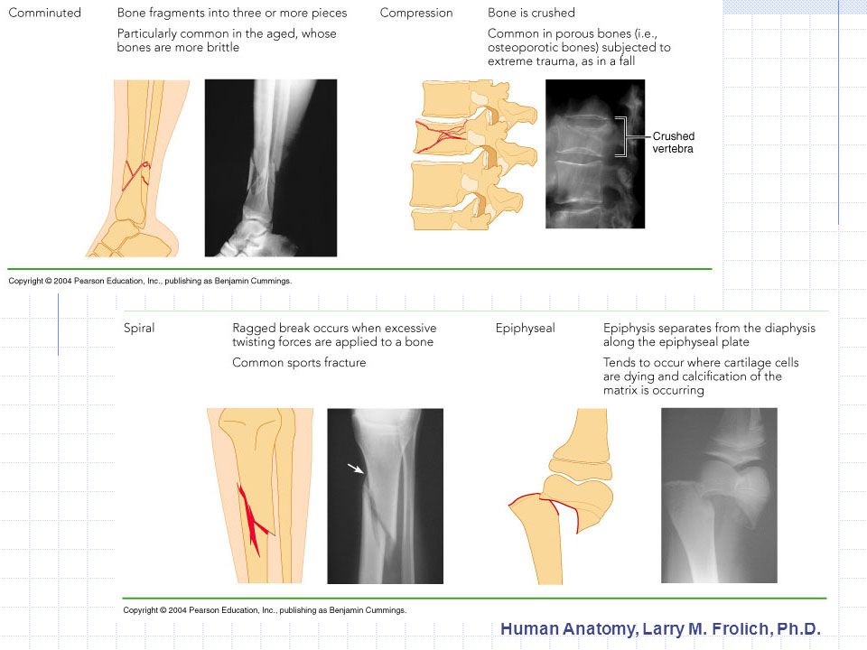
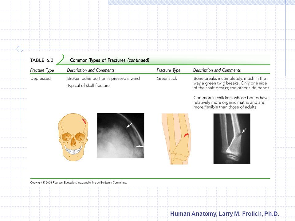
Human Anatomy, Larry M. Frolich, Ph.D. Bone Fractures Treatment is reduction Closed--set in place by physical manipulation from outside body Open--surgical placement of pins or screws Healing Hematoma Fibrocartilaginous callus Bony calllus Remodeling by osteoclasts/osteoblasts Types of Fractures

20
Human Anatomy, Larry M. Frolich, Ph.D.

23
Fracture repair

24
Human Anatomy, Larry M. Frolich, Ph.D. Calcium regulation is negative feedback mechanism

25
Human Anatomy, Larry M. Frolich, Ph.D. Osteoporosis Affects elderly, especially women Bone resorption proceeds faster than deposition Low estrogen levels implicated but estrogen replacement now considered risky Importance of calcium in diet??? Leads to fractures Compression fractures of vertebrae Neck of femur

26
Human Anatomy, Larry M. Frolich, Ph.D. Bone grafts and artificial bone Widely used cutting-edge technologies Bone cells highly regenerative and move into any suitable matrix Use bone pieces from same body—fibula Use crushed bone from cadavers Use bone substitutes—coral, synthetics— ”nanotechnology” Applications are numerous Jaw bone filler for dental work Birth defects Osteoporosis Bone repair

Similar presentations


 or an organ –Bone referred to as a connective tissue consists of: cells extracellular.>")
 - hematopoeisis.>")
 Joints ► Cartilages Ligaments ► Divided.>")


 Joints Cartilages Ligaments Divided into two divisions Axial skeleton –>")


