Download presentation
Presentation is loading. Please wait.

3
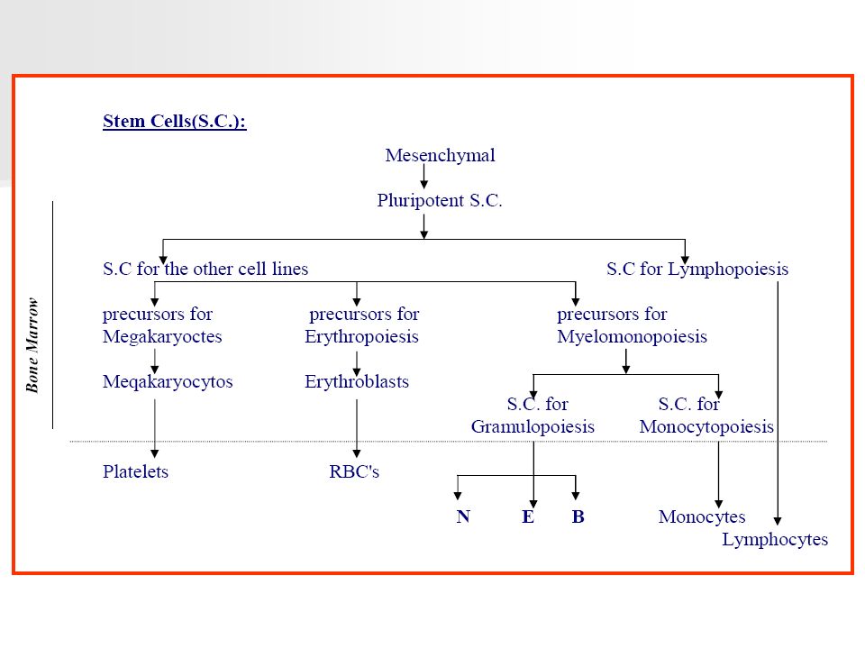
Composition: suspension of cells in a complex liquid (plasma ) Plasma:* H2O + organic molecules + mineral salls * After coagulation (- Fibrinogen) = serum Blood Cells: * R.B. C* W. B. C. * platelets. Origin: Site: * In the embryo (Liver, Spleen, Bone marrow) * After birth (only in the Bone marrow) * Lymmphoid tissues (B.M., L.N., Spleen, Payer's patches)
 * After birth (only in the Bone marrow) * Lymmphoid tissues (B.M., L.N., Spleen, Payer s patches).")
5
HEMOPOIESIS 1-Erythropoiesis: The erythroblastic line represents 10 -30% of the bone marrow cells. 2-Steps of maturation: It take 7 days under normal condition. 1-Erythroblast (pronormoblast) 2-Basophilic Normoblast 3- Polychromatic normoblast. 4-Orthochromatic normoblast. 5-Reticulocyte 6- R. B. C
 2-Basophilic Normoblast 3- Polychromatic normoblast. 4-Orthochromatic normoblast. 5-Reticulocyte 6- R. B. C.")
7
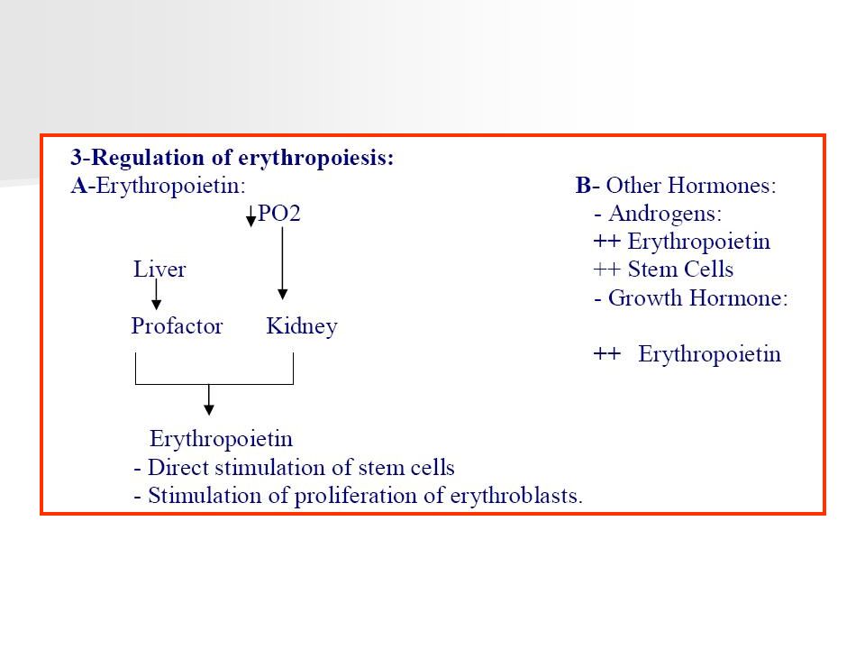
4-In Cases or Urgent Blood Loss: E.g.: - after haemolytic attack - after Hge 1- Hypersecretion of erythropoietin 2- Augmentation of no. of erythroblasts in B. M. resulting in: 3- Acceleration of Hb. synthesis normal corpuscular conc. or Hb. occurs early decrease the number of cell divisions between stage of erythroblast and reticulocytes -- reduction of duration of erythropoiesis to 3-4 days. N.B as a result of Hyper secretion of the erythropoietin and acceleration of Hb. synthesis. Hypererythropoiesis occurs.

8
HYPERERYTHROPOIESIS a- Increase in number or reticulocytes in the peripheral blood b- presence of immature R.B.CS (polvchromatophilia ) c- Macrocytosis. (duo to reduction of number of mitoses between proerthroblasts and Reticulocytes) d- Some orthochromatic normoblasts may be found in the peripheral Blood
 d- Some orthochromatic normoblasts may be found in the peripheral Blood.")
9
5-Exogenous Factors necessary for Erythropoiesis: 1-Iron 2-Folic acid and Vitamin B12

10
EXAMINATION OF THE BLOOD: COMPLETE BLOOD PICTURE (C.B.C.) A-QUANTITATIVE EXAMINATION: 1-Quantitative Exam. of the RBCS with its constituents: a-R.B.Cs no./mm3: Male4.5 – 6.2 x 10 6 Female & Child:4 – 5.4 x 10 6 Infant3.6 – 5 x 10 6 at Birth5 – 6 x 10 6

11
b- Hematocrite: (relative volume of R.B.CS in the blood) -By centrifugation of a small column of blood in a standard tube: Male 40 - 54 % Female 35 - 47 %
 -By centrifugation of a small column of blood in a standard tube: Male % Female %")
12
c- Hb. Content of 100 cc blood: Male13 - 18 gm/100cc Female & Child:12 - 16 gm/100cc Infant12 - 16 gm/100cc at Birth14 - 20 gm/100cc

15
f- Reticulocytic count: - Normal life span of RBCs = 120 days -Reticulocytes are the newly fabricated RBCs within the 1st 24 hours (still containing cytoplasmic organelles) i.e. --- by special stain --- counting the no. of RBCs containing such organells among 1000 RBCs in %. - Absolute no. of reticutocytes: = RBCs count X reticulocytic %. * normal 25.000----- 100.000/mm3 (l-2 %) For Hb content within the normal range. * with anaemia increase in no. of the reticulocytes, if there is a normal ability of the B.M. to synthesis RBCs i.e., If Hb 100.000. If < 100.000 this means aregnerative, i.e., ( hypoplastic or aplastic ) anaemia.
 For Hb content within the normal range. * with anaemia increase in no. of the reticulocytes, if there is a normal ability of the B.M. to synthesis RBCs i.e., If Hb If < this means aregnerative, i.e., ( hypoplastic or aplastic ) anaemia..")
16
2 - Quantitative Exam. of WBCS: Normal 4.000--- 10.000/mm 3 in adults Infants1st year 5.000- 20.000 /mm 3 2nd year6.000 – 17.000 / mm 3 10 years 5.000 --- 13.000/mm 3 N.B. In adult. * > 11.000 indicates leucocytosis * < 4.000 indicates leucopenia.

17
3- QUANTITATIVE ESTIMATION OF PLATELETS: Normal l50.000 - 450. 000/mm 3 < 150,000 indicates thrombocytopenia. >450.000indicates thrombocytosis

18
B- MORPHOLOGIC EXAMINATION: RBCsMorphological exam. of RBCs. WBCS Differential white cell count. I- Morphologic Examination of RBCs: Normally : RBCs are of * same form * same colour * same diameter. Any abnormality Pathological condition: * in size : anisocytosis * in form: poikilocytosis

19
II- Differential white cell Count: -The absolute no. more important than the %

20
Polymorphonuclear Neutrophils: * 40 – 70% of WBCS (1700 - 7000) * Function: The main Function is phagocytosis of foreign bodies esp. Bacteria

21
Granulopoiesis: It takes 10 days under normal conditions 50 -70 % of B. M. cells - Myeloblasts 2.3 % - Promyelocytes 4-8% - Myelocytes 10-15% - Metamyelocytes 15-20% - Polynuclear cells 20-30% * Life span: in the circulation is very short within 12 hours. 50% of formed neutrophils leave the blood to the tissues and don't turn back again.

22
In cases of excess demand: Rapid passage of neutrophils to the blood from the marrow resulting in the release of band forms, even metamyelocytes and myelocytes.

23
Polymorphonuclear leucocytosis: Due to increase polymorphonuclear neut. i.e.> 7000/ mm 3 1- Reactional: a- physiologic: -Neonates -Severe exercise -Menestruation -Pregnancy

24
b- Pathologic : - Bacterial infections -Inflammatory diseases : (Arthritis - Allergic ) - Tissue necrosis e.g., rnyocardial Infraction, pancreatitis -Heavy smokers. -Acute hemorrhage or hemolysis -Intoxication (radiation, benzene) -Start of chronic myeloid leukemia -Without cause
 -Start of chronic myeloid leukemia -Without cause.")
25
2- Myeloproliferative disorders: as Chronic Myeloid 1eukemia Neutropenia: means that Neutrophils < 1700 / mm 3, It is due to either: 1- Insufficient production (central): A-As a part of Pancytopenia: e.g., aleukemic leukemia or aplastic amenia B-Isolated: 1- Hereditary form. 2- Allergy to certain drugs 3- Direct central inhibition e.g., henothiazine 4- Constitutional agranulocytosis.

26
2- Hyperdestruction (peripheral): - Auto immunisation - Lupus Erythromatosis. Usually with thrombocytopenia - Felty's syndrome # But espe. in moderate cases (i.e. 800- 1700) we must exclude : -Some parasitic infections (kala Azar) - Some bacterial infections (typhoid, Brucellosis) - Some viral infections (Viral hepatitis) - May be normal esp. in black races.
 we must exclude : -Some parasitic infections (kala Azar) - Some bacterial infections (typhoid, Brucellosis) - Some viral infections (Viral hepatitis) - May be normal esp. in black races..")
27
Eosinophiles: * Functions: (not exactly known) - phagocytosis : Ag- Ab complex - Transport the plasminogen - Play a role in destruction of certain larvae - Has a role in allergy
 - phagocytosis : Ag- Ab complex - Transport the plasminogen - Play a role in destruction of certain larvae - Has a role in allergy")
28
Eosinophilia: means eosinophils > 300/mm 3 but more significant if > 500/mm 3 causes: 1- Allergic conditions: - Asthma, - Allergy to certain drugs as penicillin's. 2- parasitic infections: Filariasis, Ascaris, Oxyuris, Taeniasis. 3- Polyarteritis nodosa 4- Dermatosis 5- Malignancy: - H.D - certain cancers (Liver, Ovary)
.")
29
Basophils: * Function: - Unknown. - Rich in histamine and heparin - Play a role in delayed hypersensitivity reaction If >1 % = pathologic conditions: 1- Myeloproliferative syndromes esp. C.M.L 2-Severe hyperlipidaemia 3- Hypothyroidism

30
Monocytes : - The largest circulatory blood cells, have: * characteristic Nuclear shape, * Longer life span than other WBCs (2-3 days) Function : 1- phagocytosis :Play a role in defence mechanism In tissues --- Macrophages 2- Play a role in immunologic reactions
 Function : 1- phagocytosis :Play a role in defence mechanism In tissues --- Macrophages 2- Play a role in immunologic reactions")
31
Monocytosis: means monocytes > 1000 /mm3: 1- reactional: - infections: (bacteria, virus parasites) - At the recovery of agranulocytosis 2- Acute monocytic leukemia, chronic myelomonocytic leukemia 3- Isolated chronic Monocytosis : rare as in refractory anaemias at the start of acute monocytic Leukemia, and chronic myelomonocytic eukemia.
 - At the recovery of agranulocytosis 2- Acute monocytic leukemia, chronic myelomonocytic leukemia 3- Isolated chronic Monocytosis : rare as in refractory anaemias at the start of acute monocytic Leukemia, and chronic myelomonocytic eukemia.")
32
Lymphocytosis: Means increased number of lymphocytes above the upper limit of normal (> 4000 – 4500/cumm in adults, > 6000- 7000, cu mm in neonates and infants. Causes: 1.Viral infections : hepatitis, measles, infective mononucleosis 2.Bacterial infections : brucellosis, T.B., whooping cough. 3.Malignant lymphocytosis : Chronic lymphatic leukemia and Waldestron’s macroglobulinemia.

Similar presentations


. Complete Blood Count ( CBC)>")


.>")


>")


 –Formed elements 45%– rbc’s, wbc’s, platelets –Buffy coat – wbc and platelets.>")
