Download presentation
Presentation is loading. Please wait.

1
CH 4: Tissues

2
Tissues Tissues are groups of cells that are similar in structure and function. Tissues can be solid, semi-solid or liquid. Histology is the study of tissues. There are 4 main types of tissues in the human body: Epithelial Connective Muscular Nervous

3
Epithelial Tissue (Epithelium)
Covers body surfaces and lines cavities, hollow organs and tubes. It also forms glands. Cells are closely packed and arranged in sheets of either single or multiple layers. Bottom surface of epithelium is attached to the basement membrane- fibers that are located between the epithelium and the connective tissue below it.

4
Epithelium continued…..
Avascular- without blood vessels. Epithelium gets nutrients from the adjacent connective tissue. Has a nerve supply. Is constantly being regenerated because it suffers so much wear and tear. There are 2 main types of epithelial tissue: Covering/Lining epithelium Glandular epithelium

6
Covering/Lining Epithelium
Outer covering of the skin and some internal organs. Inner lining of blood vessels, ducts, and body cavities. Interior of the digestive, respiratory, reproductive and urinary systems.

7
Glandular Epithelium Consists of glands that secrete substances.
Glands are either endocrine or exocrine. Endocrine- Secrete hormones that enter the fluid around the cells (interstitial) and eventually the bloodstream (Ex. thyroid) Exocrine- Secrete substances into ducts that empty onto the surface of a covering/lining epithelium (Ex. Sweat gland)
 and eventually the bloodstream (Ex. thyroid) Exocrine- Secrete substances into ducts that empty onto the surface of a covering/lining epithelium (Ex. Sweat gland)")
8
Epithelial Tissue Classification
Epithelial tissue is classified based on the number of cell layers present and the shape of the cells of the tissue. 4/21/2017 Bio

9
Number of Layers Simple- Stratified- one layer 2 or more layers
usually involved in absorption and/or diffusion Stratified- 2 or more layers Protects underlying tissues from wear and tear

10
Number of Layers Continued..
Pseudostratified Looks like many layers because not all cells reach the surface and the nuclei lie at different levels

11
Cell Shape Squamous Thin and flat and attach to each other like tiles
Easily allow substances to pass through Cuboidal Shaped like cubes Function in secretion or absorption

12
Cell Shape Continued… Columnar taller than wide
protect underlying tissues Transitional change shape due to stretching or movement of body parts

13
Other modifications Cilia- Tiny hairs that move substances around outside the cell Microvilli- Folds that increase surface area for absorption Goblet cells- embedded in between epithelium and secrete mucus for lubrication or to trap particles

14
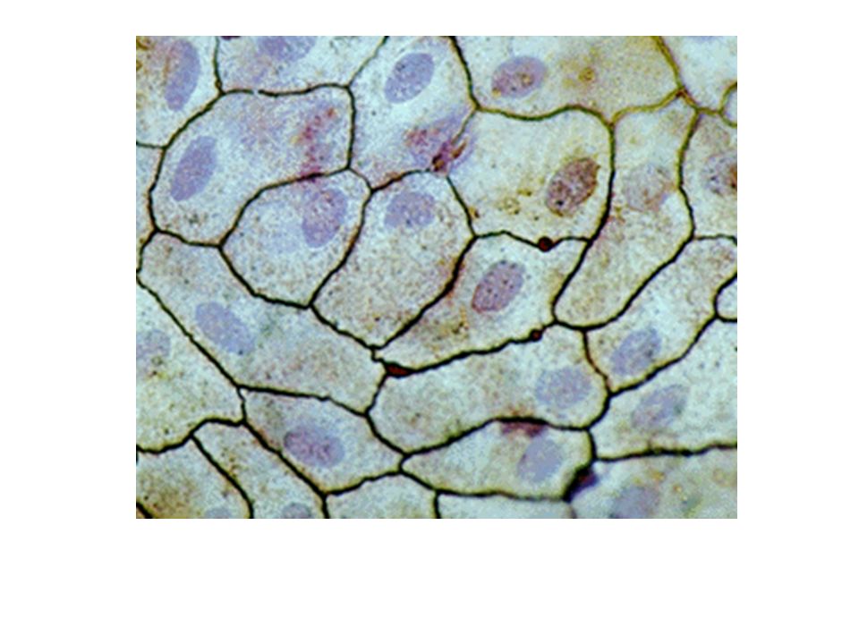
Simple Squamous A single layer of tightly packed thin, flat, irregularly shaped cells Location: Lines the heart, blood vessels, and lymphatic vessels Forms the peritoneum, pericardium and pleural linings Found in the air sacs of lungs (alveoli) Function: Allows for easy diffusion of gases and blood components
 Function: Allows for easy diffusion of gases and blood components.")
15
Simple Squamous – Top View – found in the walls of capillaries and alveoli of lungs

17
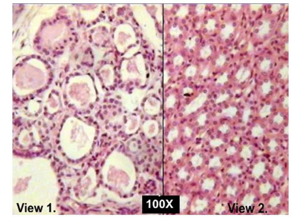
Simple Cuboidal A single layer of tightly packed cube shaped cells.
Location: Lines kidney tubules, ducts of glands, and secreting parts of glands Covers the surface of the ovary Function: Secretion and absorption

18
Simple Cuboidal Epithelial Tissue – this is of ducts of the kidney

21
Simple Columnar A single layer of tightly packed rectangular cells
Can be non-ciliated or ciliated Goblet cells are modified columnar cells that secrete mucus Non-ciliated have microvilli on the surface of the cells Non-ciliated Location: Lines the gastrointestinal tract (from stomach to anus), ducts of many glands, and gall bladder
, ducts of many glands, and gall bladder.")
22
Simple Columnar Continued…
Non-ciliated Function: Secretion and absorption Ciliated Location: Lines tubes of the respiratory tract, fallopian tubes, uterus, and ventricles of the brain Ciliated Function: The cilia beat in unison to move substances outside the cell like mucus and egg cells.

23
Simple Columnar Non-Ciliated Epithelial Tissue - This is a cross section through the small intestine – for absorption – microvilli on surface of cells

26
Pseudostratified Columnar
A single layer of tightly packed columnar cells that appear to be multi-layered Location: Ciliated- Lines respiratory tract Non-ciliated- Male urethra Function: Ciliated- moves materials across the apical surface of a tissue. Non-ciliated- Absorb and protect

28
Stratified Squamous Multiple layers of tightly packed squamous epithelial cells. Location: Areas that are subject to much “wear and tear”. Upper layers are designed to be shed and replaced. Function: Protection

30
Stratified Cuboidal Two layers of cube shaped epithelial cells Rare
Location: Lining the ducts of glands found in the esophagus Function: Moderate protection from the stomach acid that refluxes

32
Stratified Columnar Multiple layers of columnar epithelial cells Rare
Only the layer closest to the lumen (opening in a duct) is columnar in shape. The outer layers are somewhat squashed. Location: Lining the ducts of glands lining the esophagus Function: Moderate protection from stomach acid that refluxes
 is columnar in shape. The outer layers are somewhat squashed. Location: Lining the ducts of glands lining the esophagus. Function: Moderate protection from stomach acid that refluxes.")
34
Transitional Epithelial tissue that has a variable shape Location:
Can stretch and relax multiple times without being damaged Location: Lines the urinary bladder Function: Protection

36
Let’s Practice For the following, indicate the type of epithelial tissue and where you would find it.

48
4/21/2017 Bio

49
4/21/2017 Bio

51
Connective Tissue The most abundant and widely distributed type of tissue in the whole body. General characteristics: With the exception of 3 types, connective tissues have a great blood supply. Reproduce quickly Classified as being “connective” because at least 50% of the tissue is non-living.

52
Connective Tissue continued….
Function: Designed to bind tissues together Helps protect the body Store energy in the form of fat Transport materials throughout the body (blood, lymph) 2 main components of connective tissue: Matrix Cells
 2 main components of connective tissue: Matrix. Cells.")
53
Matrix of Connective Tissue
A base material that contains protein fibers and is found between the cells of connective tissue. Fibers are proteins found in the matrix that strengthen and support connective tissue. Non-living The structure of the matrix determines the characteristics of the connective tissue Functions to support the cells of the connective tissue

54
Fibers in Connective Tissue
The properties of a connective tissue can change if the types/amounts of fibers in the matrix change. Types of fibers: Collagen- Strong and flexible and add strength to connective tissue Elastic- Strong and add flexibility to connective tissue Reticular- Strong and highly branching fibers that help form the framework of a connective tissue

56
Cells of Connective Tissue
Not all of the possible types are found in all connective tissues Fibroblasts: Make the fibers found in the matrix Found in most connective tissues Phagocytes: Macrophages and neutrophils- Phagocytic cells that “eat and eliminate” foreign materials.

57
Cells of Connective Tissue continued…
Plasma cells: Found in most connective tissues Produce antibodies Antibodies- Molecules that help protect us from foreign particles Mast cells: Found in all connective tissues Release histamine Histamine- Causes swelling because it signals for blood vessels to become more “leaky” and signals for blood to rush to an area

59
Cells of Connective Tissue continued…
Adipocytes: Found in some connective tissues Contain fat

60
Types of Connective Tissue
There are 6 types of mature connective tissue. They include: loose connective tissue, dense connective tissue, cartilage, bone tissue, blood tissue and lymph.

61
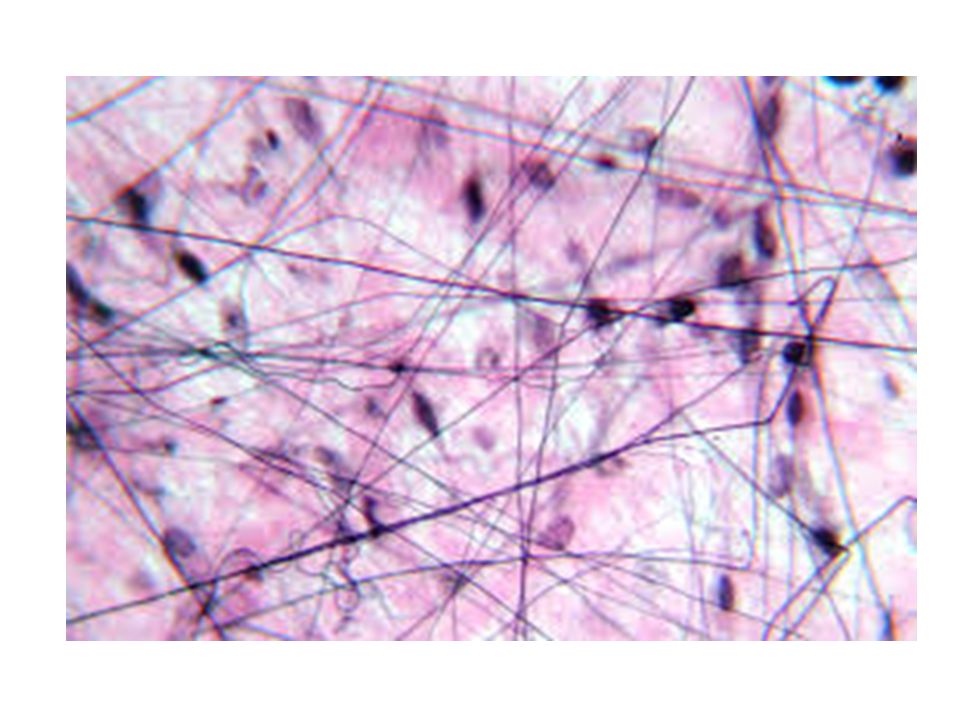
Loose Connective Tissue
Loosely arranged fibers in the matrix. Areolar- Loose arrangements of collagen, elastic and reticular fibers present in the matrix Location: Between the skin and muscle and holds the skin in place Function: Binds structures together, provides strength (collagen), and provides flexibility (elastic) Adipose- Lipids (fats) present in the matrix Location: Below the skin and surrounding organs Function: Stores fat, insulates against heat loss, and protects organs
, and provides flexibility (elastic) Adipose- Lipids (fats) present in the matrix. Location: Below the skin and surrounding organs. Function: Stores fat, insulates against heat loss, and protects organs.")
64
Loose Connective Tissue
Reticular- Loose arrangements of reticular fibers and reticulocytes in the matrix Function: Form the framework for larger organs (like the framework of a house)
")
66
Dense Connective Tissue
Large amounts of thick, dense fibers and fewer cells than loose connective tissue Dense regular- Parallel bundles of collagen fibers present in the matrix Connects muscle to bone (tendon) or bone to bone (ligament) Can resist lots of force along the length of the fibers A force against the fibers can tear them (torque) Poor blood supply so it takes a long time to heal
 or bone to bone (ligament) Can resist lots of force along the length of the fibers. A force against the fibers can tear them (torque) Poor blood supply so it takes a long time to heal.")
68
Dense Connective Tissue
Dense irregular- Random arrangements of collagen fibers present in the matrix. Good blood supply Makes up the dermis (middle layer) of the skin Resists pulling forces in multiple directions
 of the skin. Resists pulling forces in multiple directions.")
70
Dense Connective Tissue
Elastic: Freely branching elastic fibers present in the matrix. Good blood supply Makes up the wall of an artery Allows a hollow structure to expand and recoil freely This keeps blood moving along in arteries

72
Cartilage Consists of a dense network of collagen and elastic fibers in a semi-solid matrix Poor blood supply Chondrin- Gel-like protein found making up the matrix of cartilage tissue Types of cartilage Hyaline Cartilage cells (chondrocytes) suspended in a chondrin matrix. Acts as a shock absorber Covers the ends of bones at a joint
 suspended in a chondrin matrix. Acts as a shock absorber. Covers the ends of bones at a joint.")
74
Types of cartilage continued…
Fibrocartilage Large bundles of collagen fibers and few chondrocytes suspended in a chondrin matrix Forms the larger tendons of the body Acts as a shock absorber Elastic cartilage Elastic fibers and few chondrocytes suspended in a chondrin matrix

77
Bone Characterized by a matrix composed of inorganic minerals.
Minerals are embedded in rings Support and protect Histology of bone will be explained later in the Bone unit

79
Blood Liquid connective tissue
Transports materials throughout the body 2 basic components: Plasma- 55% of total blood mostly water supports the formed elements Formed elements (cells)- Erythrocytes- red blood cells- transport oxygen- outnumber white cells 700:1 Leukocytes- white blood cells- fight infection Thrombocytes- platelets- cell fragments- help form blood clots
- Erythrocytes- red blood cells- transport oxygen- outnumber white cells 700:1. Leukocytes- white blood cells- fight infection. Thrombocytes- platelets- cell fragments- help form blood clots.")
80
Lymph Liquid connective tissue
Located in lymphatic vessels- separate from blood vessels No red blood cells nor platelets Fights infection

82
Fluid Connective Tissue

83
Let’s Practice Connective Tissues

102
Muscle Tissue Muscle tissue consists of cells that are highly specialized to generate force. This force, produces motion, maintains posture, and generates heat. Muscle tissue cells have lots of mitochondria. Why? There are 3 types of muscle tissue. They are skeletal muscle, smooth muscle and cardiac muscle. 4/21/2017 Bio

104
Skeletal Muscle Skeletal muscle is named for its location. It is attached to bones of the skeleton. Made of long cells called muscle fibers that attach to bones by connective tissue (tendon). Do you remember what kind of connective tissue? These cells are multinucleated. Proteins within skeletal muscle cells are very organized creating striations (alternating light and dark bands). In most cases, this type of muscle tissue is voluntary (consciously controlled). Can you think of a case when it is involuntary?
. Do you remember what kind of connective tissue These cells are multinucleated. Proteins within skeletal muscle cells are very organized creating striations (alternating light and dark bands). In most cases, this type of muscle tissue is voluntary (consciously controlled). Can you think of a case when it is involuntary")
105
Skeletal Muscle

106
Smooth Muscle Smooth muscle is located in the walls of hollow internal structures such as blood vessels, airways to the lungs, the stomach, intestines, and bladder. It constricts blood vessels, physically breaks down and moves food and moves fluid through the body. Smooth muscle tissue is not striated and involuntary The cells of smooth muscle have 1 nucleus.

107
Smooth Muscle

108
Cardiac Muscle Cardiac muscle forms the bulk of the wall of the heart.
Cardiac tissue is striated and involuntary. Cardiac muscle fibers are y-branched and have 1 nucleus. Cardiac muscle fibers are attached end to end by thickened membranes called intercalated disks. These hold the fibers together during contractions.

109
Cardiac Muscle intercalated disk nucleus striations

111
Skeletal Tissue

114
Smooth Muscle Tissue

115
Cardiac Muscle Tissue

117
Nervous Tissue Nervous tissue functions to convert stimuli into nerve impulses (electricity) and conduct these impulses to other nerve cells, muscle fibers or glands. The 2 types of nerve cells are neurons and neuroglia. Neurons conduct nerve impulses and neuroglia do not.
 and conduct these impulses to other nerve cells, muscle fibers or glands. The 2 types of nerve cells are neurons and neuroglia. Neurons conduct nerve impulses and neuroglia do not.")
118
Neuron structure

120
Nervous Tissue

122
ACT-UP

123
ACT - UP The characteristics of tissue allow it to perform specific functions. Choose 5 different tissue types from this unit and answer the following for each: Where can this tissue be found in the body? What is/are the functions of this tissue? What are 2 physical characteristics of this tissue? How do both of the physical characteristics help the tissue perform its functions?

Similar presentations