Download presentation
Presentation is loading. Please wait.

1
Rowan University Spring Semester Mrs. Patricia Sidelsky 2008 Rowan University Spring Semester Mrs. Patricia Sidelsky 2008

2
Regulatory RNAs

3
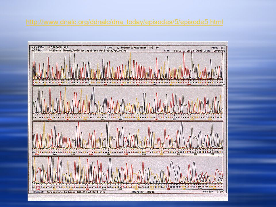
http://www.dnalc.org/ddnalc/dna_today/episodes/5/episode5.html
9
Molecular Genetics Molecular genetics and molecular biology are almost synonomous terms A “ hybrid” science The change in the understanding of life has led to a revolution in the field of Biology Molecular genetics and molecular biology are almost synonomous terms A “ hybrid” science The change in the understanding of life has led to a revolution in the field of Biology

10
Molecular Genetics The result of an amalgam of a variety of physical and biological sciences Genetics, microbiology, biochemistry, physical chemistry, and physics Driven by the need to understand the underlying principles of life and the reactions of life The result of an amalgam of a variety of physical and biological sciences Genetics, microbiology, biochemistry, physical chemistry, and physics Driven by the need to understand the underlying principles of life and the reactions of life

11
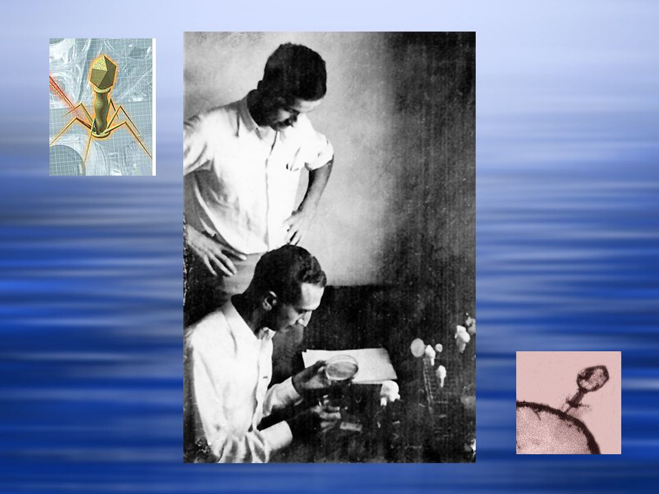
Max Delbruck Ilustrates the blend in scientific disciplines German immigrant Originally trained in physical chemistry and theoretical physics Converted to molecular genetics Collaborated with Salvador Luria on the characterization and genetics of bacteriophages German immigrant Originally trained in physical chemistry and theoretical physics Converted to molecular genetics Collaborated with Salvador Luria on the characterization and genetics of bacteriophages

13
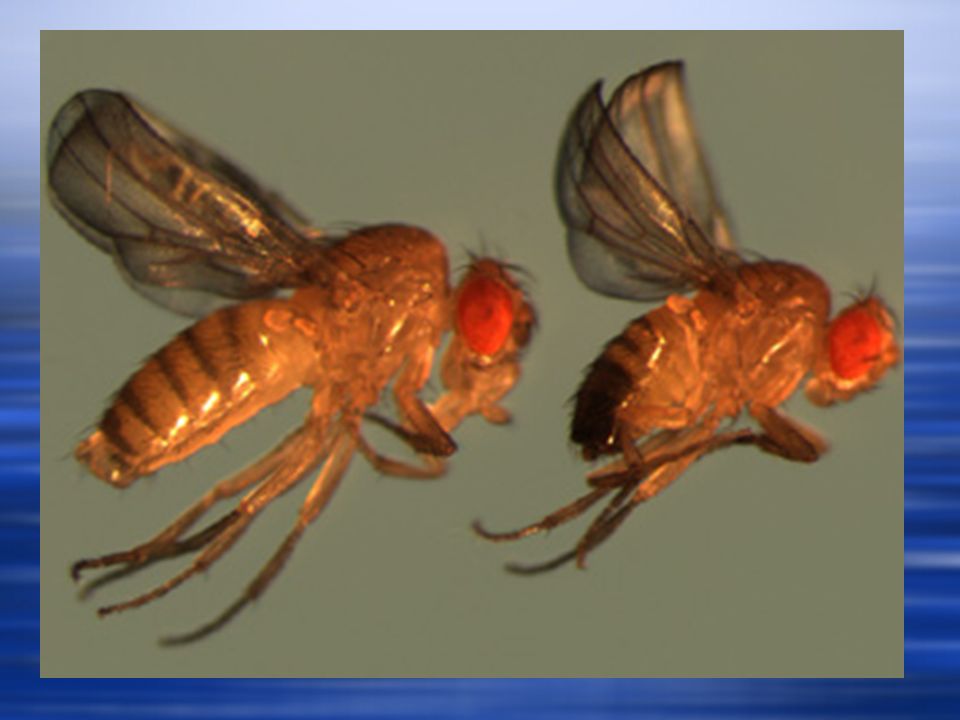
Molecular Genetics - Origins Thomas Hunt Morgan- Columbia University The physical nature of the gene A discovery in 1910 changed the course of genetics Developed experimental model for the study of modern genetics- the fruit fly – Drosophila melanogaster The white eyed male mutant appeared in a culture of flies in the fly room and this was the beginning of a search for mutants Thomas Hunt Morgan- Columbia University The physical nature of the gene A discovery in 1910 changed the course of genetics Developed experimental model for the study of modern genetics- the fruit fly – Drosophila melanogaster The white eyed male mutant appeared in a culture of flies in the fly room and this was the beginning of a search for mutants

14
White and Wild type

15
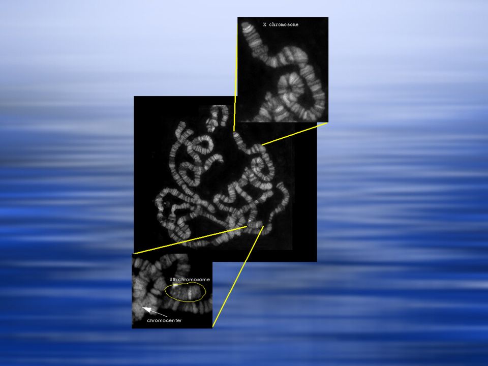
Easy to cultivate Prolific progeny Small and inexpensive Large polytene chromosomes Diploid number 8 Many mutations Easy to cultivate Prolific progeny Small and inexpensive Large polytene chromosomes Diploid number 8 Many mutations

16
Hermann Joseph Muller X rays cause mutations Produced a variety of flies with phenotypes such asvestigial

17
Alfred Sturdevant produced the first genetic map from linkage experiments Genes were related to position on the chromosome map Mutants were related to differences in the appearance of the polytene chromosomes due to staining

20
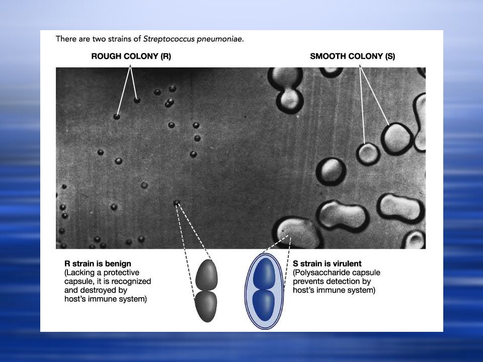
DNA as Genetic Material Transformation Griffith in 1928 observed the change of non-virulent organisms into virulent ones as a result of “transformation” MacLeod and McCarty in 1944 showed that the transforming principle was DNA Griffith in 1928 observed the change of non-virulent organisms into virulent ones as a result of “transformation” MacLeod and McCarty in 1944 showed that the transforming principle was DNA

21
Figure 11.1

23
Transforming principle

24
Avery, McLeod, and McCarty

25
Proof of the Transforming Principle Chemical analysis of sample containing the transforming principle showed that the major component was a deoxyribose -containing nucleic acid Physical measurements show that the sample contained a highly viscous substance having the properties of DNA Incubatyion with trypsin or chymotrypsin, enzymes that catalzye protein hydrolysis or with ribonuclease( RNase), an enzyme that catalyzes RNA hydrolysis did not affect the transforming principle Incubation with DNase, an enzyme that catalyzes DNA hydrolysis inactivates the transforming principle Chemical analysis of sample containing the transforming principle showed that the major component was a deoxyribose -containing nucleic acid Physical measurements show that the sample contained a highly viscous substance having the properties of DNA Incubatyion with trypsin or chymotrypsin, enzymes that catalzye protein hydrolysis or with ribonuclease( RNase), an enzyme that catalyzes RNA hydrolysis did not affect the transforming principle Incubation with DNase, an enzyme that catalyzes DNA hydrolysis inactivates the transforming principle
, an enzyme that catalyzes RNA hydrolysis did not affect the transforming principle Incubation with DNase, an enzyme that catalyzes DNA hydrolysis inactivates the transforming principle Chemical analysis of sample containing the transforming principle showed that the major component was a deoxyribose -containing nucleic acid Physical measurements show that the sample contained a highly viscous substance having the properties of DNA Incubatyion with trypsin or chymotrypsin, enzymes that catalzye protein hydrolysis or with ribonuclease( RNase), an enzyme that catalyzes RNA hydrolysis did not affect the transforming principle Incubation with DNase, an enzyme that catalyzes DNA hydrolysis inactivates the transforming principle")
26
Transfection

27
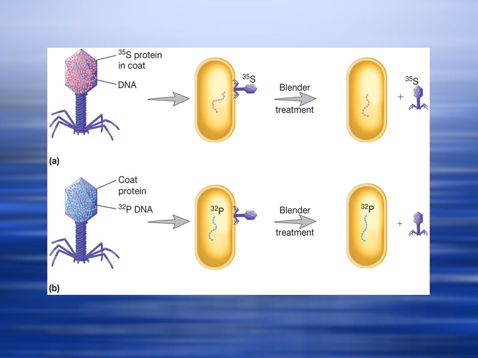
DNA as Genetic Material ( of viruses) Hershey and Chase, 1952 used bacteriophage T2 infection as model DNA labeled with 32 P;protein coat labeled with 35 S Only DNA entered cell but both new DNA and protein coats synthesized and incorporated into new viruses indicating that DNA had the genetic information for synthesis of both of these viral components Hershey and Chase, 1952 used bacteriophage T2 infection as model DNA labeled with 32 P;protein coat labeled with 35 S Only DNA entered cell but both new DNA and protein coats synthesized and incorporated into new viruses indicating that DNA had the genetic information for synthesis of both of these viral components
 Hershey and Chase, 1952 used bacteriophage T2 infection as model DNA labeled with 32 P;protein coat labeled with 35 S Only DNA entered cell but both new DNA and protein coats synthesized and incorporated into new viruses indicating that DNA had the genetic information for synthesis of both of these viral components Hershey and Chase, 1952 used bacteriophage T2 infection as model DNA labeled with 32 P;protein coat labeled with 35 S Only DNA entered cell but both new DNA and protein coats synthesized and incorporated into new viruses indicating that DNA had the genetic information for synthesis of both of these viral components")
28
T2 phage

30
Chargaff’s Rule Analyzed DNA from a variety of sources and improved both the separation and quantitation of the DNA bases [C] = [G] and [A] = [T] Today this is applied to the G=C or G.C pairs. Scientists describe the G+C content in organisms Analyzed DNA from a variety of sources and improved both the separation and quantitation of the DNA bases [C] = [G] and [A] = [T] Today this is applied to the G=C or G.C pairs. Scientists describe the G+C content in organisms
![Chargaff’s Rule Analyzed DNA from a variety of sources and improved both the separation and quantitation of the DNA bases [C] = [G] and [A] = [T] Today this is applied to the G=C or G.C pairs.](http://images.slideplayer.com/21/6303547/slides/slide_30.jpg "Scientists describe the G+C content in organisms Analyzed DNA from a variety of sources and improved both the separation and quantitation of the DNA bases [C] = [G] and [A] = [T] Today this is applied to the G=C or G.C pairs. Scientists describe the G+C content in organisms.")
31
G+C Now used as a means of classifying bacteria G+C content varies in Gram Positive Bacteria G+C content ranges from.27 in Clostridium to.76 for Sarcina Most Eukaryotes have a value close to 50% Now used as a means of classifying bacteria G+C content varies in Gram Positive Bacteria G+C content ranges from.27 in Clostridium to.76 for Sarcina Most Eukaryotes have a value close to 50%

32
G+C content G+C content = [G] +[C] / all bases in DNA
![G+C content G+C content = [G] +[C] / all bases in DNA](http://images.slideplayer.com/21/6303547/slides/slide_32.jpg "G+C content G+C content = [G] +[C] / all bases in DNA")
33
The Race for the Double Helix Rosalind Franklin and Maurice Wilkins at Kings College Studied the A and B forms of DNA Rosalind’s famous x- ray crystallography picture of the B form held the secret, but she didn’t realize its significance Rosalind Franklin and Maurice Wilkins at Kings College Studied the A and B forms of DNA Rosalind’s famous x- ray crystallography picture of the B form held the secret, but she didn’t realize its significance

34
Rosalind Franklin Technically and scientifically a gifted scientist Focused on the A form of DNA and missed the double helix Technically and scientifically a gifted scientist Focused on the A form of DNA and missed the double helix

35
The Race for the Double Helix Watson and Crick formed an unlikely partnership A 22 year old PhD and a thirty + PhD “want to be” embarked on a model making venture at Cambridge Used the research of other scientists to determine the nature of the double helix Watson and Crick formed an unlikely partnership A 22 year old PhD and a thirty + PhD “want to be” embarked on a model making venture at Cambridge Used the research of other scientists to determine the nature of the double helix

36
Nucleic Acid Composition DNA and RNA DNA – Basic Molecules a.Purines – adenine and guanine b.Pyrmidines – cytosine and thymine c.Sugar – Deoxyribose d.Phosphate phosphate group http://www.dnai.org/index.htmhttp://www.dnai.org/index.htm - DNA background DNA – Basic Molecules a.Purines – adenine and guanine b.Pyrmidines – cytosine and thymine c.Sugar – Deoxyribose d.Phosphate phosphate group http://www.dnai.org/index.htmhttp://www.dnai.org/index.htm - DNA background

37
Nucleotides Sugar Phosphate Base Adenine and guanine are purines Thymine and Cytosine are pyrimidines

38
Deoxyribose in DNA

39
Double Helix Two polynucleotide strands joined by phosphodiester bonds( backbone) Complementary base pairing in the center of the molecule A= T and C G – base pairing. Two hydrogen bonds between A and T and three hydrogen bonds between C and G. A purine is bonded to a complementary pyrimidine Bases are attached to the 1’ C in the sugar by a glycosidic linkage At opposite ends of the strand – one strand has the 3’hydroxyl, the other the 5’ hydroxyl of the sugar molecule Two polynucleotide strands joined by phosphodiester bonds( backbone) Complementary base pairing in the center of the molecule A= T and C G – base pairing. Two hydrogen bonds between A and T and three hydrogen bonds between C and G. A purine is bonded to a complementary pyrimidine Bases are attached to the 1’ C in the sugar by a glycosidic linkage At opposite ends of the strand – one strand has the 3’hydroxyl, the other the 5’ hydroxyl of the sugar molecule
 Complementary base pairing in the center of the molecule A= T and C G – base pairing. Two hydrogen bonds between A and T and three hydrogen bonds between C and G. A purine is bonded to a complementary pyrimidine Bases are attached to the 1’ C in the sugar by a glycosidic linkage At opposite ends of the strand – one strand has the 3’hydroxyl, the other the 5’ hydroxyl of the sugar molecule.")
40
DNA Structure http://www.johnkyrk.com/DNAanatomy.htmlhttp://www.johnkyrk.com/DNAanatomy.html - DNA structure

41
Double helix( continued) The double helix is right handed – the chains turn counter-clockwise. As the strand turn around each other they form a major and minor groove. The is a distance of.34nm between each base The distance between two major grooves is 3.4nm or 10 bases The diameter of the strand is 2nm The double helix is right handed – the chains turn counter-clockwise. As the strand turn around each other they form a major and minor groove. The is a distance of.34nm between each base The distance between two major grooves is 3.4nm or 10 bases The diameter of the strand is 2nm

42
Complementary Base Pairing Adenine pairs with Thymine Cytosine pairs with Guanine Adenine pairs with Thymine Cytosine pairs with Guanine

43
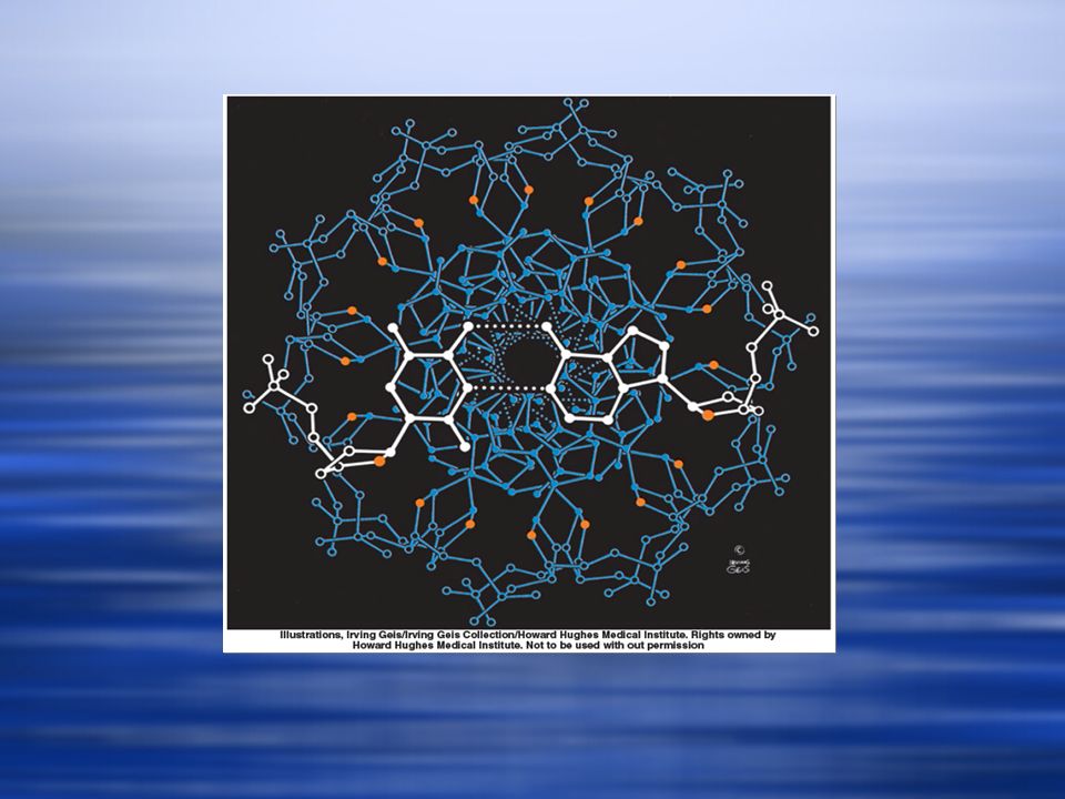
The end view of DNA This view shows the double helix and the outer backbone with the bases in the center. An AT base pair is highlighted in white This view shows the double helix and the outer backbone with the bases in the center. An AT base pair is highlighted in white

45
Double helix and anti-parallel DNA is a directional molecule The complementary strands run in opposite directions One strand runs 3’-5’ The other strand runs 5’ to 3’ ( the end of the 5’ has the phosphates attached, while the 3’ end has a hydroxyl exposed) DNA is a directional molecule The complementary strands run in opposite directions One strand runs 3’-5’ The other strand runs 5’ to 3’ ( the end of the 5’ has the phosphates attached, while the 3’ end has a hydroxyl exposed)
 DNA is a directional molecule The complementary strands run in opposite directions One strand runs 3’-5’ The other strand runs 5’ to 3’ ( the end of the 5’ has the phosphates attached, while the 3’ end has a hydroxyl exposed)")
46
Prokaryote DNA Tightly coiled Coiling maintained by molecules similar to the coiling in eukaryotes Circular ds molecule Borrelia burgdoferi ( Lyme Disease )has a linear chromosome Other bacteria have multiple chromosomes Agrobacterium tumefaciens ( Produces Crown Gall disease in plants) has both circular and linear Tightly coiled Coiling maintained by molecules similar to the coiling in eukaryotes Circular ds molecule Borrelia burgdoferi ( Lyme Disease )has a linear chromosome Other bacteria have multiple chromosomes Agrobacterium tumefaciens ( Produces Crown Gall disease in plants) has both circular and linear
has a linear chromosome Other bacteria have multiple chromosomes Agrobacterium tumefaciens ( Produces Crown Gall disease in plants) has both circular and linear Tightly coiled Coiling maintained by molecules similar to the coiling in eukaryotes Circular ds molecule Borrelia burgdoferi ( Lyme Disease )has a linear chromosome Other bacteria have multiple chromosomes Agrobacterium tumefaciens ( Produces Crown Gall disease in plants) has both circular and linear")
47
Prokaryote chromosomes Circular DNA

48
Mitochondria Mitochondrial DNA( mt DNA) 16,500 base pairs 37 genes 24 encode RNA Defects lead to diseases that are related to energy Mitochondrial DNA( mt DNA) 16,500 base pairs 37 genes 24 encode RNA Defects lead to diseases that are related to energy
 16,500 base pairs 37 genes 24 encode RNA Defects lead to diseases that are related to energy Mitochondrial DNA( mt DNA) 16,500 base pairs 37 genes 24 encode RNA Defects lead to diseases that are related to energy")
49
Chloroplast DNA Chloroplast DNA( cp DNA) is larger than mitochondrial DNA 195,000 bp Genes for photosynthesis Cp ribosomal RNAs Chloroplast DNA( cp DNA) is larger than mitochondrial DNA 195,000 bp Genes for photosynthesis Cp ribosomal RNAs
 is larger than mitochondrial DNA 195,000 bp Genes for photosynthesis Cp ribosomal RNAs Chloroplast DNA( cp DNA) is larger than mitochondrial DNA 195,000 bp Genes for photosynthesis Cp ribosomal RNAs")
50
Heavy and Light N Meselson and Stahl experiment

51
In the first generation of E. coli, all the DNA was heavy After one generation, the DNA was half heavy and half light In the first generation of E. coli, all the DNA was heavy After one generation, the DNA was half heavy and half light

52
DNA Replication –Semi Conservative

53
DNA Replication DNA opens at an Ori ( origin of replication) Combination of many enzymes coordinate the replicative process Template strand used to make the copy DNA polymerases read the template and match the complementary base DNA opens at an Ori ( origin of replication) Combination of many enzymes coordinate the replicative process Template strand used to make the copy DNA polymerases read the template and match the complementary base
 Combination of many enzymes coordinate the replicative process Template strand used to make the copy DNA polymerases read the template and match the complementary base DNA opens at an Ori ( origin of replication) Combination of many enzymes coordinate the replicative process Template strand used to make the copy DNA polymerases read the template and match the complementary base")
54
Degradation of DNA Endonucleases cleave DNA and RNA, by cutting between individual bonds Some endonucleases cleave one strand some cleave both strands at a specific point or sequence( restriction nucleasess) Endonucleases cleave DNA and RNA, by cutting between individual bonds Some endonucleases cleave one strand some cleave both strands at a specific point or sequence( restriction nucleasess)
 Endonucleases cleave DNA and RNA, by cutting between individual bonds Some endonucleases cleave one strand some cleave both strands at a specific point or sequence( restriction nucleasess)")
55
The Flow of Genetic Information from one generation to the next DNA stores genetic information Information is duplicated by replication and is passed on to next generation from one generation to the next DNA stores genetic information Information is duplicated by replication and is passed on to next generation

56
The Flow of Genetic Information within a single cell Process called gene expression DNA divided into genes transcription yields a ribonucleic acid (RNA) copy of specific genes translation uses information in messenger RNA (mRNA) to synthesize a polypeptide Also involves activities of transfer RNA (tRNA) and ribosomal RNA (rRNA) Process called gene expression DNA divided into genes transcription yields a ribonucleic acid (RNA) copy of specific genes translation uses information in messenger RNA (mRNA) to synthesize a polypeptide Also involves activities of transfer RNA (tRNA) and ribosomal RNA (rRNA)
 copy of specific genes translation uses information in messenger RNA (mRNA) to synthesize a polypeptide Also involves activities of transfer RNA (tRNA) and ribosomal RNA (rRNA) Process called gene expression DNA divided into genes transcription yields a ribonucleic acid (RNA) copy of specific genes translation uses information in messenger RNA (mRNA) to synthesize a polypeptide Also involves activities of transfer RNA (tRNA) and ribosomal RNA (rRNA)")
57
Flow of Genetic Information in Cells

59
Nucleic Acid Structure Ribonucleic Acid (RNA) Polymer of nucleotides Contains the bases adenine, guanine, cytosine and uracil Sugar is ribose Most RNA molecules are single stranded Polymer of nucleotides Contains the bases adenine, guanine, cytosine and uracil Sugar is ribose Most RNA molecules are single stranded
 Polymer of nucleotides Contains the bases adenine, guanine, cytosine and uracil Sugar is ribose Most RNA molecules are single stranded Polymer of nucleotides Contains the bases adenine, guanine, cytosine and uracil Sugar is ribose Most RNA molecules are single stranded")
60
RNA Types of RNA a.Messenger b.Transfer c.Ribosomal d.micro RNAs ( regulatory RNAs) Types of RNA a.Messenger b.Transfer c.Ribosomal d.micro RNAs ( regulatory RNAs)
 Types of RNA a.Messenger b.Transfer c.Ribosomal d.micro RNAs ( regulatory RNAs)")
61
Messenger RNA

62
16s rRNA

63
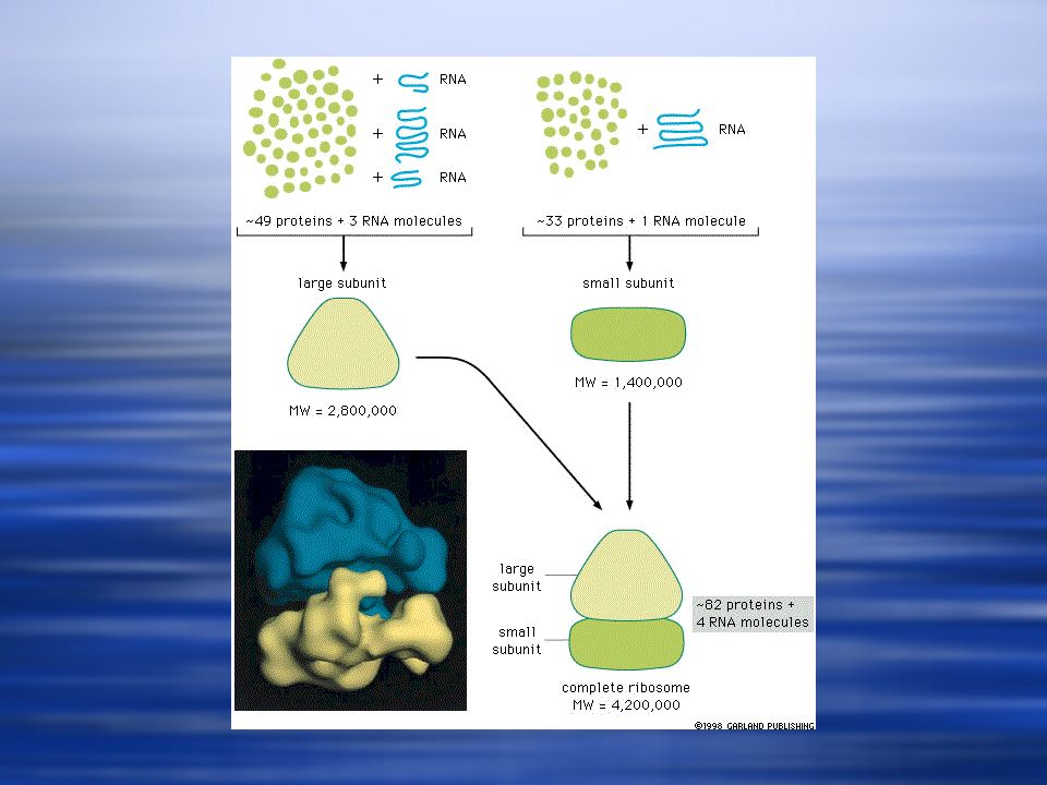
Ribosomal RNA

65
tRNA

66
RNA viruses Reoviruses Retroviruses Enteroviruses Reoviruses Retroviruses Enteroviruses

67
Genomics of RNA viruses Genomes - + RNA - RNA segmented RNA Ds RNA Genomes - + RNA - RNA segmented RNA Ds RNA

68
Polio virus

69
Polio Virus- + ss RNA virus

70
Viroids Infectious agents that causes disease in higher plants Small circular loops of RNA The viroid RNA is infectious and its is not surrounded by a capsid Viroids RNA replicates autonomously

71
Viroids PSTV Potato SpindleTuber Viroid PSTV Potato SpindleTuber Viroid

72
Proteins are polymers Proteins are polymers of amino acids. They are molecules with diverse structures and functions. Polymers are made up of units called monomers The monomers in proteins are the 20 amino acids Proteins are polymers of amino acids. They are molecules with diverse structures and functions. Polymers are made up of units called monomers The monomers in proteins are the 20 amino acids

73
Protein Facts Proteins: Polymers of Amino Acids Proteins are polymers of amino acids. They are molecules with diverse structures and functions. Each different type of protein has a characteristic amino acid composition and order. Proteins range in size from a few amino acids to thousands of them. Folding is crucial to the function of a protein and is influenced largely by the sequence of amino acids. Proteins: Polymers of Amino Acids Proteins are polymers of amino acids. They are molecules with diverse structures and functions. Each different type of protein has a characteristic amino acid composition and order. Proteins range in size from a few amino acids to thousands of them. Folding is crucial to the function of a protein and is influenced largely by the sequence of amino acids.

74
Proteins: Polymers of Amino Acids Each different type of protein has a characteristic amino acid composition and order. Proteins range in size from a few amino acids to thousands of them. Folding is crucial to the function of a protein and is influenced largely by the sequence of amino acids. Each different type of protein has a characteristic amino acid composition and order. Proteins range in size from a few amino acids to thousands of them. Folding is crucial to the function of a protein and is influenced largely by the sequence of amino acids.

75
Proteins are complex molecules They have levels of structure Structure based upon the sequence of the amino acids They have levels of structure Structure based upon the sequence of the amino acids

76
Polar side chains

77
Non Polar Hydrophobic side chains

78
Electrical charged hydrophilic

79
Function of Proteins - continued Enzymes – Biological catalysts Transport of small molecules – Albumin and haptoglobin Transport of oxygen – hemoglobin and myoglobin Membrane proteins – to assist in support Channels in membranes – to allow the passage of molecules or ions Electron carriers in electron transport in the production of ATP Enzymes – Biological catalysts Transport of small molecules – Albumin and haptoglobin Transport of oxygen – hemoglobin and myoglobin Membrane proteins – to assist in support Channels in membranes – to allow the passage of molecules or ions Electron carriers in electron transport in the production of ATP

80
Functions( continued)i Clotting proteins Immune proteins to fight infectious agents Histones – DNA binding proteins Toxins to repel or kill other organisms Bacteriocins – molecules produced by bacteria against bacteria Clotting proteins Immune proteins to fight infectious agents Histones – DNA binding proteins Toxins to repel or kill other organisms Bacteriocins – molecules produced by bacteria against bacteria
i Clotting proteins Immune proteins to fight infectious agents Histones – DNA binding proteins Toxins to repel or kill other organisms Bacteriocins – molecules produced by bacteria against bacteria Clotting proteins Immune proteins to fight infectious agents Histones – DNA binding proteins Toxins to repel or kill other organisms Bacteriocins – molecules produced by bacteria against bacteria")
81
Functions of proteins Hormones – Growth hormone Receptors – to Receive information so that cell can communicate with other cells Neurotransmitters – messenger molecules – to send information between neurons Cytoskeleton – actin, myosin, and collagen – the structure of connective tissue and muscles Antibodies – Immunoglobulins to fight disease Hormones – Growth hormone Receptors – to Receive information so that cell can communicate with other cells Neurotransmitters – messenger molecules – to send information between neurons Cytoskeleton – actin, myosin, and collagen – the structure of connective tissue and muscles Antibodies – Immunoglobulins to fight disease

82
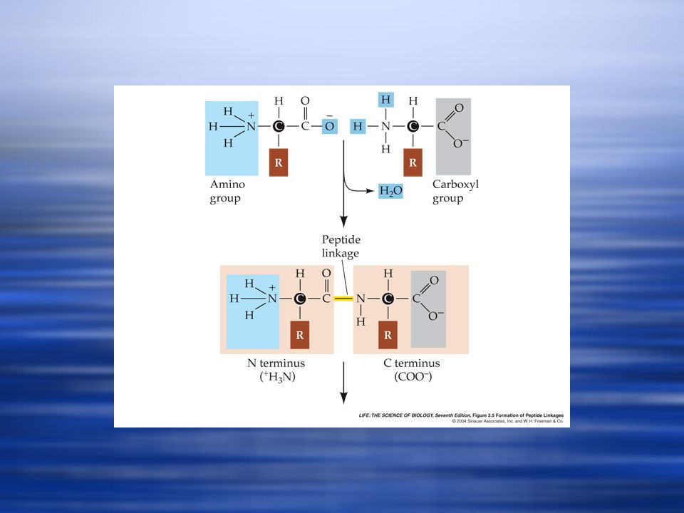
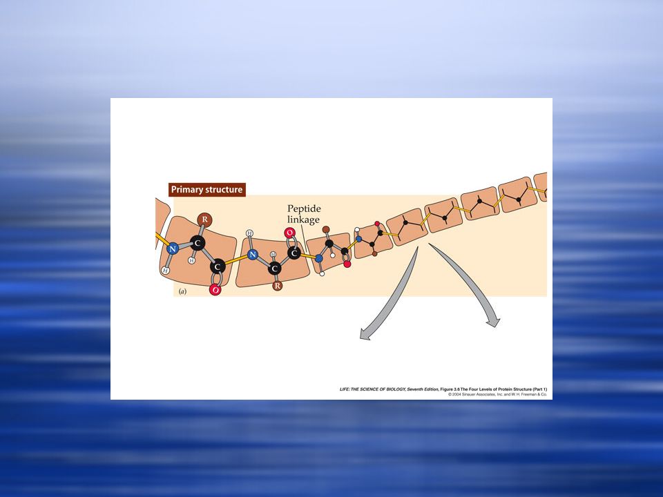
Four levels of Protein Structure There are four levels of protein structure: primary, secondary, tertiary, and quaternary. The precise sequence of amino acids is called its primary structure. The peptide backbone consists of repeating units of atoms: N—C—C—N—C—C. Enormous numbers of different proteins are possible. There are four levels of protein structure: primary, secondary, tertiary, and quaternary. The precise sequence of amino acids is called its primary structure. The peptide backbone consists of repeating units of atoms: N—C—C—N—C—C. Enormous numbers of different proteins are possible.

86
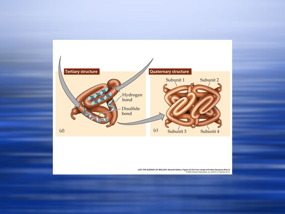
The causes of Tertiary structure

Similar presentations


 Harmless bacteria (rough colonies) Heat-killed, disease- causing bacteria (smooth colonies) Control (no growth)>")


