Download presentation
Presentation is loading. Please wait.

1
Gram Negative Bacteria

2
Enterobacteriaceae

3
Infections associated with Enterobacteriaceae
Fig. 30-1

4
Enterobacteriaceae Gram negative rods
most are motile (peritrichous flagella) most are encapsulated LPS is a virulence factor many have “serum resistance” inhibitions of complement proteins (Ab’s can’t attack) Associated with: Enteric (GI) infections Bacteremia (bacteria in the blood) UTIs (urinary tract infections)
 most are encapsulated. LPS is a virulence factor. many have serum resistance inhibitions of complement proteins (Ab’s can’t attack) Associated with: Enteric (GI) infections. Bacteremia (bacteria in the blood) UTIs (urinary tract infections)")
5
Diarrhea ~1 billion people worldwide suffer from acute diarrhea at least once/year 5-8 million deaths/year primarily in developing nations ~100 million infections in U.S. 250,000 require hospitalization 3000 die 90% of acute diarrhea caused by infectious agents fecal-oral contamination

6
High risk groups in U.S. Travelers – 40% of tourists to Latin America, Africa, Asia develop “traveler’s diarrhea” ETEC – enterotoxigenic E. coli Shigella Salmonella Campylobacter Giardia (Russia, camper’s, swimmers) Cyclospora (Nepal) Consumers of certain foods Chicken, mayonnaise, creams, eggs: picnic, banquet, restaurant (Salmonella, Campylobacter, Shigella) Hamburger: undercooked (EHEC – enterohemorrhagic E. coli) Fried rice (B. cereus) Seafood (Salmonella, Vibrio, hepatitis A) Fermented tofu (C. botulinum) Immunocompromised
 Cyclospora (Nepal) Consumers of certain foods. Chicken, mayonnaise, creams, eggs: picnic, banquet, restaurant (Salmonella, Campylobacter, Shigella) Hamburger: undercooked (EHEC – enterohemorrhagic E. coli) Fried rice (B. cereus) Seafood (Salmonella, Vibrio, hepatitis A) Fermented tofu (C. botulinum) Immunocompromised.")
7
High risk groups in U.S. (cont.)
Daycare participants and their family Shigella Giardia Cryptosporidium rotavirus Institutionalized persons Nosocomial (acquired in a hospital) infections of hospital patients Clostridium difficile
 infections of hospital patients. Clostridium difficile.")
8
Treatment - gastrointestinal disease
Fluid/electrolyte replacement fluid alone for mild cases dehydration most common cause of death due to diarrheal disease Antibiotics not used unless systemic/severe e.g. enteric fever immunosuppressed Antibiotic prophylaxis for those traveling to high-risk countries (esp. immunocompromised)
")
9
Enteric infections Overview of symptoms non-inflammatory inflammatory
nausea vomiting diarrhea inflammatory Dysentery (severe diarrhea containing mucus and/or blood) Invasive (systemic) Typhoid Fever (enteric fever)
 Invasive (systemic) Typhoid Fever (enteric fever)")
10
“Common” organisms associated with enteric infections
II III Mechanism: Non-inflammatory (enterotoxin) Inflammatory (invasive, cytotoxin) Penetrating (invasive, spread) Location: proximal small bowel colon distal small bowel Illness: Diarrhea Dysentery Enteric fever Stool exam: no fecal leukocytes blood, fecal PMNs (polymorphonuclear leukocytes = neutrophils) fecal mononuclear leukocytes (monocytes, lymphocytes) Example organisms: V. cholerae E. coli Salmonella Campylobacter Giardia Cryptosporidium Rotavirus Norwalk-like agents Shigella Invasive E. coli S. enteritidis C. difficile E. histolytica B. coli S. typhi Y. enterocolitica
 Inflammatory (invasive, cytotoxin) Penetrating. (invasive, spread) Location: proximal small bowel. colon. distal small bowel. Illness: Diarrhea. Dysentery. Enteric fever. Stool exam: no fecal leukocytes. blood, fecal PMNs. (polymorphonuclear leukocytes = neutrophils) fecal mononuclear leukocytes (monocytes, lymphocytes) Example organisms: V. cholerae. E. coli. Salmonella. Campylobacter. Giardia. Cryptosporidium. Rotavirus. Norwalk-like agents. Shigella. Invasive E. coli. S. enteritidis. C. difficile. E. histolytica. B. coli. S. typhi. Y. enterocolitica.")
11
Pathogenicity of enteric bacteria
Host factors personal hygiene fecal-oral contamination gastric acidity enteric microflora specific immunity

12
Pathogenicity of enteric bacteria (cont.)
Microbial factors Toxins Neurotoxins usually ingested as preformed toxins Staphylococcal toxins (Staph. aureus) Botulinum toxin (Clostridium. botulinum) Enterotoxins having a direct effect on intestinal mucosa (elicit fluid secretions) Cholera toxin (Vibrio. cholerae) E. coli toxins Cytotoxins mucosal destruction (often see dysentery) Shigella dysenteriae Clostridium perfringens S. aureus Clostridium difficile
 Botulinum toxin (Clostridium. botulinum) Enterotoxins. having a direct effect on intestinal mucosa (elicit fluid secretions) Cholera toxin (Vibrio. cholerae) E. coli toxins. Cytotoxins. mucosal destruction (often see dysentery) Shigella dysenteriae. Clostridium perfringens. S. aureus. Clostridium difficile.")
13
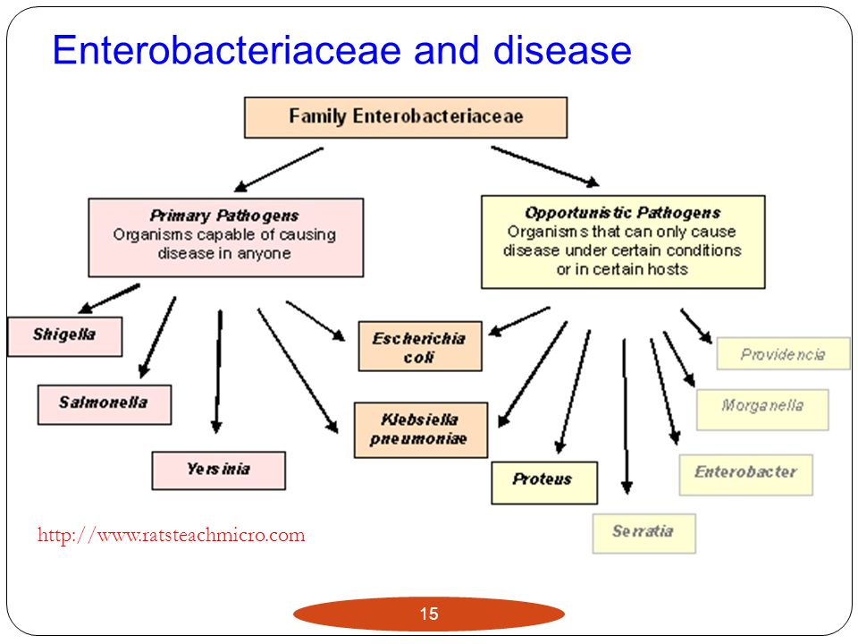
Enterobacteriaceae Ubiquious (they are everywhere) - soil, water, vegetation, normal intestinal flora ~40 genera, 150 species Gram negative, facultative anaerobic rods oxidase negative - no cytochrome oxidase members of family commonly associated with human disease: Escherichia Salmonella Shigella Yersinia Klebsiella Serratia Proteus

14
Enterobacteriaceae pathogens
associated with opportunistic infections septicemia pneumonia meningitis urinary tract infections (UTI) can be primary pathogens (unrelated to immune status)
 can be primary pathogens (unrelated to immune status)")
15
Enterobacteriaceae and disease
16
Lab identification of the enterics
Green sheen; black nucleated centers MacConkey agar selective and differential EMB agar selective and differential

17
TSI Yellow = sugar fermentation Black = H2S positive
Air bubble = gas production

18
Salmonella-Shigella differentiation
Salmonella-Shigella agar (SS agar) Hecktoen agar – inhibits most Gram +, various sugars to judge fermentation (fermenters – red/orange; non-fermenters – green), H2S production (black) Note: Shigella is a non-fermenter, H2S- K. pneumoniae (L); M. luteus (R) S. typhimurium (L); P. vulgaris (R) H2S production (black ppt)
 Hecktoen agar – inhibits most Gram +, various sugars to judge fermentation (fermenters – red/orange; non-fermenters – green), H2S production (black) Note: Shigella is a. non-fermenter, H2S- K. pneumoniae (L); M. luteus (R) S. typhimurium (L); P. vulgaris (R) H2S production (black ppt)")
19
E. coli and the serotypes
Lactose positive note: many intestinal pathogens are lactose negative ex. Salmonella, Shigella, Yersinia grouped based on surface antigens (serotypes) O antigen (lipopolysaccharide) H antigen (flagellar) K antigen (capsular) O157:H7 (EHEC – enterohemorrhagic E. coli) O148:H28 (ETEC – enterotoxigenic E. coli)
 O antigen (lipopolysaccharide) H antigen (flagellar) K antigen (capsular) O157:H7 (EHEC – enterohemorrhagic E. coli) O148:H28 (ETEC – enterotoxigenic E. coli)")
20
E. coli serotype differentiation
1. immunologic assay 2. growth on MacConkey agar with sorbitol (S- Mac) most E. coli can ferment sorbitol (form pink colonies) E. coli O157:H7 does not ferment sorbitol (colonies are clear/colorless)
 most E. coli can ferment sorbitol (form pink colonies) E. coli O157:H7 does not ferment sorbitol (colonies are clear/colorless)")
21
E. coli pathology most strains of the pathogenic E. coli are capable of pathology only within the intestinal tract (some exceptions) most pathogenic strains associated with disease in developing countries (except EHEC is common in the USA) dependent upon strain, different disease severity/symptoms (e.g. pathotype)
 dependent upon strain, different disease severity/symptoms (e.g. pathotype)")
22
E. coli pathology (cont.)
pathogenic strains produce virulence factors found on: Plasmids (a DNA molecule that is separate from, and can replicate independently of, the chromosomal DNA) Bacteriophages (viruses that infect bacteria) virulence factors include: Fimbriae (allow bacteria to stack up on each other to shelter themselves from immune system secretion systems (the process of toxin release) toxins
 Bacteriophages (viruses that infect bacteria) virulence factors include: Fimbriae (allow bacteria to stack up on each other to shelter themselves from immune system. secretion systems (the process of toxin release) toxins.")
23
E. coli strains/serotypes
most normal flora E. coli are non-pathogenic in intestinal tract pathogenic strains: EPEC (enteropathic) ETEC (enterotoxic) EHEC (enterohemorrhagic) EIEC (enteroinvasive) EAEC (enteroaggregative) UPEC (uropathogenic)
 ETEC (enterotoxic) EHEC (enterohemorrhagic) EIEC (enteroinvasive) EAEC (enteroaggregative) UPEC (uropathogenic)")
24
Enteropathogenic E. coli (EPEC)
destruction of surface microvilli (small intestines) fever diarrhea (infantile) malabsorption of fluids vomiting/nausea hard to replace fluids non-bloody stools common in developing countries (rare in U.S.)
 fever. diarrhea (infantile) malabsorption of fluids. vomiting/nausea. hard to replace fluids. non-bloody stools. common in developing countries (rare in U.S.)")
25
EPEC pathology - diarrhea
since this is primarily a disease of the young (less the 6 months old), fluid replacement is important intense vomiting - i.v. fluids are usually required disease self-limiting (antibiotics usually not required) breast feeding seems to have a strong protective effect IgA and other factors decrease bacterial attachment
, fluid replacement is important. intense vomiting - i.v. fluids are usually required. disease self-limiting (antibiotics usually not required) breast feeding seems to have a strong protective effect. IgA and other factors decrease bacterial attachment.")
26
Enterotoxigenic E. coli (ETEC) “Traveler’s diarrhea”
primarily in developing nations ~650 million cases/year ~80,000 in travelers from the U.S. Two types of toxins heat labile toxins (LT) similar to cholera toxin (although not as severe) lack of absorption of fluids = watery diarrhea heat stabile toxins (ST) no inflammation, self-limiting
 similar to cholera toxin (although not as severe) lack of absorption of fluids = watery diarrhea. heat stabile toxins (ST) no inflammation, self-limiting.")
27
Enterotoxigenic E. coli (ETEC)
many different ETEC strains disease is self-limiting watery diarrhea common symptom exposure provides immunity adults living in endemic areas, often immune children, through exposure to the many strains, eventually develop immunity therapy fluid replacement bismuth subsalicylate tablets (Pepto-Bismol, etc.) provide antibiotics to travelers in the event they get sick while abroad
 provide antibiotics to travelers in the event they get sick while abroad.")
28
Enterohemorrhagic E. coli (EHEC)
usually O157:H7 many different types of E. coli identifying O157:H7…finding a slightly different hay in a large haystack. strain must have virulence/toxin genes. Vero toxin (VTEC) = “shiga-like” toxin (cytotoxin) aka Shiga toxin-producing E. coli (STEC) AB toxin “A” inactivates 28S rRNA = stop protein synthesis death of epithelial cells
 = shiga-like toxin (cytotoxin) aka Shiga toxin-producing E. coli (STEC) AB toxin. A inactivates 28S rRNA = stop protein synthesis. death of epithelial cells.")
29
EHEC ~75,000 cases in U.S./year ~60 deaths estimated that only ~100 cells can cause infection many EHEC serotypes (~50) in U.S., most diseases due to 0157:H7 disease can be mild to severe (hemorrhagic colitis = bloody diarrhea) depends on strain patient status (age, physiological status)
 in U.S., most diseases due to 0157:H7. disease can be mild to severe (hemorrhagic colitis = bloody diarrhea) depends on strain. patient status (age, physiological status)")
30
EHEC symptoms Hemorrhagic (hemorrhagic coilitis)
bloody, copious diarrhea few leukocytes afebrile (no fever) usually self limiting (in ~1 week) Hemolytic Uremic Syndrome (HUS) hemolytic anemia thrombocytopenia kidney failure 5-10% of kids infected with EHEC
 usually self limiting (in ~1 week) Hemolytic Uremic Syndrome (HUS) hemolytic anemia. thrombocytopenia. kidney failure. 5-10% of kids infected with. EHEC.")
31
EHEC cattle seem to be the major reservoir
humans become infected by ingesting undercooked meat (beef), unpasteurized milk, fruits and fruit juices (fecal-contaminated fruit), uncooked vegetables detection: 0157 strains do not ferment sorbitol (or do so slowly) follow up with serological/biochemical testing to confirm therapy supportive therapy
, unpasteurized milk, fruits and fruit juices (fecal-contaminated fruit), uncooked vegetables. detection: 0157 strains do not ferment sorbitol (or do so slowly) follow up with serological/biochemical testing to confirm. therapy. supportive therapy.")
32
Enteroinvasive E. coli (EIEC )
dysentery resembles shigellosis (Shigella dysenteriae) relatively rare in U.S. common strains: O124, O143, O164 need relatively large inoculum: invade and destroy colonic epithelium usually causing watery diarrhea some patients will progress to dysentery organism replicates within cytoplasm of cell
 relatively rare in U.S. common strains: O124, O143, O164. need relatively large inoculum: invade and destroy colonic epithelium. usually causing watery diarrhea. some patients will progress to dysentery. organism replicates within cytoplasm of cell.")
33
Enteroaggregative E. coli (EAEC )
associated with persistent watery diarrhea > 14 days (especially infants) traveler’s diarrhea – maybe as important as ETEC fimbriae allow for bacteria to stack up on each other bacteria stimulate mucous production called biofilm formation (bacterial community)
 traveler’s diarrhea – maybe as important as ETEC. fimbriae allow for bacteria to stack up on each other. bacteria stimulate. mucous production called. biofilm formation. (bacterial community)")
34
Uropathogenic E. coli (UPEC)
most common cause of UTIs females more than males some serotypes have pili that preferentially binds to uroepithelial cells

35
Salmonella ~2500 serotypes!
Gram negative bacilli, lactose negative motile, H2S gas production (some exceptions) ~2500 serotypes! often, the serotypes are considered to be individual species nomenclature is a “mess”
 ~2500 serotypes! often, the serotypes are considered to be individual species. nomenclature is a mess")
36
Salmonella S. choleraesuis is the “major” species
organism that causes enteric fever: S. choleraesuis ssp. choleraesuis, serovar typhi often just called: S. typhi Better designation: Salmonella Typhi S. enterica ssp. enterica serotype typhimurium is shortened to S. typhimurium (Salmonella Typhimurium)
")
37
Salmonella subtyping Serotypes based on: O antigen (LPS outer sugars)
surface Vi antigen (only in some sub- types) capsule antigens (vi = virulence antigens) flagella H antigens most clinical labs divide Salmonella into serogroups (A, B, C1, C2, D, and E) based on O-antigen antisera
 capsule antigens (vi = virulence antigens) flagella H antigens. most clinical labs divide Salmonella into serogroups (A, B, C1, C2, D, and E) based on O-antigen antisera.")
38
Salmonella alters host cells:
Salmonella infection human intestinal disease due to ingestion of bacteria (contaminated food/water) organism gets to small intestines macrophages often ingest bacteria bacteria are then protected from host responses (e.g. complement, antibodies, etc) Salmonella alters host cells: changes host cell to allow for “bacteria-mediated endocytosis (absorbing a substance from outside the cell)” prevents lysosomal enzymes of macrophage from degrading bacteria
 organism gets to small intestines. macrophages often ingest bacteria. bacteria are then protected from host responses (e.g. complement, antibodies, etc) Salmonella alters host cells: changes host cell to allow for bacteria-mediated endocytosis (absorbing a substance from outside the cell) prevents lysosomal enzymes of macrophage from degrading bacteria.")
39
Salmonella pathology bacteria is disseminated by macrophages to:
liver, spleen, lymph nodes, bone marrow systemic symptoms likely due to host response against pathogen inflammatory cytokines secreted by activated macrophages. Cytokines are chemicals that call other WBCs to come to the area.

40
Salmonella pathology (cont.)
non-typhoid and typhoid Salmonella infections typhoid relatively rare in U.S., although 21 million infections worldwide (~200,000 deaths) non-typhoid Salmonella much more common human acquire infections from poultry/eggs, dairy, and contaminated work surfaces (cutting boards) in U.S., ~40,000 reported cases (estimated 2 million)
 non-typhoid Salmonella much more common. human acquire infections from poultry/eggs, dairy, and contaminated work surfaces (cutting boards) in U.S., ~40,000 reported cases (estimated 2 million)")
41
Enteric (typhoid) fever
systemic disease caused by S. Typhi or S. Paratyphi originally called typhoid fever because of some similarities to typhus (fever, nausea, rash, and other systemic symptoms) different bacteria, different mechanism of spread “better name” is enteric fever disease from ingesting contaminated food humans only known hosts of these strains endemic (commonly occuring) in developing nations not common in U.S. (food/water/sewage care)
 different bacteria, different mechanism of spread. better name is enteric fever. disease from ingesting contaminated food. humans only known hosts of these strains. endemic (commonly occuring) in developing nations. not common in U.S. (food/water/sewage care)")
42
Enteric (typhoid) fever (cont.)
~70% of U.S. cases obtained from international travel infectious dose is low (~103 versus for infections with other species of Salmonella) Clinical manifestations febrile illness disease more severe by S. typhi as compared to S. paratyphi after days of initial infection, patients have gradually increasing fever, headache, myalgia (muscle pain), malaise (fatigue). at around 21 days after infection, GI symptoms present (not seen in all patients) – diarrhea
 Clinical manifestations. febrile illness. disease more severe by S. typhi as compared to S. paratyphi. after days of initial infection, patients have gradually increasing fever, headache, myalgia (muscle pain), malaise (fatigue). at around 21 days after infection, GI symptoms present (not seen in all patients) – diarrhea.")
43
Went back to work as a cook in 1914 at a NYC hospital.
Mary Mallon (right) in 1931. First diagnosed as carrier in Quarantined and then released in 1910. Went back to work as a cook in 1914 at a NYC hospital. Quarantined permanently to North Brother island in 1915.
 in First diagnosed as carrier in Quarantined and then released in Went back to work as a cook in 1914 at a NYC hospital. Quarantined permanently to North Brother island in")
44
Diagnosis and treatment of typhoid fever
culture of S. Typhi/S. Paratyphi from patient blood problem because of variable numbers of bacteria throughout the infection positive diagnosis can be accomplished from stool, urine, or bone marrow culture stool culture is often negative in 60-70% early in infection some strains of S. Typhi have been shown to be MDR (multidrug resistant) check for antibiotic susceptibility carrier state requires ~6 week therapy if patient has kidney/gall stones, need surgery as well as antibiotic therapy
 check for antibiotic susceptibility. carrier state requires ~6 week therapy. if patient has kidney/gall stones, need surgery as well as antibiotic therapy.")
45
Gastroenteritis Gastroenteritis caused by Salmonella:
acute gastritis is characterized by vomiting, abdominal pain, fever, and diarrhea (many causes) Gastroenteritis caused by Salmonella: S. Typhimurium, S. enteritidis, etc. (~200 serovariants) many animal reservoirs (hard to control) symptoms often within 8-24hr after ingestion often self limiting diarrhea can be mild to very severe (watery, green, offensive) symptoms usually last 2-3 days (can be up to 1 week)
 Gastroenteritis caused by Salmonella: S. Typhimurium, S. enteritidis, etc. (~200 serovariants) many animal reservoirs (hard to control) symptoms often within 8-24hr after ingestion. often self limiting. diarrhea can be mild to very severe (watery, green, offensive) symptoms usually last 2-3 days (can be up to 1 week)")
46
Salmonellosis outbreaks in U.S.
attributed to chicken eggs, processed foods, and vegetables and fruits (fruit juices) fecal contaminants exposure to pets (especially reptiles) ~90% of reptiles carry the bacteria 1970s – 14% of human cases of salmonellosis attributed to exposure to turtles birds, rodents, dogs, and cats are also potential reservoirs
 fecal contaminants. exposure to pets (especially reptiles) ~90% of reptiles carry the bacteria. 1970s – 14% of human cases of salmonellosis attributed to exposure to turtles. birds, rodents, dogs, and cats are also potential reservoirs.")
47
Salmonellosis diagnosis and treatment
stool culture sent to public health departments for phage typing mechanism to identify serotype/serovar disease generally self-limiting replace fluids/electrolytes if needed antibiotics for infants, elderly, immunocompromised, and those with bacteremia many antibiotic resistant strains vaccine for those traveling to endemic areas (especially those going camping)
")
48
Shigella causes acute infectious inflammatory colitis (colon infection) aka – bacillary dysentery not all infected develop dysentery Gram negative rod (bacillus), non-motile lactose negative (S. sonnei is a weak fermenter) H2S negative genetically similar to Esherichia Shigella are thought to be serotypes of E. coli historically names have not been changed
, non-motile. lactose negative (S. sonnei is a weak fermenter) H2S negative. genetically similar to Esherichia. Shigella are thought to be serotypes of E. coli. historically names have not been changed.")
49
Shigella 4 main species, different serotypes within each species (47 serotypes) S. dysenteriae (Group A) – most pathogenic S. flexneri (Group B) most common cause of shigellosis in developing nations S. boydii (Group C) - India S. sonnei (Group D) – U.S. most common cause of shigellosis in industrial world mildest
 most common cause of shigellosis in developing nations. S. boydii (Group C) - India. S. sonnei (Group D) – U.S. most common cause of shigellosis in industrial world. mildest.")
50
Shigella ~200 million cases worldwide
~1 million deaths (especially among children) ~15,000 cases reported/year in U.S. (real number is higher - ~500,000/year?) pathogen of humans and higher primates infection from fecal-oral transmission from infected humans highly communicable (need only ~200 cells to produce disease) high rate of secondary household transmission
 ~15,000 cases reported/year in U.S. (real number is higher - ~500,000/year ) pathogen of humans and higher primates. infection from fecal-oral transmission from infected humans. highly communicable (need only ~200 cells to produce disease) high rate of secondary household transmission.")
51
Shigella pathology Clinical manifestation Diagnosis Therapy
abdominal cramps, diarrhea, fever, bloody stools large numbers of WBC in stool inflammatory damage to intestinal epithelium Diagnosis standard microbiological testing (selective/differential media: MacConkey followed by SS or Hektoen-enteric, etc) Therapy most cases self-limiting antibiotic therapy for those with severe symptoms because of high rate of spread, all patients should be treated
 Therapy. most cases self-limiting. antibiotic therapy for those with severe symptoms. because of high rate of spread, all patients should be treated.")
52
Shigella pathology virulence proteins cause “ruffling” of epithelial cells allows for endocytosis of the bacteria actin rearrangement allows for cell-to-cell spread S. dysenteriae produces shiga toxin (similar to EHEC)
")
53
Yersinia Enteric pathogens: Y. enterocolitica, Y. pseudotuberculosis
Y. pestis – plague (fleas and rats) Gram negative, pleomorphic rods primarily found in animals (rodents, swine, cattle, etc) – all are zoonotic diseases Y. enterocolitica
 Gram negative, pleomorphic rods. primarily found in animals (rodents, swine, cattle, etc) – all are zoonotic diseases. Y. enterocolitica.")
54
Y. pestis Pneumonic plague – high mortality (90% of untreated)
Bubonic and Pneumonic plague formation of bubos bacteria resists phagocytosis painful inflammatory lesions Pneumonic plague – high mortality (90% of untreated) highly infections
 highly infections.")
55
Y. enterocolitica (and Y. pseudotuberculosis)
Y. enterocolitica more common than other enteric Yersinia sp. acute enterocolitis mesenteric lymphadenitis (can mimic appendicitis) over 60 different serotypes serotypes 3, 8 and 9 account for most human infections ingestion of contaminated food/milk (can grow at lower temperatures, 4°C) associated with a blood transfusion septicemia
 over 60 different serotypes. serotypes 3, 8 and 9 account for most human infections. ingestion of contaminated food/milk (can grow at lower temperatures, 4°C) associated with a blood transfusion septicemia.")
56
Y. enterocolitica transmission
bold lines = common spread

57
Yersinia Diagnosis for Yersinia Therapy
isolation of organism from stool or blood sample may need to do “cold enrichment” growth at 4-7°C for 28 days with weekly subculture on SS agar Therapy Plague antibiotics (control rodent population) Enteric infections: often self-limiting (except if progress to septicemia)
 Enteric infections: often self-limiting (except if progress to septicemia)")
58
Diarrhea pathobiology, #1
Agent Incubationperiod Vomiting Abdominal pain Fever Diarrhea Toxin producers B. cereus S. aureus C. perfringens 1-8h 8-24h 3-4+ 1-2+ 0-1+ 3-4+, watery Enterotoxin V. cholera ETEC K. pneumoniae 8-72h 2-4+ Enteroadherent EPEC EAEC Giardia Cryptosporidium Helminths 1-8d 1-3+ Harrison’s principles of internal medicine, 2005

59
Diarrhea pathobiology, #2
Agent Incubation period Vomiting Abdominal pain Fever Diarrhea Cytotoxin producers C. difficile EHEC 1-3d 12-72h 0-1+ 3-4+ 1-2+ 1-3+, usually watery, occasional bloody 1-3+, initially watery, quickly bloody

60
Diarrhea pathobiology, #3
Agent Incubation period Vomiting Abdominal pain Fever Diarrhea Invasive minimal inflammation Rotavirus Norwalk agent 1-3d 1-2+ 2-3+ 3-4+ 1-3+, watery moderate inflammation Salmonella Camylobacter V. parahaemolyticus Yersinia 12h-11d 0-3+ 2-4+ 1-4+, watery or bloody severe inflammation Shigella EIEC E. histolytica 12h-8d 0-1+ 1-2+, bloody

61
Neisseria two major pathogenic species N. gonorrheae N. meningitidis
associated with STDs N. meningitidis associated with respiratory and CNS infections

62
Microbiology/Pathology
Gram-negative intracellular diplococcus infects mucus-secreting epithelial cells Oxidase positive evades host response through alteration of surface structures

63
Neisseria gonorrhoeae with pili

64
Oxidase Positive

65
In vitro growth Obligate aerobes
Sensitive to drying (“delicate”) and some products in blood (that is why one uses “Chocolate agar” for culture called fastidious Out-competed by normal flora so grow in presence of select antibiotics (Thayer-Martin agar) Need 5% CO2
 and some products in blood (that is why one uses Chocolate agar for culture. called fastidious. Out-competed by normal flora so grow in presence of select antibiotics (Thayer-Martin agar) Need 5% CO2.")
66
Endotoxin LPS - lipopolysaccharide LOS - lipooligosaccharide
Lipid A, core sugars, outer sugars LOS - lipooligosaccharide Lipid A, core sugars present in Neisseria

67
NG: Incidence and Prevalence
Significant public health problem in U.S. Number of reported cases underestimates incidence incidence remains high in some groups defined by geography, age, race/ethnicity, or sexual risk behavior Increasing proportion of gonococcal infections caused by resistant organisms

68
Gonorrhea — Rates by state: United States and outlying areas, 2006
Note: The total rate of gonorrhea for the United States and outlying areas (Guam, Puerto Rico and Virgin Islands) was per 100,000 population. The Healthy People 2010 target is 19.0 cases per 100,000 population.
 was per 100,000 population. The Healthy People 2010 target is 19.0 cases per 100,000 population.")
69
Gonorrhea — Age- and sex-specific rates: United States, 2006

70
Transmission Efficiently transmitted by:
Male to female via semen Female to male urethra Rectal intercourse Fellatio (pharyngeal infection) Perinatal transmission (mother to infant) Gonorrhea associated with increased transmission of and susceptibility to HIV infection
 Perinatal transmission (mother to infant) Gonorrhea associated with increased transmission of and susceptibility to HIV infection.")
71
Virulence Factors of Gonococcus
Pilus Phase variation and Antigenic variation (of pilus and opacity protein) phase variation – differences in colony appearance antigenic variation – varying pili antigenic type development of a vaccine will be difficult Endotoxin (LOS) IgA protease – cleaves at hinge region Serum resistance
 phase variation – differences in colony appearance. antigenic variation – varying pili antigenic type. development of a vaccine will be difficult. Endotoxin (LOS) IgA protease – cleaves at hinge region. Serum resistance.")
72
Gonorrhea: Gram Stain of Urethral Discharge
Source: CDC/NCHSTP/Division of STD Prevention, STD Clinical Slides

73
Neutrophils Containing Neisseria
biology.clc.uc.edu/Fankhauser/Labs/Microbiolo...

74
Genital Infection in Men
Urethritis – inflammation of urethra typically purulent or mucopurulent urethral discharge asymptomatic in 10% of cases Epididymitis – inflammation of the epididymis unilateral testicular pain and swelling infrequent

75
Genital Infection in Women
most infections are asymptomatic Cervicitis – inflammation of the cervix non-specific symptoms: abnormal vaginal discharge, intermenstrual bleeding, dysuria, lower abdominal pain, or dyspareunia clinical findings: mucopurulent or purulent cervical discharge, easily induced cervical bleeding 50% of women with clinical cervicitis have no symptoms Urethritis – inflammation of the urethra

76
Complications in Women
Pelvic Inflammatory Disease (PID) may be asymptomatic may present with lower abdominal pain, discharge, dyspareunia, irregular menstrual bleeding and fever Fitz-Hugh-Curtis Syndrome Perihepatitis
 may be asymptomatic. may present with lower abdominal pain, discharge, dyspareunia, irregular menstrual bleeding and fever. Fitz-Hugh-Curtis Syndrome. Perihepatitis.")
77
Syndromes in Men and Women
Conjunctivitis usually autoinoculation in adults symptoms/signs: eye irritation with purulent conjunctival exudate Disseminated gonococcal infection (DGI) systemic gonococcal infection occurs infrequently. More common in women than in men associated with gonococcal strain that produce bacteremia without associated urogenital symptoms clinical manifestations: skin lesions, arthralgias, arthritis, hepatitis, myocarditis, endocarditis, meningitis
 systemic gonococcal infection. occurs infrequently. More common in women than in men. associated with gonococcal strain that produce bacteremia without associated urogenital symptoms. clinical manifestations: skin lesions, arthralgias, arthritis, hepatitis, myocarditis, endocarditis, meningitis.")
78
Gonococcal Ophthalmia
Source: CDC/NCHSTP/Division of STD Prevention, STD Clinical Slides

79
Disseminated Gonorrhea— Skin Lesion
Source: CDC/NCHSTP/Division of STD Prevention, STD Clinical Slides

80
Septic Arthritis

81
Gonorrhea Infection in Children
Perinatal: infections of the conjunctiva, pharynx, respiratory tract ophthalmia neonatorum silver nitrate, antibiotics Older children (>1 year): considered possible evidence of sexual abuse
: considered possible evidence of sexual abuse.")
82
Diagnostic Methods Culture tests
Advantages: antimicrobial susceptibility can be performed (Chocolate agar/Thayer-Martin)
")
83
Reporting Laws and regulations in all states require that persons diagnosed with gonorrhea are reported to public health authorities by clinicians, labs, or both.

84
Meningococcus Capsule
meningitisuk.org

85
Diseases caused by N. meningitidis
Meningococcal meningitis Meningococcemia, sepsis

86
Virulence Factors of Meningococcus
Polysaccharide capsule Endotoxin (LOS) IgA protease Serum resistance
 IgA protease. Serum resistance.")
87
Control of Meningococcus
Vaccine does not display same types of phase/antigenic variation as seen in NG Antimicrobials somewhat susceptible to penicillins (although some degree of resistance reported)
")
88
Vibrio Members of this genus share many characteristics with enteric bacteria such as Escherichia and Salmonella Found in water environments worldwide Vibrio cholerae is the most common species to infect humans Causes cholera Humans become infected with V. cholerae by ingesting contaminated food and water Found most often in communities with poor sewage and water treatment

89
Vibrio A large inoculum is required to cause disease because the bacteria are susceptible to the acidic stomach environment Cholera toxin is the most important virulence factor of V. cholerae

90
Cholera Pathology Some infections are asymptomatic or cause mild diarrhea Can cause severe disease resulting in abrupt watery diarrhea and vomiting “Rice-water stool” is characteristic Results in severe fluid and electrolyte loss Can progress to coma and death

91
Diagnosis, Treatment, and Prevention
Usually based on the characteristic diarrhea Treatment Fluid and electrolyte replacement Antimicrobial drugs are not as important because they are lost in the watery stool Prevention Adequate sewage and water treatment can limit the spread of V. cholerae

92
Campylobacter jejuni Likely the most common cause of gastroenteritis in the United States 5-7% of cases Many animals serve as reservoirs for the bacteria Humans become infected by consuming contaminated food, milk, or water Poultry is the most common source of infection Infections produce dysenteri and frequent diarrhea that is self- limiting Spread of the bacteria can be reduced by proper food handling and preparation

93
Pathophysiology Transmission infectious dose is 1000-10,000 bacteria
fecal-oral, person-to-person sexual contact, unpasteurized raw milk and poultry ingestion, and waterborne Exposure to sick pets, especially puppies infectious dose is ,000 bacteria incubation period of up to a week

94
Disease Patients may have a history of ingesting inadequately cooked poultry, unpasteurized milk, or untreated water. The incubation period is 1-7 days and is probably related to the dose of organisms ingested. A brief prodrome of fever as high as 40°C headache, and myalgias lasting up to 24 hours crampy abdominal pain (abdominal pain and tenderness may be localized) Pain in the right lower quadrant may mimic acute appendicitis (pseudoappendicitis). Up to 10 watery, frequently bloody, bowel movements per day Patients with C jejuni infection who report vomiting, bloody diarrhea, or both tend to have a longer illness and require hospital admission.
 Pain in the right lower quadrant may mimic acute appendicitis (pseudoappendicitis). Up to 10 watery, frequently bloody, bowel movements per day. Patients with C jejuni infection who report vomiting, bloody diarrhea, or both tend to have a longer illness and require hospital admission.")
95
Helicobacter pylori Slightly helical, highly motile bacterium that colonizes the stomach of its hosts Causes most (if not all) peptic ulcers H.pylori produces numerous virulence factors that enable it to colonize the stomach Many people have this organism in their stomach, but don't get an ulcer or gastritis. Coffee drinking, smoking, and drinking alcohol increase your risk for an ulcer
 peptic ulcers. H.pylori produces numerous virulence factors that enable it to colonize the stomach. Many people have this organism in their stomach, but don t get an ulcer or gastritis. Coffee drinking, smoking, and drinking alcohol increase your risk for an ulcer.")
96
Symptoms If you are a carrier of H. pylori, you may have no symptoms.
May cause cancer If you have an ulcer or gastritis, you may have some of the following symptoms: Abdominal pain Bloating and fullness Dyspepsia or indigestion Feeling very hungry 1 to 3 hours after eating Mild nausea (may be relieved by vomiting)
")
97
Diagnosis Simple blood, breath, and stool tests can determine if you are infected with H. pylori. The most accurate way to diagnose is through upper endoscopy of the esophagus, stomach, and duodenum. Because this procedure is invasive, it is generally only done on people suspected to have an ulcer, or who are at high risk for ulcers or other complications from H. pylori, such as stomach cancer. Risk factors include being over 45 or having symptoms such as: Anemia Difficulty swallowing Gastrointestinal bleeding Unexplained weight loss

98
Treatment Patients who have H. pylori and also have an ulcer are most likely to benefit from being treated. Patients who only have heartburn or acid reflux and H. pylori are less likely to benefit from treatment. The treatment does not work in all patients. Treatment must be taken for 10 to 14 days. Medications may include: Two different antibiotics Proton-pump inhibitor, Bismuth subsalicylate (Pepto-Bismol)
")
99
Haemophilus Small, pleomorphic bacilli
Obligate parasites due to their requirement of heme and NAD+ for growth Colonize the mucous membranes of humans and some animals

100
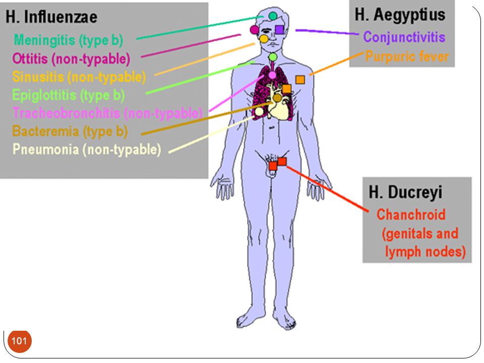
Haemophilus influenzae
Most strains have a polysaccharide capsule that resists phagocytosis and is used in classification of the bacteria H.influenzae type b is the most significant Was the most common form of meningitis in infants prior to the use of an effective vaccine Can cause a number of other diseases in young children Use of the Hib vaccine has eliminated much of the disease caused by H.influenzae b Other strains still cause a variety of diseases

102
Other Species of Haemophilus
H. aegypticus Causes conjunctivitis with pus H.ducreyi Causes a sexually transmitted disease Results in the formation of a genital ulcer called a chancroid Often asymptomatic in women but in men the chancroid is often painful H.aphrophilus causes a rare type of endocarditis Other species primarily cause opportunistic infections

103
Bordetella Small, aerobic, nonmotile coccobacillus
B. pertussis is the most important Causes pertussis, also called whopping cough Most cases of disease are in children Produce various adhesins and toxins, including pertussis toxin, that mediate the disease Bacteria are first inhaled in aerosols and multiply in epithelial cells Then progress through three stages of disease

104
Stages Catarrhal Paroxymal Convalescence

105
Bordetella Clinical significance B. pertussis – causes whooping cough
Acquired by inhalation of droplets containing the organism The organism attaches to the ciliated cells of the respiratory tract. During an incubation period of 1-2 weeks, the organism multiplies and starts to liberate its toxins.

106
Catarrhal Pertussis toxin
Has one A subunit (toxic part), plus five different kinds of B subunits (involved in binding).
, plus five different kinds of B subunits (involved in binding).")
107
Catarrhal The increase in cAMP from the combined effects of pertussis toxin and bacterial adenylate cyclase inhibits host cell phagocytic cell responses and the inhibition of natural killer cell activity. Dermonecrotic toxin –released upon cell lysis causing strong vasoconstrictive effects.

108
Catarrhal Trachael cytotoxin – is related to the B. pertussis peptidoglycan. might contribute to the killing and sloughing off of ciliated cells in the respiratory tract. Lipooligosaccharide has potent endotoxin activity.

109
Paroxymal Lasts 4-6 weeks.
The patient has rapid, consecutive coughs with a rapid intake of air between the coughs (has a whooping sound). mucous has accumulated, and the patient is trying to cough up the mucous accumulations. The coughs are strong enough to break ribs! Other symptoms due to the activity of the released toxins include: Increased peripheral lymphocytes Metabolic alteration such as increased insulin release and the resulting hypoglycemia Increased capillary permeability and increased susceptibility to histamine, serotonin, and endotoxin shock
. mucous has accumulated, and the patient is trying to cough up the mucous accumulations. The coughs are strong enough to break ribs! Other symptoms due to the activity of the released toxins include: Increased peripheral lymphocytes. Metabolic alteration such as increased insulin release and the resulting hypoglycemia. Increased capillary permeability and increased susceptibility to histamine, serotonin, and endotoxin shock.")
110
Convalescence Symptoms gradually subside. This can last for months.
B. pertussis rarely spreads to other sites, but a lot of damage may occur, such as CNS dysfunction which occurs in ~10 % of the cases and is due to an unknown cause. Secondary infections such as pneumonia and otitis media are common.

111
Bordetella Current treatment
B. parapertussis – causes a mild form of whooping cough B. bronchoseptica Widespread in animals where it causes kennel cough. Occasionally causes respiratory or wound infections in humans. Current treatment Erythromyin – only effective in early stages of the disease before the toxin(s) have been released Vaccination P part of DPT (killed, encapsulated organism); a subunit vaccine has also been developed (purified pertussis toxin).
 have been released. Vaccination P part of DPT (killed, encapsulated organism); a subunit vaccine has also been developed (purified pertussis toxin).")
112
Diagnosis, Treatment, and Prevention
Symptoms of pertussis are usually diagnostic Treatment Primarily supportive Antibacterial drugs have little effect on the course of the disease Prevention Immunization with the DPT vaccine Cases in the United States have increased due to a refusal by some parents to have their children immunized

113
Francisella Classification – only 1 pathogenic species – F. tularensis
Morphology and cultural characteristics Minute, pleomorphic g- rod that stains poorly Staining may be bipolar Nonmotile Nonencapsulated Won’t grow on ordinary media – requires cysteine or cystine for growth

114
Francisella tulerensis
Found living in water as an intracellular parasite of animals Causes the zoonotic disease tuleremia Spread to humans occurs mainly through the bite of an infected Dermacentor or by contact with an infected animal The bacteria can spread through unbroken skin and mucous membranes, making it highly infectious Tuleremia produces symptoms common to other bacterial and viral diseases and may be misdiagnosed

115
Francisella Entry through skin abrasions (ulceroglandular form of the disease) - after ~ 48 hours a lesion occurs at the inoculated site. Symptoms Ulcer Headaches, Pain and Fever Adjacent lymph nodes become enlarged. If not contained, this can progress to septicemia, pneumonia, and abscesses throughout the body. The organism survives for long periods of time inside phagocytic cells).
.")
116
Francisella Antimicrobial susceptibility
Ingestion (typhoidal form of the disease) the focus of infection is the mouth, throat, and GI tract. Inhalation (pneumonic form of the disease) This is the most severe form of the disease and it manifests as a pneumonia with a high mortality rate of 30% in untreated cases. Antimicrobial susceptibility Streptomycin or tetracycline An attenuated, live vaccine that protects against the inhalation form of the disease is available for those exposed to the organism.
 the focus of infection is the mouth, throat, and GI tract. Inhalation (pneumonic form of the disease) This is the most severe form of the disease and it manifests as a pneumonia with a high mortality rate of 30% in untreated cases. Antimicrobial susceptibility. Streptomycin or tetracycline. An attenuated, live vaccine that protects against the inhalation form of the disease is available for those exposed to the organism.")
117
Prevention A vaccine is available to at risk individuals
Preventing infection is done by avoiding the major reservoirs of the bacteria

118
Brucella Classification Are all intracellular organisms
4 species can infect humans B. abortus B. suis B. melitensis B. canis Morphology and cultural characteristics Small g-cb that stain poorly

119
Brucella Antigenic structure Virulence factors Clinical significance
2 antigens that are part of the LPS are recognized: A and M B. melitensis has the highest concentration of M and causes the most serious infections Virulence factors Endotoxin Clinical significance Has a tropism for erythritol Animal fetal tissues and placenta, other than those in humans, are rich in erythritol and, therefore, the organisms often cause abortions in these animals.

120
Brucella Causes Brucellosis or undulent fever in man following ingestion of contaminated milk or cheese from goats (B. melitensis), cows (B. abortus), pigs (B. suis), or canines (B. canis). Man can also acquire the organism via contact with infected animals. Clinical manifestations range from subclinical, to chronic with low grade symptoms of low fever and muscular stiffness, to acute with fever and chills. The fever typically spikes each evening and this coincides with the release of organisms from phagocytes (hence the name undulent fever). The patient may also experience malaise, weakness, enlarged lymph nodes, weight loss, and arthritis.
, cows (B. abortus), pigs (B. suis), or canines (B. canis). Man can also acquire the organism via contact with infected animals. Clinical manifestations range from subclinical, to chronic with low grade symptoms of low fever and muscular stiffness, to acute with fever and chills. The fever typically spikes each evening and this coincides with the release of organisms from phagocytes (hence the name undulent fever). The patient may also experience malaise, weakness, enlarged lymph nodes, weight loss, and arthritis.")
121
Brucella Antibiotic susceptibility
Chemotherapy is difficult because of the intracellular survival of the organism. Tetracycline for 21 days, sometimes combined with streptomycin.

122
Pseudomonads Gram-negative, aerobic bacilli
Ubiquitous in soil, decaying organic matter, and almost every moist environment Problematic in hospitals because they can be found in numerous locations Opportunistic pathogens

123
Pseudomonas aeruginosa
Rarely part of the normal microbiota Opportunistic pathogen of immunocompromised patients Can colonize almost every organ and system and result in various diseases Often infects the lungs of cystic fibrosis patients The bacteria form a biofilm that protects them from phagocytosis Increases the likelihood of death in these patients

124
Pseudomonas aeruginosa
Diagnosis can be difficult as the presence of bacteria may represent contamination of the sample Treatment is difficult because P. aeruginosa is resistant to many antibacterial drugs

125
Miscellanous Bacterial Pathogens
Stain pink in a Gram stain but differ from typical Gram-negative organisms Have different morphology, growth habits, or reproductive strategies Traditionally discussed separately due to their unique features

126
Spirochetes Thin, tightly coiled, helically shaped bacteria
Moves in a corkscrew fashion through its environment This movement is thought to enable pathogenic spirochetes to burrow through their hosts’ tissues 3 genera cause human disease Treponema, Borrelia, and Leptospira

127
Treponema pallidum pallidum
Cannot survive in the environment Lives naturally only in humans as an obligate parasite Causative agent of syphilis Syphilis occurs worldwide Transmission is almost solely via sexual contact Endemic among sex workers, men who have sex with men, and users of illegal drugs Can also be spread from an infected mother to her fetus Often results in the death of the fetus or in mental retardation and malformation

128
Treponema pallidum pallidum
Syphilis can proceed through four stages Primary-symptoms associated with the initial infection Secondary-related to spread of the organisms away from the site of the original infection Latent Tertiary syphilis

129
Primary Syphilis Enlarged lymph nodes near the chancre the chancre
Symptoms include: Chancre that should heal by itself in 3-6 weeks painless genitals Mouth Skin rectum Enlarged lymph nodes near the chancre the chancre

130
Secondary Syphilis Spotted rash all over Fever general ill feeling
loss of appetite muscle aches joint pain enlarged lymph nodes hair loss may occur.

131
Tertiary Syphilis Cardiovascular syphilis causes aneurysms or valve disease Central nervous system disorders (neurosyphilis) Infiltrative tumors of skin, bones, or liver (gumma)
")
132
Diagnosis, Treatment, and Prevention
Primary, secondary, and congenital can be readily diagnosed with antibody tests against bacterial antigens Tertiary syphilis is difficult to diagnose Treatment Penicillin is the drug of choice except with tertiary syphilis which is a hyperimmune response and not an active infection Prevention Abstinence and safe sex are the primary ways to avoid contracting syphilis

133
Borrelia Lightly staining, Gram-negative spirochetes
Cause two diseases in humans Lyme disease Relapsing fever

134
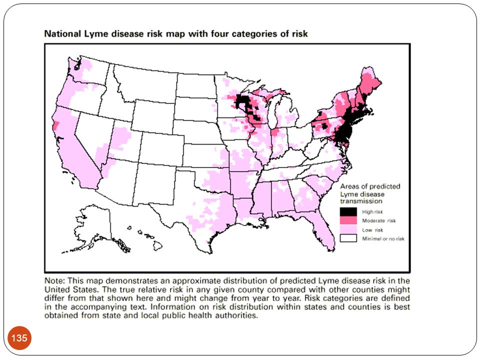
Lyme Disease Borrelia burgdorferi is the causative agent
Bacteria are transmitted to humans via a tick bite Hard ticks of the genus Ixodes are the vectors of Lyme disease

136
Lyme Disease Pathology
Shows a broad range of signs and symptoms 3 phases of disease in untreated patients In most cases an expanding red “bull’s eye” rash occurs at the site of infection Neurological symptoms and cardiac dysfunction Severe arthritis that can last for years Pathology of this stage is largely a result of the body’s immune response

137
Lyme Disease Pathology
The increase of cases is a result of humans coming in closer association with ticks infected with Borrelia Antimicrobial drugs can effectively treat the first stage of Lyme disease Treatment of later stages is difficult because symptoms result from the immune response rather than the presence of bacteria Prevention is best achieved by taking precautions to avoid ticks

138
Relapsing Fever 2 types of relapsing fever Epidemic relapsing fever
Endemic relapsing fever

139
Epidemic Relapsing Fever
Mortality rate is 1% with treatment; 30-70% without treatment Transmitted by lice Mortality rate is 1% with treatment; % without treatment

140
Endemic Relapsing Fever
Several Borrelia species can cause this disease Transmitted to humans by soft ticks of the genus Ornithodoros

141
Relapsing Fever Both types of relapsing fever are characterized by recurring episodes of fever and septicemia separated by symptom free intervals Pattern results from the body’s repeated efforts to remove the spirochetes,which continually change their antigenic surface components

142
Relapsing Fever Observation of the spirochetes is the primary method of diagnosis Successful treatment is with antimicrobial drugs Prevention involves avoidance of ticks and lice, good personal hygiene, and use of repellent chemicals

143
Mycoplasmas Smallest free-living microbes
Lack cytochromes, enzymes of the Krebs cycle, and cell walls Often have sterols in their cytoplasmic membranes which other prokaryotes lack Require various growth factors that must be acquired from a host or supplied in laboratory media Can colonize the mucous membranes of the respiratory and urinary tracts

144
Mycoplasma pneumoniae
Attaches specifically to receptors located at the bases of cilia on epithelial cells lining the respiratory tracts of humans Causes primary atypical pneumonia, or walking pneumonia Symptoms such as fever, headache, and sore throat are not typical of other types of pneumonia Not usually severe enough to require hospitalization or to cause death Spread by nasal secretions among people in close contact

145
Mycoplasma pneumoniae
Diagnosis is difficult because mycoplasmas are small and grow slowly Prevention can be difficult because patients can be infective for long periods of time without signs or symptoms

146
Rickettsias Extremely small (not much bigger than a smallpox virus)
Appear almost wall-less due to the small amount of peptidoglycan present Obligate intracellular parasites Unusual because they have functional genes for protein synthesis, ATP production, and reproduction Three genera cause disease in humans Rickettsia, Orienta, and Ehrlichia

147
Characteristics of Rickettsias
Table 21.1

148
Rocky Mountain Spotted Fever
Symptoms usually develop about 2 to 14 days after the tick bite. They may include: Chills & Fever Severe headache Muscle pain Mental confusion & Hallucinations Rash Abnormal sensitivity to light Diarrhea Excessive thirst Loss of appetite Nausea &Vomiting Spread by ticks

149
Endemic Typhus Chills Cough Delirium
High fever (104 degrees Fahrenheit) Joint pain (arthralgia) Light may hurt the eyes Low blood pressure Rash that begins on the chest and spreads to the rest of the body (except the palms of the hands and soles of the feet) Severe headache Severe muscle pain Stupor Spread by fleas
 Joint pain (arthralgia) Light may hurt the eyes. Low blood pressure. Rash that begins on the chest and spreads to the rest of the body (except the palms of the hands and soles of the feet) Severe headache. Severe muscle pain Stupor. Spread by fleas.")
150
Epidemic Typhus Abdominal pain Spread by lice Backache
Dull red rash that begins on the middle of the body and spreads Extremely high fever ( degrees Fahrenheit), which may last up to 2 weeks Hacking, dry cough Headache Joint pain (arthralgia) Nausea Vomiting
, which may last up to 2 weeks. Hacking, dry cough. Headache. Joint pain (arthralgia) Nausea. Vomiting.")
151
Chlamydias Do not have cell walls
Have two membranes but without any peptidoglycan between them Grow and multiply only within the vesicles of host cells Have a unique developmental cycle involving two forms Both forms can occur within the phagosome of a host cell

152
Chlamydia trachomatis
Has a limited host range One strain infects mice, all others infect humans Infect the conjunctiva, lungs, urinary tract, or genital tract Enters the body through abrasions and lacerations Clinical manifestations result from the destruction of infected cells at the infection site, and from the resulting inflammatory response

153
Chlamydia trachomatis
Causes two main types of disease Sexually transmitted diseases Causes the most common sexually transmitted disease in the United States Ocular disease called trachoma Occur particularly in children Endemic in crowded, poor communities with poor hygiene, inadequate sanitation, and inferior medical care

154
Chlamydia—Rates by Sex, United States, 1990–2009
2008 2006 2004 2002 2000 1998 1996 1994 1992 1990 Total Women Men 100 200 300 400 500 600 Rate (per 100,000 population) Year NOTE: As of January 2000, all 50 states and the District of Columbia had regulations that required chlamydia cases to be reported.
 Year. NOTE: As of January 2000, all 50 states and the District of Columbia had regulations that required chlamydia cases to be reported.")
155
Sexually Transmitted Diseases
Lymphogranuloma veneruem Characterized by a transient genital lesion and swollen, painfully inflamed, inguinal lymph nodes Occurs in three stages Initial stage Produces a lesion at the infection site that is small painless, and heals rapidly Second stage Buboes develop at the infection site

156
Sexually Transmitted Diseases
Third stage Only some cases progress to this stage Characterized by genital sores, constriction of the urethra, and genital elephantiasis Most infections in women are symptomatic but men may or may not have symptoms Women can develop pelvic inflammatory disease if reinfected with C. trachomatis

157
Trachoma Disease of the eye
Leading cause of nontraumatic blindness in humans Bacteria multiply in the conjunctival cells resulting in scarring The scarring causes the eyelashes to turn inwards and abrade the eye that can eventually result in blindness

158
Trachoma Typically a disease of children who have been infected during birth Infection of the eye with bacteria from the genitalia can also result in disease

159
Diagnosis, Treatment, and Prevention
Demonstration of the bacteria inside cells from the site of infection Treatment Antibiotics can be administered for genital and ocular infections Surgical correction of eyelid deformities from Trachoma may prevent blindness

160
Diagnosis, Treatment, and Prevention
Abstinence and safe sex can prevent sexually transmitted chlamydial infection Blindness can only be prevented by prompt treatment with antibacterial agents and preventing reinfections

161
Legionella pneumophila
Aerobic, slender, pleomorphic bacteria Universal inhabitants of water Humans acquire the disease by inhaling the bacteria in aerosols from various water sources Intracellular parasites

162
Legionella pneumophila
Causes Legionnaires’ disease Results in pneumonia Immunocompromised individuals are more susceptible Elimination of the bacteria is not feasible but reducing their number is a successful control measure

163
Bartonella Gram-negative aerobic bacilli
Found in animals but only cause disease in humans 3 species are pathogenic Bartonella bacilliformis Bartonella quintana Bartonella henselae

164
Bartonella bacilliformis Bartonellosis -Carrión’s Disease Transmitted by blooding-sucking sand flies
Acute phase: (Carrion's disease) fever pallor, malaise, nonpainful hepatomegaly, jaundice, lymphadenopathy, splenomegaly. This phase is characterized by severe hemolytic anemia and transient immunosuppression. The case fatality ratios of untreated patients exceeded 40% but reach around 90% when opportunistic infection with Salmonella occurs.
 fever. pallor, malaise, nonpainful hepatomegaly, jaundice, lymphadenopathy, splenomegaly. This phase is characterized by severe hemolytic anemia and transient immunosuppression. The case fatality ratios of untreated patients exceeded 40% but reach around 90% when opportunistic infection with Salmonella occurs.")
165
Bartonella quintana Trench fever
Spread person to person by human body lice Also causes disease in immunocompromised patients The disease is classically a five-day fever of the relapsing type

166
Bartonella henselae Cat scratch fever
Introduced into humans through cat scratches or bites

Similar presentations


 Prof. Dr. Ebtisam.F. El Ghazzawi Medical Research Institute (MRI) Alexandria University.>")


![BY RANJEET RAMAN GRAM NEGATIVE BACILLI- MICRO {ST1]](/15/4636473/big_thumb.jpg "BY RANJEET RAMAN GRAM NEGATIVE BACILLI- MICRO {ST1]>")
