Download presentation
Presentation is loading. Please wait.

1
Genitourinary Trauma TIM EVANS VIRGINIA COMMONWEALTH UNIVERSITY
January 28, 2015

2
Background If injury to GU system identified, multi-organ injury is the rule. Examples: If renal injury found following penetrating trauma, 80-95% chance of other significant injury If renal injury found following blunt trauma, 75% chance of other significant injury found Other injuries may be more immediately life threatening and therefore GU injury diagnosis may be overlooked or delayed

3
Case Patient #1 is a 25 year old male is struck in the flank with a baseball bat. His systolic blood pressure is always above 100 mm Hg and his exam is only remarkable for a flank hematoma without abdominal tenderness. His urinalysis shows no RBCs. Patient #1 got pissed off at the guy who hit him so he shot Patient #2 in the flank. Patient #2 is hemodynamically stable and does not have any RBCs in his urine Two Questions: Do either of these reprobates need imaging? Do we need more bat control legislation?

4
Renal Trauma Most common GU injury—65% of GU injuries
10% of abdominal injuries involve the kidneys Mechanism 80-95% due to blunt force—MVC, falls, assaults, sporting events

5
Renal Anatomy Retroperitoneal
Adjacent to lower two thoracic and first four lumbar vertebrae Upper poles protected by ribs so lower poles more commonly injured Right kidney inferior to left and more commonly injured Kidney mobile, hilum more fixed—concern with shearing injury with deceleration

6
When are you concerned about renal injuries?
Mechanism of Injury Penetrating injuries of abdomen, back or flank Deceleration injuries Physical exam Tenderness of abdomen or flank Ecchymosis of abdomen or flank Xray Fractures of lower ribs, thoraco-lumbar spine

7
When are you concerned about renal injuries?
Hematuria—over 95% of patients with renal trauma will have some degree of hematuria (>5 rbc/hpf) THE PRESENCE OR DEGREE OF HEMATURIA DOES NOT CORRELATE WITH THE SEVERITY OF THE INJURY 25% of patients with gross hematuria have minor injuries 40% of the most serious renal injuries do not have any hematuria
 THE PRESENCE OR DEGREE OF HEMATURIA DOES NOT CORRELATE WITH THE SEVERITY OF THE INJURY. 25% of patients with gross hematuria have minor injuries. 40% of the most serious renal injuries do not have any hematuria.")
8
Indications for imaging for renal trauma
Penetrating trauma in proximity to kidneys—the presence or absence of hematuria in penetrating trauma not predictive of injury, location of wound is most important factor Gross hematuria Microscopic hematuria (>3-5 RBC/HPF) with hemodynamic instability—systolic BP<90 at any time Persistent microscopic hematuria ?Significant deceleration mechanisms ?Proximal injuries with blunt mechanisms Mee SL, et al: Radiographic Assessment of Renal Trauma: A ten-year prospective study of patient selection. J Urol 141:1095, 1989
 with hemodynamic instability—systolic BP<90 at any time. Persistent microscopic hematuria. Significant deceleration mechanisms. Proximal injuries with blunt mechanisms. Mee SL, et al: Radiographic Assessment of Renal Trauma: A ten-year prospective study of patient selection. J Urol 141:1095,")
9
When not to image in patients with concern for renal trauma
Patients with microscopic hematuria who have always been hemodynamically stable Patients who are not hemodynamically stable

10
No significant renal injuries missed
Microhematuria and no shock Gross hematuria or Microhematuria and shock (SBP<90 mmHg) all imaged-422 Imaged- Significant injury 3 Significant renal injuries 78 Imaged- Contusion 581 Without Imaging 1004 1 renal repair Renal repair 34 No significant renal injuries missed Miller KS, McAninch JW: Radiographic assessment of renal trauma. Our 15-year experience. J Urol 1995;154:
 all imaged-422. Imaged- Significant injury 3. Significant renal injuries. 78. Imaged- Contusion Without. Imaging renal repair. Renal repair. 34. No significant renal injuries missed. Miller KS, McAninch JW: Radiographic assessment of renal trauma. Our 15-year experience. J Urol 1995;154:")
11
Imaging techniques Contrast enhanced CT—the best test, up to 98% accurate, not great for renal vein injuries IVP—perhaps useful in the OR to determine function of contralateral kidney before contemplated nephrectomy Angiography—better than CT for defining injuries to renal artery and vein, also used therapeutically to embolize or stent artery injury Ultrasound—30% false negative rate for injury, used to look for two kidneys, free fluid Contrast Enhanced Ultrasound—perhaps MRI—not first line due to time, sensitivity similar to CT, can be used for follow up studies

12
AAST Kidney Injury Severity Scale

13
AAST Kidney Injury Severity Scale—Revision 2011
Grade IV - originally encompassed contained injuries to the main renal artery and vein, and collecting system injuries. Revision: adds segmental arterial and venous injury, and laceration to the renal pelvis or ureteropelvic junction. Multiple lacerations into the collecting system used to be considered a shattered kidney (Grade V), but now remains Grade IV. Grade V - originally included main renal artery or vein laceration or avulsion, and multiple collecting system lacerations (shattered kidney). The revised classification includes only vascular injury (arterial or venous) and includes laceration, avulsion or thrombosis.
, but now remains Grade IV. Grade V - originally included main renal artery or vein laceration or avulsion, and multiple collecting system lacerations (shattered kidney). The revised classification includes only vascular injury (arterial or venous) and includes laceration, avulsion or thrombosis.")
14
Grade I-Renal contusion

15
Grade I-Subcapsular Hematoma

16
Grade II-Small Cortical Laceration

17
Grade III-Major Renal Laceration

18
Grade IV-Major Laceration involving Collecting System

19
Grade IV- Multiple Renal Lacerations

20
Grade IV-“Shattered” Kidney

21
Grade V- Avascular Left Kidney

22
Injury in proximity to kidney Hematuria (Gross or microscopic)
Trauma Penetrating Blunt Injury in proximity to kidney Hematuria (Gross or microscopic) Associated with shock (SBP <90) Hematuria Microscopic (>5 RBC/HPF) No shock Unstable Stable Image with concern for other organs Abdominal exploration CT scan with IV contrast Single-shot IVP on table Grades III-V Clinical follow-up Abnormal or inconclusive Selective renal exploration Renal exploration
 Associated with shock. (SBP <90) Hematuria. Microscopic (>5 RBC/HPF) No shock. Unstable. Stable. Image with concern for other organs. Abdominal exploration. CT scan with IV contrast. Single-shot. IVP on table. Grades III-V. Clinical. follow-up. Abnormal or inconclusive. Selective renal. exploration. Renal exploration.")
23
Management of Renal Injuries
Grade I—home Grade II-IV—admit, observe Grade V—observe, vascular repair/stent, or nephrectomy Only absolute indications for surgery are persistent renal bleeding with hemodynamic instability, active extravasation of IV contrast, expanding or pulsatile perirenal hematoma suggesting Grade V vascular injury

24
Complications of Renal Injuries
Mortality 3% Complications First six weeks Hemorrhage/shock Sepsis/abscess ATN Late Renovascular HTN 1-4%

25
CASE 30 year old s/p cystoscopic removal of distal ureteral stone. Now with flank pain and nausea. T 39 C, diffuse abdominal and flank tenderness noted. U/A--negative Diagnosis? Studies?

26
Ureteral Trauma Accounts for 1% of urologic trauma
Most commonly iatrogenic following GU, gynecologic, vascular or colorectal surgery If following external trauma, 80-95% due to penetrating mechanism, usually GSW

27
Ureteral Anatomy Thin, mobile tubes running between renal pelvis and posterior superior angle of bladder Retroperitoneal in abdomen Protected from injury by size and mobility

28
When are you concerned about ureteral injuries?
Recent GU, gynecologic, vascular or colorectal procedure Penetrating (usually GSW) trauma to abd, back, flank Deceleration mechanisms Suspicion raised with injuries to iliac vessels, urinary bladder, sigmoid colon, thoracolumbar dislocations, lumbar spine (including process) fractures
 trauma to abd, back, flank. Deceleration mechanisms. Suspicion raised with injuries to iliac vessels, urinary bladder, sigmoid colon, thoracolumbar dislocations, lumbar spine (including process) fractures.")
29
Hematuria following ureteral injuries
Ureteral injury following iatrogenic cause—10-15% of patients with hematuria Hematuria absent in 30-60% of identified ureteral injuries from external violence Hematuria following penetrating trauma—a study of 71 ureteral injuries 32% without hematuria 40% with gross hematuria 28% with microscopic hematuria Brandes SB, et al: Ureteral injuries from penetrating trauma, J Trauma 36:766, 1994.

30
IMAGING FOR URETERAL INJURIES
Most injuries diagnosed during laparotomy and no imaging ever done Contrast CT with delayed imaging—most common findings are extravasation of contrast into medial perirenal space and absence of contrast in distal ureter if transected Retrograde pyelogram IVP—one shot IVP done in OR for penetrating trauma

31
Delayed CT images showing extravasation of urine from ureteral injury

32
Blunt trauma Penetrating trauma Gross hematuria, or microhematuria with deceleration or hypotension or associated injuries Stable, to CT + contrast + delayed films Unstable, to OR Gross or micro-hematuria Unstable, to OR Yes No Potential ureteral injury (ureteral nonopacification or extravasation) Abnormal Intraoperative One-shot IVP Normal Intraoperative one-shot IVP Normal Consider other sources for hematuria (bladder, urethra, kidney) Stent removal 6 weeks After stent removal consider periodic renogram or surveillance ultrasound (defect hydronephrosis to rule out recurrence Explore ureter and repair Bullet/knife wound in vicinity of ureter
 Abnormal. Intraoperative. One-shot IVP. Normal. Intraoperative one-shot IVP. Normal. Consider other sources for. hematuria (bladder, urethra, kidney) Stent removal. 6 weeks. After stent removal consider periodic renogram or surveillance ultrasound (defect hydronephrosis to rule out recurrence. Explore ureter and repair. Bullet/knife wound in vicinity of ureter.")
33
I Hematoma Contusion or hematoma without devascularization
American Association for the Surgery of Trauma (AAST) Ureter Injury Severity Scale Grade Description I Hematoma Contusion or hematoma without devascularization II Laceration <50% transection III Laceration >50% transection IV Laceration Complete transection with <2 cm devascularization V Laceration Avulsion with >2 cm devascularization
 Ureter Injury Severity Scale Grade Description. I Hematoma Contusion or hematoma without devascularization. II Laceration <50% transection. III Laceration >50% transection. IV Laceration Complete transection with <2 cm devascularization. V Laceration Avulsion with >2 cm devascularization.")
34
MANAGEMENT OF URETERAL INJURIES
Treatment Stents—Grade 1 Surgery—Grade 2 and above Complications Ureteral stricture Fistula Retroperitoneal fibrosis Abscess/Sepsis

35
Intraoperative recognition
Minor ureteral injury Major ureteral injury Stent Primary stented ureterourostony, psoas hitch, or flap with or without kidney mobilization Consider placement of percutaneous nephrostomy in rare case of extremely long injury Ureteral stent 6 weeks Follow-up retrograde pyelography and stent removal or replacement as needed Consider endoscopic methods (laser, balloon) Primary stented ureteroureterostomy After stent removal consider periodic renogram or surveillance ultrasound to rule our recurrence
 Primary stented ureteroureterostomy. After stent removal consider periodic renogram or surveillance ultrasound to rule our recurrence.")
36
Postoperative recognition
CT with contrast (+ delayed films) + retrograde pyelography Minor ureteral injury Major ureteral injury Ureteral stent 6 weeks Attempted retrograde stent placement Follow-up retrograde pyelography and stent removal or replacement as needed Fail Success Success Percutaneous nephrostomy and anterograde stent placement, if possible Consider endoscopic methods (laser, balloon) Primary stented ureteroureterostomy, psoas hitch or flap Consider autotransplant or ileal loop in rare case of extremely long injury After stent removal consider renogram or surveillance ultrasound to rule out recurrence Fail, wait 6 weeks
 + retrograde pyelography. Minor ureteral injury. Major ureteral injury. Ureteral stent 6 weeks. Attempted retrograde stent placement. Follow-up retrograde pyelography and stent removal or replacement as needed. Fail. Success. Success. Percutaneous nephrostomy and anterograde stent placement, if possible. Consider endoscopic methods (laser, balloon) Primary stented ureteroureterostomy, psoas hitch or flap. Consider autotransplant or ileal loop in rare case of extremely long injury. After stent removal consider renogram or surveillance ultrasound to rule out recurrence. Fail, wait. 6 weeks.")
37
Case

39
Urinary Bladder Trauma
Mechanisms of Injury Blunt—up to 85% of cases 70-95% of patients with bladder injuries will have pelvic fractures 6-10% of patients with pelvic fractures will have bladder injuries Penetrating—up to 15% of cases Surgical/Cystoscopy

40
Urinary Bladder Anatomy
Empty bladder is a pelvic organ and protected by pelvic bones With distention, becomes an abdominal organ and more prone to injury due to direct trauma Peritoneum covers superior surface of bladder

41
When are you concerned about a bladder injury?
Clinical Presentation Suprapubic pain Difficulty voiding Gross Hematuria—incidence approaches 100% Microscopic Hematuria possible with penetrating trauma, spontaneous bladder rupture X-ray Widened symphysis pubis is stongest predictor Pelvic, sacrum, iliac, ramus fractures Widening of SI joint

42
Diagnostic Studies Retrograde cystogram Retrograde CT cystogram
Either one follows urethogram if concern for urethral injury exists

43
Indications for Cystography
Blunt Trauma in close proximity to bladder with gross hematuria Pelvic fractures from blunt mechanism with any degree of hematuria Penetrating Trauma in proximity to the bladder Penetrating trauma with any degree of hematuria

44
Technique for Cystogram
Retrograde urethrogram if indicated Urinary catheter 100 cc contrast Plain film cc contrast (5cc/kg) Empty bladder Sensitivity for bladder rupture near 100% if each step performed
 Empty bladder. Sensitivity for bladder rupture near 100% if each step performed.")
45
Retrograde Cystogram--Normal

46
Retrograde Cystogram—Post-Void, Normal

47
CT Cystogram Same technique as for plain cystogram, no need to do post void study Sensitivity also approaches 100%

48
Extraperitoneal Bladder Rupture
50-90% of bladder ruptures Usually associated with pelvic fracture Usually treated with urethral/suprapubic catheter

49
Retrograde Cystogram—Extraperitoneal Rupture

50
Retrograde Cystogram—Extraperitoneal Rupture

51
CT Cystogram—Extraperitoneal Rupture

52
CT Cystogram with Extraperitoneal Rupture

53
CT Cystogram with Extraperitoneal Rupture with Sagittal View

54
Intraperitoneal Bladder Rupture
15-35% of bladder ruptures Bladder usually distended at time of trauma Historically treated surgically Conservative management possible

55
Retrograde Cystogram—Intraperitoneal Rupture

56
Retrograde Cystogram—Intraperitoneal Rupture

57
Retrograde Cystogram—Intraperitoneal Rupture

58
Retrograde Cystogram—Intraperitoneal Rupture

59
CT Cystogram-Intraperitoneal Rupture

60
CT Cystogram—Intraperitoneal Rupture

61
American Association for the Surgery of Trauma (AAST) Bladder Injury Severity Scale Grade Description I Hematoma Contusion, intramural hematoma Laceration Partial thickness II Laceration Extraperitoneal bladder wall laceration <2 cm III Laceration Extraperitoneal (>2 cm) or intraperitoneal (<2 cm) bladder wall laceration IV Laceration Intraperitoneal bladder wall laceration >2 cm V Laceration Intraperitoneal or extraperitoneal bladder wall laceration extending into the bladder neck or ureteral orifice (trigone)
 or intraperitoneal (<2 cm) bladder wall laceration. IV Laceration Intraperitoneal bladder wall laceration >2 cm. V Laceration Intraperitoneal or extraperitoneal bladder wall laceration extending into the bladder neck or ureteral. orifice (trigone)")
62
Urinary Bladder Ruptures
Patients may have both intra- and extra-peritoneal bladder ruptures 20-40% Mortality for Associated Injuries Hemorrhage Sepsis

63
Case 22 year old male engaging in sexual activity
Hears and feels snap, crack and pop No more sex Diagnosis?

64
Penile Fracture

65
Urethral Injuries 10% of all injuries to GU system
Potentially most debilitating GU injury due to complications Rare in women Mechanism of Injury Blunt trauma such as mvc, bike accidents, straddle mechanisms Often associated with pelvic fractures Rarely penetrating trauma Occasionally iatrogenic

66
Urethral Anatomy Anatomy based on relation to urogenital diaphragm
Posterior Prostatic Membranous Anterior Bulbous Penile

67
Posterior Urethral Injuries
80-90% occur in combination with pelvic fracture 10-25% of pelvic ring fractures disrupt posterior urethra as puboprostatic ligaments are torn or stretched Associated with bladder injuries and vaginal lacerations

68
Anterior Urethral Disruption
Usually due to direct blunt force trauma such as saddle injury Does not cause high riding prostate as injury is below the urogenital diaphragm Ureteral injury present in 10-38% of penile fractures (rupture of one or both tunica albuginea, fibrous covering of corpus cavernosa)
")
69
When do you worry about urethral injuries?
Symptoms Abdominal/Perineal Pain Difficulty urinating—females can present with incontinence Posterior—unable to urinate Anterior—dysuria, small amounts Signs Gross hematuria Blood at urethral meatus Perineal swelling/ecchymosis Vaginal lacerations Inability to pass urinary catheter (gentle attempt) Abnormal prostate exam Absent High riding Boggy X rays Pelvic Fractures
 Abnormal prostate exam. Absent. High riding. Boggy. X rays. Pelvic Fractures.")
70
Retrograde Urethrogram
If urethral injury suspected, you may try one gentle attempt at passing urinary catheter—if it does not pass easily, don’t push Perform urethrogram—instill cc of contrast retrograde through urethra Complete disruption—contrast extravasates and none reaches bladder Partial disruption—contrast extravasates and some reaches bladder

71
Grade* Injury Type Description
American Association for the Surgery of Trauma (AAST) Urethra Injury Severity Scale Grade* Injury Type Description I Contusion Blood at urethral meatus; urethrography normal II Stretch Injury Elongation of urethra without extravasation on urethrography III Partial Extravasation of urethrography contrast at injury site with Disruption contrast visualized in the bladder IV Complete Extravasation of urethrography contrast at injury site Disruption without contrast visualization in the bladder; <2 cm of urethral separation V Complete Complete transection with >2 cm urethral separation, or Disruption extension into the prostate or vagina
 Urethra Injury Severity Scale. Grade* Injury Type Description. I Contusion Blood at urethral meatus; urethrography normal. II Stretch Injury Elongation of urethra without extravasation on urethrography. III Partial Extravasation of urethrography contrast at injury site with Disruption contrast visualized in the bladder. IV Complete Extravasation of urethrography contrast at injury site Disruption without contrast visualization in the bladder; <2 cm. of urethral separation. V Complete Complete transection with >2 cm urethral separation, or Disruption extension into the prostate or vagina.")
72
Normal Urethrogram

73
Grade III-Partial Urethral Disruption

74
Grade III Partial Urethral Disruption

75
Grade IV or V Complete Urethral Disruption

76
Grade V Complete Urethral Disruption

77
Urethral Trauma Diagnosis Treatment Complications
Retrograde Urethrogram Treatment Catheter, Stent, Primary anastomosis Complications Stricture Impotence Incontinence

78
Case

79
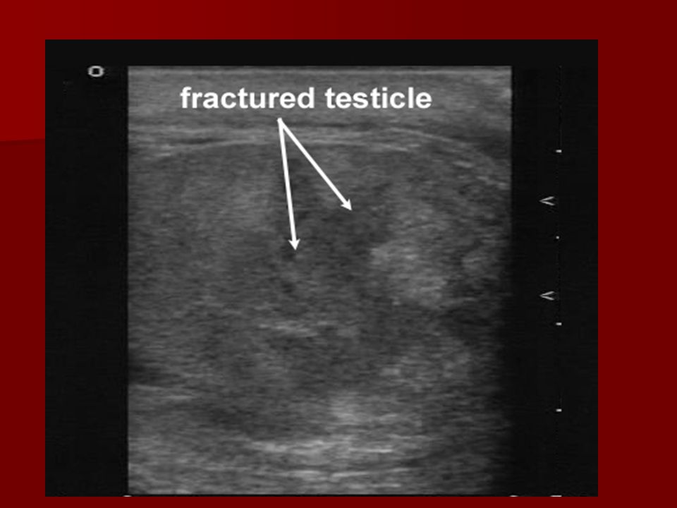
Testicular Trauma Mechanism—fall, kick, sports
Symptoms—pain, N/V, lightheaded, remorse Diagnosis—laceration, contusion, fracture, dislocation

80
Testicular Trauma Diagnosis—Color flow Doppler ultrasound Management
Contusion—rest, ice, analgesia, F/U Laceration, dislocation, rupture--operative

85
Penile Amputation

86
Penile Amputation

87
Penile Resurrection!

Similar presentations


>")
