Download presentation
Presentation is loading. Please wait.

1
Amit Sarnaik MD Scottish Rite Pediatric Emergency Department
Urogenital Trauma Amit Sarnaik MD Scottish Rite Pediatric Emergency Department

2
Genitourinary trauma In multiple trauma patients, GU trauma is second in frequency (#1 CNS)- 10% MOI: Blunt (90%) vs. Penetrating MVC: most common Falls, Sport related and direct blow Most common Injury is to the kidney – 47% Associated intra-peritoneal injuries Penetrating – 80% vs. Blunt

3
Pediatric considerations
Renal injury more likely in children Larger proportion of kidney to abdominal size Retained fetal lobulations: Easier parenchymal disruption Weaker abdominal muscles Less ossified thoracic cage Less developed perirenal fat and fascia

4
Renal trauma - Presentation
Localized signs: flank tenderness, flank hematoma, or palpable flank mass. Non specific: Abdominal tenderness, rigidity, paralytic ileus or hypovolemic shock Gross hematuria is the hallmark of severe injury: absent in 50% of patients with vascular pedicle injuries and 30% penetrating injuries Most common injuries Parenchymal contusions and hematomas (60-90%) Lacerations are less common (10%)
 Lacerations are less common (10%)")
5
Classification of renal trauma
Grade I : Contusion or subcapsular nonexpanding hematoma Grade II : Nonexpanding hematoma confined to the retroperitoneum or lac <1 cm Grade III : Lac >1 cm into the renal cortex without collecting system rupture or urinary extravasation Grade IV : Lac extending into the collecting system or renal vascular injuries with contained hemorrhage Grade V : Shattered kidneys or avulsions of renal hilum with devascularized kidneys

6
Grade 1,2 and 3 renal injuries

7
Grade 4 and 5 injuries

8
Renal Trauma: Management
Evaluate GU system only after life threatening conditions have been indentified A urinalysis should be obtained in all patients with multisystem trauma or suspected isolated renal injury Pediatric renal trauma patient – order a CXR, Abdominal and Pelvis X-rays

9
Renal trauma: Hematuria and Kidney injury
Gross hematuria Microscopic hematuria with major mechanisms or signs of renal injury Hematuria of > 20 RBC per hpf Microscopic hematuria with shock ( relied upon in adult EM)
")
10
Imaging of renal trauma
CT with contrast is preferred study at most trauma centers - 98% sensitivity Detection of associated injuries 3-D views and no dependence on renal vascularity Ultrasound – 70% sensitivity Not accepted for the staging of renal trauma Has been used for long term follow up Alternative modality for the evaluation of the pregnant trauma patient

11
Imaging in renal trauma
IVP : used only if CT is not readily available Provides rapid information about the overall functional and anatomic integrity of both kidneys It can be obtained in the ED in an unstable patient or in the OR prior to surgery IVP will only diagnose 5% contusions, 50% lacs, 29% pedicle injuries Renal injury: non visual of calyceal, extravasation of contrast

12
Blunt Uro-genital trauma: Diagnostic evaluation
Blunt and Unstable: Limited IVP Blunt & Stable, major renal injury, none lower CT scan Blunt and Stable, Findings of lower tract injury Cystourethrogram +/- upper tract evaluation Blunt and Stable, Minor renal injury (Microscopic hematuria) No CT, serial UA, delayed imaging Tenderness, contusion, mass, gross hematuria, major mechanism of injury or associated injuries
 No CT, serial UA, delayed imaging. Tenderness, contusion, mass, gross hematuria, major mechanism of injury or associated injuries.")
13
Management of blunt renal trauma
Grades 1,2 and 3 Strict bed rest, analgesia, prophylactic antibiotics. Limited activity on resolution of gross hematuria Grades 4,5 – Management is controversial. Depends upon hemodynamic status, degree of urinary extravasation, renal bleeding, associated injuries. Many patients are being managed with serial hematocrit, vital signs and broad spectrum antibiotics

14
Complications from non operative management of Grade 4 and 5 renal trauma
Patients managed nonoperatively have a 50% complication rate Persistent/recurrent hemorrhage Extravasation and urinoma formation Infection Infarction Segmental hydronephrosis Neprectomy upto 20% immediate surgery, 10% in delayed surgical intervention

15
Penetrating Uro-genital trauma: Diagnostic evaluation
Penetrating and Unstable Limited IVP Penetrating & Stable, upper tract injury suspected CT scan Penetrating & Stable, findings of lower tract injury Cystourethrogram +/- upper tract evaluation

16
Management of Penetrating renal trauma
Surgical Vascular injury Hemodynamic instability Urinary extravasation Non surgical Hemodynamically stable + Isolated Low grade Delayed bleeding may occur in 24% with grade 3-4

17
Complications of renal trauma
Short Term Delayed hemorrhage Urinary extravasation Abscess formation Obstruction secondary to clot formation Long Term HTN (<5%) Hydronephrosis Arteriovenous fistulas Renal intestinal fistula Stone formation
 Hydronephrosis. Arteriovenous fistulas. Renal intestinal fistula. Stone formation.")
18
Ureteral Trauma Ureteral injuries are uncommon, <1% of all urologic trauma Blunt trauma usually involves the UPJ Suspect ureter trauma if fracture of the transverse process of lumbar vertebra Penetrating injuries along the ureter – 90% association with other intra-abdominal injuries Stab wounds rarely cause ureteral injury, but 50% of GSW to abdomen have injury to the ureter

19
Ureteral trauma: Diagnosis
Diagnosis is difficult, >50% not diagnosed in 1st 24h PE may be unremarkable, urinalysis is unreliable Delayed diagnosis may manifest as fever, chills, lethargy, leukocytosis, pyuria, bacteriuria, flank mass/pain, fistulas, strictures

20
Ureteral Trauma CT and IVP has low sensitivity (33%)
Retrograde pyelogram may be more reliable

21
Bladder Trauma Blunt trauma secondary to MVC is most common cause
80% of injuries associated with pelvic fracture Mortality rate 40% with bladder rupture (from assoc head injury)
")
22
Classification of bladder trauma
Extraperitoneal: associated with pelvic fractures. Intraperitoneal: caused by blunt trauma to distended bladder. Combined: GSW.

23
Bladder trauma: Diagnosis
Hematuria and dysuria typically seen at presentation >90% with bladder rupture have gross hematuria Diagnostic evaluation is indicated in patients who sustain pelvic or lower abdominal trauma with gross hematuria inability to void abnormal GU exam multiple associated injuries

24
Evaluation of bladder trauma
Pelvic X-rays Retrograde cystogram High suspicion and normal X-rays No catheterization if blood at the urethral meatus or high-riding prostate CT cystography is recommended over plain cystogram for patients undergoing CT for associated injuries

25
Management of bladder injuries
Extra peritoneal Contusion = conservative management, +/- catheter Manage with urethral cath or suprapubic drainage for 7-10 days. Large tear = OR Intraperitoneal - Go to OR. Combined – Go to OR

26
Urethral trauma Mechanisms More common in males Urethral injuries MVC
straddle injuries Instrumentation More common in males Urethral injuries Anterior: Pendulous and Bulbar Posterior: Membranous and Prostatic

27
Blunt Anterior Urethral trauma
Due to direct trauma, usually isolated, low mortality Bulbar injuries : common in straddle injury Blood at the urethral meatus is present in 90% of anterior injuries Perineal ecchymosis (butterfly), inability/difficulty voiding also possible Retrograde urethrogram is diagnostic Manage with 7-10 days of catheterization plus antibiotics Severe injuries need urinary diversion
, inability/difficulty voiding also possible. Retrograde urethrogram is diagnostic. Manage with 7-10 days of catheterization plus antibiotics. Severe injuries need urinary diversion.")
28
Blunt Posterior urethral Trauma
Occur with severe trauma and are associated with other injuries (pelvic fx) Signs are blood at the meatus, hematuria, perineal ecchymosis (butterfly), inability/difficulty voiding Retrograde urethrogram is diagnostic Urology consultation Higher rate of complications Ct scan not useful for urethral injuries Initial management is controversial…they vary from immediate exploration to placement of tube with delayed urethroplasty
 Signs are blood at the meatus, hematuria, perineal ecchymosis (butterfly), inability/difficulty voiding. Retrograde urethrogram is diagnostic. Urology consultation. Higher rate of complications. Ct scan not useful for urethral injuries Initial management is controversial…they vary from immediate exploration to placement of tube with delayed urethroplasty.")
29
Female urethral trauma
Urethral injuries in girls Rare, due to mobile short urethra Associated with pelvic fractures or instrumentation Managed with suprapubic drainage and elective repair

30
Penile Trauma Blunt trauma from toilet seat is common
Managed with warm soaks. Tourniquet injuries Exposure and removal of hair Urethrocutaneous fistula and penile loss Zipper entrapment.

31
Penile Trauma Penis fracture. Lacerations:
Traumatic rupture of corpus cavernosum. Erect penis vs. hard surface. Patient may hear a cracking sound with pain and edema. Most required surgical evacuation of hematoma, ice packs, pressure dressing Lacerations: Involving the corporal bodies or the urethra require urologic consult Superficial: simple repair

32
Perineal trauma Most common is straddle injury
Vulvar hematomas = ice packs and rest Superficial lacerations treat with sitz baths Deep lacerations: Extension into rectum or urethra

33
Straddle injury Injury is caused by the compression of soft tissues against the bony margins of the pelvic outlet Mechanisms: Bicycle riding Falls Monkey bars

34
Straddle injury: Appearance
Straddle injuries typically are unilateral and superficial Anterior portion of genitalia involved Girls: Mons, clitoral hood and labia minora anterior and lateral to hymen Straddle injury to hymen and posterior fourchette is rare Boys: Injury to penis or scrotum

35
Straddle injury vs Abuse
Infant younger than nine months Perianal, rectal, vaginal, or hymenal injury without history of penetrating trauma Extensive or severe trauma Presence of non-urogenital trauma Lack of correlation between history and physical findings Abnormal genital secretions

36
Straddle injury: Treatment principles
Visibility of injury Physician must be assured that the injury is properly inspected Ability to void Inability to void Pain Large hematoma Urethral disruption

37
Treatment : Girls Vulvar hematoma: size dependant Vulvar lacerations
Ice packs, analgesia, sitz baths Increasing size: Surgical drainage Vulvar lacerations Heal by secondary intention ( lateral wall of vestibule) Repair of perineal lacerations under sedation Vaginal injury: suspect if hymenal tear Lacerations: superficial or deep - Repair Hematomas: Observation Small, medium and large” egg, orange, grapefruit Superficial vag lac –ED repair Deep vag lac – GYN or Urology, Intact hymen: vag injury is unlikely. Torn hymen- detailed vag exam
 Repair of perineal lacerations under sedation. Vaginal injury: suspect if hymenal tear. Lacerations: superficial or deep - Repair. Hematomas: Observation. Small, medium and large egg, orange, grapefruit. Superficial vag lac –ED repair. Deep vag lac – GYN or Urology, Intact hymen: vag injury is unlikely. Torn hymen- detailed vag exam.")
38
Treatment: Boys Urethral injury: Anterior vs. posterior
Testicular injury: Depends on severity Assessment with US and Urology Scrotal injury Hematoma, ecchymosis: Ice packs Superficial lacerations: Repair in ED Hematocele and scrotal swelling Deep ( extension through Dartos): Urology Penile injuries
: Urology. Penile injuries.")
39
Penile Trauma: Direct Injury
Causes and management Falling toilet seat Significant penile edema Injury to corporal bodies or urethra is rare Treatment: warm soaks, void in bath tub, Observation Blunt trauma: Blood at urethral meatus Urethral injury Diagnosis: Retrograde urethrogram Laceration to penile shaft R/O urethral injury and injury to corporal bodies Consult urology, urethrogram, exploration in ?? Cases Simple laceration: Repair with chromic catgut

40
Penile Trauma: Zipper Injury
Most common genital injuries in prepubertal boys. Typically involve the foreskin or redundant penile skin and may occur during the zipping or unzipping process Localized edema and pain are the most common complications Significant injury, including skin loss or necrosis, is unusual.

41
Zipper Injury: Treatment
Mineral oil: Allows tissue to slide freely Entrapment release — The procedure for entrapment release depends upon the site of entrapment within the zipper. Entrapment of penile skin between the zipper teeth (and not the zipper mechanism) Release by cutting the cloth of the zipper - results in separation of the zipper teeth Local anesthesia or sedation usually is not necessary for this procedure.
 Release by cutting the cloth of the zipper - results in separation of the zipper teeth. Local anesthesia or sedation usually is not necessary for this procedure.")
42
Zipper Injury : Treatment
Entrapment of penile skin in the zipper mechanism (which consists of two faceplates connected with a median bar)- More difficult to release. Sedation may be necessary to complete procedures Local anesthesia usually is adequate for older children.
- More difficult to release. Sedation may be necessary to complete procedures. Local anesthesia usually is adequate for older children.")
43
Zipper injury: Treatment
Recommended technique: The median bar may be cut with wire cutters, bone cutters, or a mini hacksaw Allows the mechanism to fall apart and leads to release of the entrapped skin Alternate technique: Thin blade of a small flathead screwdriver Placed between the faceplates on the side of the mechanism in which the penile skin is not entrapped. The blade is then rotated toward the median bar This widens the gap between the faceplates, releasing the skin

46
Penile Injury: Strangulation
Constriction ring: Hair, fiber, thread Pitfall: Local edema may hide the ring of hair Treatment: Division of hair &release of constriction May require GA and urologic consultation Complication Urethrocutaneous fistula Penile loss: case report Occasional report as form of sexual abuse

47
Scrotal Trauma Mechanisms of trauma Direct blow
Straddle injury: Impingement of testis against the pubic bone Penetrating injuries: Rare Spectrum of scrotal trauma Minimal scrotal swelling to testicular rupture with blood filled scrotum Suspicion of testicular rupture: surgical exploration Best salvage of ruptured testis Rare presentation of testicular torsion

48
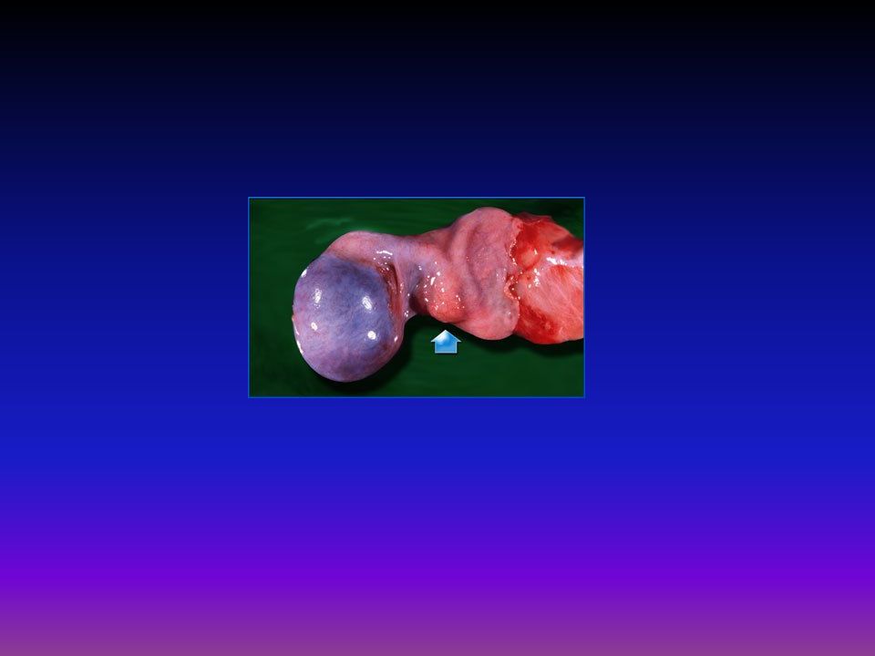
Scrotal trauma: hematocele
Hematocele: Blood within tunica vaginalis May represent severe testicular injury Ecchymosis of scrotal wall in setting of trauma Sonography: Identifies fluid collection in the tunica Blood more echogenic than hydrocele fluid Treatment: Surgical exploration to drain large hematoceles as well as testicular repair if ruptured

49
Scrotal trauma spectrum
Intratesticular hematoma or laceration of tunica Ultrasound : Assists to determine location of blood Intact Tunica: Surgery not necessary ? Testicular laceration: surgical exploration Traumatic epididymitis Results from blunt trauma Initial pain, then pain free, then pain returns Scrotal erythema, edema, epididymal tenderness Ultrasound: rules out severe injury Treatment: Supportive Scrotal laceration Evaluate testis and spermatic cord for injury Simple laceration: Hemostasis and chromic sutures

50
Scrotal injuries: Urology intervention
Large testicular hematoma may need drainage Delay in surgery may lead to ischemic necrosis, secondary infections, disruption of testicular function Testicular rupture with tear of the tunica albuginea requires surgical exploration. Salvage more likely if repaired within 24h Laceration to scrotum through the dartos All penetrating testicular injuries

51
Testicular torsion Scrotal pain and swelling – common presenting symptom in ED Acute scrotum Acute testicular torsion – rapid pickup is vital for salvage Salvage rate drops when repair delayed beyond 6-8 hours after acute event Acute scrotum – Testicular torsion is the working diagnosis until proven otherwise

52
Testicular torsion: Stats
1 out of every 4000 males before age of 25 Peak incidence: 13 years of age Another peak: Perinatal period. Newborn born with hard, necrotic testis. Hard or discolored scrotum Salvage not possible Rare: after age of 30 yrs

53
Testicular torsion: Cause
Basic mechanism – Movement of testis that is abnormally fixed in tunica vaginalis Infants: Lack of fixation of tunics in the scrotum. Extravaginal torsion Bell Clapper deformity: Tunica vaginalis has abnormally high attachment to spermatic cord Testis not fixed. Prone to torsion Allows testis to lie transversely and rotate Found in most cases. Commonly bilateral

54
Bell Clapper deformity

55
Testicular torsion: Clinical features
PAT: Appearance: Crying, irritable, uncomfortable WOB: Normal Circulation: Normal systemic Other findings: History of pain in past Acute onset pain in groin or scrotum Nausea and vomiting High riding testis, Transverse lie of testis Diffuse testicular pain, absent cremasteric reflex

56
Testicular torsion: Complications
Delay in re-establishing blood flow – loss of testicular function Delay Patient presentation Physician taking his or her time to establish diagnosis Testicular salvage: 80-90%: within 8 hours of acute pain <20%: for delay up to 12 hrs

59
Diagnostic studies Lab studies: not sufficient to make definitive diagnosis Ultrasound with Doppler: Readily available, non invasive and highly accurate Any uncertainty: Indicates surgical exploration Testicular scintigraphy: PPV of 95% Access may not be easy at all times High index of suspicion: Do not delay surgical procedure to confirm suspicion with a diagnostic study.

60
Testicular torsion: Differential
Torsion of appendix testis or appendix epididymis Epididymitis Orchitis Incarcerated Inguinal Hernia Scrotal trauma Hydrocele Varicocele HSP/ Kawasaki disease Scrotal cellulitis Testicular tumors

61
Management Analgesia: IV narcotic
Manual detorsion: Can preserve testicular viability and provide time Twist affected testis outwardly Successful detorsion: Relief of pain and visible lengthening of cord structures More than 360o detorsion may be required Surgery Non viable testis: Orchiectomy Viable testis: Orchiopexy Exploration of unaffected testis

62
Torsion of testicular appendage: Appendix testis or Appendix epididymis
Average age: 10 years Clinical features: Sudden onset pain limited to scrotum No abdominal or urinary symptoms Point tenderness at superior aspect of testis in early stages Blue dot: visible tender nodule in 20% cases

64
Diagnosis and Management
Testicular scan and Ultrasound Increased blood flow Inflammation at superior aspect of testis Treatment Expectant Analgesics Any doubt about diagnosis Urology consultation for exploration

65
Phimosis & Paraphimosis
Phimosis: Tightness of distal foreskin Cannot withdraw to expose the glans Not to be confused with penile adhesions Paraphimosis: Foreskin is retracted behind glans and left there – swollen, retracted foreskin Venous congestion & edema: reduction to normal position is difficult

66
Paraphimosis: Treatment
Manual reduction: Application of ice and steady local compression Local anesthesia: Penile block Pressure on glans (turning a sock inside out) Surgical reduction: Failure of manual reduction (2-3 attempts) Surgical division of foreskin Circumcision: after a few weeks Prevention: Education of uncircumcised male
 Surgical reduction: Failure of manual reduction (2-3 attempts) Surgical division of foreskin. Circumcision: after a few weeks. Prevention: Education of uncircumcised male.")
67
Improving success of manual reduction
Wrap the penis in plastic and apply ice packs Use compressive elastic dressings Apply direct circumferential manual compression Granulated sugar Hyaluronidase therapy - directly into several sites of the edematous prepuce. Puncture of the edematous site Hyaluronidase breaks down hyaluronic acid in connective tissue and enhances fluid diffusion between tissue planes, decreasing preputial swelling and resulting in almost immediate resolution of the edema

69
a. Retrograde urethrogram b. Foley catheter placement
An 8 yo boy is brought to the ED after getting hit by a car while riding his bicycle. On exam, he has stable vital signs, GCS of 15, and his abdomen is soft without tenderness. Blood is noted at the urethral meatus and he is unable to void. Which of the following is the most appropriate for management? a. Retrograde urethrogram b. Foley catheter placement c. Abdominal ultrasound d. Intravenous pyelogram (IVP) e. Ice packs and ibuprofen
 e. Ice packs and ibuprofen.")
70
A 13 yo boy comes to the ED with back pain after playing ice
hockey. He was checked and hit his back onto the boards. He noted gross hematuria a few hours afterwards. On exam, he has normal vital signs. His right flank shows a small ecchymosis on inspection. His abdomen is soft without tenderness. His urinalysis shows numerous RBCs per high power field. Which of the following tests is most appropriate in this patient? a. Intravenous pyelogram (IVP) b. Ultrasonography c. Cystourethrogram d. Abdominal CT e. Serial urinalyses
 b. Ultrasonography. c. Cystourethrogram. d. Abdominal CT. e. Serial urinalyses.")
71
A 6 yo girl comes to the ED after sustaining an injury to the
perineum. The patient was climbing on a tree when she fell approximately 4 feet landing on a large rock. She complains of pain and bleeding from vaginal area. She has been refusing to urinate due to pain. A 1-cm superficial vulvar laceration is noted at 3 o’clock with small amount of oozing blood. The hymen appears intact. The most appropriate management is: a. Surgical exploration under general anesthesia b. Laceration repair under local anesthesia c. Consultation with the child protection team d. Supportive care and sitz baths e. Placing a Foley catheter and hospitalization

72
A 13 yo boy comes to the ED after sustaining a straddle
injury to his scrotum while riding his bicycle. He is able to urinate without difficulty and has no gross hematuria. There is no trauma to the abdomen. On exam, he has normal vital signs. His right hemi-scrotum is swollen and ecchymotic. There is marked tenderness on palpation. There is no ecchymosis or swelling of the penis, and no blood per meatus. The management includes: a. Pelvic x-ray b. Needle aspiration c. Retrograde urethrogram d. Scrotal ultrasound e. No intervention is needed

73
An 8 yo boy presents to the ED after his penile skin got
caught in the zipper of his pants. On exam, his foreskin is caught in the zipper mechanism. Management includes: a. Cutting the median bar of the zipper b. Dissecting the skin free c. Applying ice before unzipping over the entrapped skin d. Moving the zipper back and forth after local anesthesia e. Performing a dorsal slit procedure

74
A 16 yo boy comes to the ED with left-sided groin pain and
scrotal swelling that began 4 hours prior to arrival. He also reports mild lower abdominal pain and nausea. On exam, his left scrotum is erythematous, moderately swollen and diffusely tender on palpation. A cremasteric reflex cannot be elicited. There is mild tenderness on palpation of lower abdomen. The most appropriate management of this patient is: a. Scrotal ultrasound b. Immediate surgical exploration c. Ceftriaxone and doxycycline d. Trimethoprim/sulfamethoxazole e. Incision and drainage

75
and penis pain since the morning of presentation. There is no
A 3 yo uncircumcised boy presents to the ED with swelling and penis pain since the morning of presentation. There is no history of trauma. He is able to void without difficulty. On exam, his foreskin is retracted and swollen, and the glans appears swollen. Which of the following would be the most appropriate initial treatment? a.Oral antibiotics b. Manual reduction c. Topical antibiotic d. Circumcision e. Warm sitz baths

Similar presentations


 Waleed M. Awwad, MD. FRCSC Assistant professor and Consultant Orthopedic Surgery department.>")