Download presentation
Presentation is loading. Please wait.

1
PREPARED BY: SANDHYA KS
CASE PRESENTATION PREPARED BY: SANDHYA KS

2
DEMOGRAPHIC DATA NAME: AH AGE: 25 yrs old SEX: Male MR NO.: 189691
NATIONALITY: Bangladeshi DIAGNOSIS: Small bowel perforation with peritonitis CHIEF COMPLAINTS: complaint of severe abdominal pain with vomiting NAME OF SURGERY: Exploratory laparotomy and small bowel resection with Anastomosis DATE OF ADMISSION: 10/01/13 DATE OF SURGERY: 11/01/2013 DATE OF DISCHARGE: 18/01/2013

3
Two drainage tubes from both sides of abdomen.
GENERAL Patient is intubated. Looks weak and fatigue. Unable to mobilize. Upper teeth fracture. Two drainage tubes from both sides of abdomen.

4
SKIN Skin is warm. Post operative scar present on abdomen.
Noted abrasion on upper and lower extremities. Post operative scar on right leg.

5
HEAD and NECK Hair is equally distributed. Absence of dandruff.
Abrasions on face. Patient’s pinna is same colour as fascial skin aligned with eye level. Lips are pink but swollen. Upper teeth fracture seen. No lymph node enlargement. CVP line present.

6
CARDIOVASCULAR Old RTA with chest trauma Airway Adequate
Heart sound : s1 and s2 normal Upon auscultation his BP is 120/80mmHg Pulse rate-66/mts Lungs – bilateral vescicular sound present.

7
Thorax is sympathetic on inspection

8
With Foleys catheter FG.16present
Genito urinary system With Foleys catheter FG.16present

9
Gastrointestinal System
Patient is old RTA with abdominal trauma tenderness present. Two drainage tubes present from both sides of abdomen.

10
MUSCULOSKELETAL SYSTEM
Unable to mobilize his right lower limb Has pain during examination Cannot perform ADL Tenderness at the site of fracture Visible deformity Lower extremities appears shortened

11
NEUROLOGIC Patient is on ventilator under sedation Old RTA with spine fracture GCS 15/15

12
PATIENT HISTORY PAST MEDICAL HISTORY Poor lung condition
Patient is old RTA with polytrauma Poor lung condition Fracture tibia and thoracic spine ORIF tibia done two months ago

13
PRESENT MEDICAL HISTORY
Patient is presented with post exploratory laparotomy with small bowel resection with anastomosis.

14
PRESENT SURGICAL HISTORY
He undergone exploratory laparotomy and small bowel resection with anastomiosis done under general anesthesia on 11/01/13

15
PAST SURGICAL HISTORY He undergone ORIF tibia done under general anesthesia on 01/11/12.

16
VITAL SIGNS BP- 120/86mmhg PR- 66 bpm Temperature C SPO2- 98%

17
MEDICATION Name of the medicine Dose Route and frequency action
Inj. promosan 10mg Iv/bid Antiemetic and gastroprokinetic agent Inj risek 40mg Iv/od H2 receptor antagonist Inj. ciproxin 200mg Antibiotic Inj. flagil 500mg Iv/tid Inj.tienan Inj.vancomycin 1gm Inj.tramadol 50mg Im/tid Analgesic Inj.clexane s/c,od Anticoagulant

18
INVESTIGATIONS Investigations Patient’s Values Normal Values PH 7.417
RBS 130 PCO2 38.7 mmHg 35-45 mmHg Na 134.8 mmol/L 135 to 145 mEq/L K 3.68 mmol/L mmol/l Total Bilirubin 31.9 µmol/L Direct Bilirubin 12.9 µmol SGOT 16.6 10-38 µ/L SGPT 17.8 10-41 µ/L Alkaline Phosphate 95.6 µ/L Protein 46.2 66-87 g/L Albumin 25.4 Hb 11.6 gm/dl g/dl WBC 20.27 PLT 328 /ul

19
INTRODUCTION small intestine (or small bowel) is the part of the gastrointestinal tract following the stomach and followed by the large intestine, and is where much of the digestion and absorption of food takes place. A bowel resection is a surgical procedure in which a part of the large or small intestine is removed. It may be performed due to cancer, necrosis, enteritis, diverticular disease, or a block in the intestine due to scar tissue. Other reasons to perform bowel resection include ulcerative colitis, traumatic injuries, precancerous polyps, and familial polyposis.
 is the part of the gastrointestinal tract following the stomach and followed by the large intestine, and is where much of the digestion and absorption of food takes place. A bowel resection is a surgical procedure in which a part of the large or small intestine is removed. It may be performed due to cancer, necrosis, enteritis, diverticular disease, or a block in the intestine due to scar tissue. Other reasons to perform bowel resection include ulcerative colitis, traumatic injuries, precancerous polyps, and familial polyposis.")
20
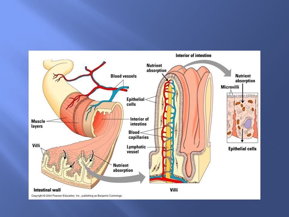
ANATOMY AND PHYSIOLOGY

21
ANATOMY AND PHYSIOLOGY

23
DISEASE CONDITION: Peritonitis
Peritonitis is an inflammation of the peritoneum, the thin tissue that lines the inner wall of the abdomen and covers most of the abdominal organs. Peritonitis may be localized or generalized, and may result from infection or from a non-infectious process.

24
The main manifestations of peritonitis are acute abdominal pain, abdominal tenderness, and abdominal guarding, which are exacerbated by moving the peritoneum, e.g., coughing (forced cough may be used as a test), flexing one's hips, or eliciting the Blumberg sign place). The presence of these signs in a patient is sometimes referred to as peritonism. The localization of these manifestations depends on whether peritonitis is localized (e.g., appendicitis or diverticulitis before perforation), or generalized to the whole abdomen. In either case, pain typically starts as a generalized abdominal pain (with involvement of poorly localizing innervations of the visceral peritoneal), and may become localized later (with the involvement of the somatically innervated parietal peritoneal layer). Peritonitis is an example of an acute abdomen.
, flexing one s hips, or eliciting the Blumberg sign place). The presence of these signs in a patient is sometimes referred to as peritonism. The localization of these manifestations depends on whether peritonitis is localized (e.g., appendicitis or diverticulitis before perforation), or generalized to the whole abdomen. In either case, pain typically starts as a generalized abdominal pain (with involvement of poorly localizing innervations of the visceral peritoneal), and may become localized later (with the involvement of the somatically innervated parietal peritoneal layer). Peritonitis is an example of an acute abdomen.")
25
COLLATERAL MNIFESTATIONS
Diffuse abdominal rigidity ("washboard abdomen") is often present, especially in generalized peritonitis Sinus tachycardia Development of ileus paralyticusi.e., intestinal paralysis), which also causes nausea, vomiting and bloating
 is often present, especially in generalized peritonitis. Sinus tachycardia. Development of ileus paralyticusi.e., intestinal paralysis), which also causes nausea, vomiting and bloating.")
26
INFECTED PERITONITIS Perforation of part of the gastrointestinal tract is the most common cause of peritonitis. Examples include perforation of the distal esophagus (Boerhaave syndrome), of the stomach (peptic ulcer, gastric carcinoma), of the duodenum (peptic ulcer), of the remaining intestine (e.g., appendicitis, diverticulitis, Meckl diverticulum, inflammatory bowel disease (IBD), intestinal infarction, intestinal strangulation, colorectal carcinoma, meconium peritonitis), or of the gallbladder (cholecystitis
, of the stomach (peptic ulcer, gastric carcinoma), of the duodenum (peptic ulcer), of the remaining intestine (e.g., appendicitis, diverticulitis, Meckl diverticulum, inflammatory bowel disease (IBD), intestinal infarction, intestinal strangulation, colorectal carcinoma, meconium peritonitis), or of the gallbladder (cholecystitis.")
27
Other possible reasons for perforation include abdominal trauma, ingestion of a sharp foreign body (such as a fish bone, toothpick or glass shard), perforation by an endoscope or catheter, and anastomotic leakage. The latter occurrence is particularly difficult to diagnose early, as abdominal pain and ileus paralyticus are considered normal in patients who have just undergone abdominal surgery. In most cases of perforation of a hollow viscous, mixed bacteria are isolated; the most common agents include Gram-negative bacilli (e.g., Escherichia coli) and anaerobic bacteria (e.g., Bacteroides fragilis). Fecal peritonitis results from the presence of feces in the peritoneal cavity. It can result from abdominal trauma and occurs if the large bowel is perforated during surgery.
 and anaerobic bacteria (e.g., Bacteroides fragilis). Fecal peritonitis results from the presence of feces in the peritoneal cavity. It can result from abdominal trauma and occurs if the large bowel is perforated during surgery..")
28
Disruption of the peritoneum, even in the absence of perforation of a hollow viscus, may also cause infection simply by letting micro-organisms into the peritoneal cavity. Examples include trauma, surgical wound, continuous ambulatory peritoneal dialysis, and intra-peritoneal chemotherapy are possible, including fungi such as Candida. Spontaneous bacterial peritonitis (SBP) is a peculiar form of peritonitis occurring in the absence of an obvious source of contamination. It occurs in patients with ascites, in particular, in children. See the article on spontaneous bacterial peritonitis for more information.
 is a peculiar form of peritonitis occurring in the absence of an obvious source of contamination. It occurs in patients with ascites, in particular, in children. See the article on spontaneous bacterial peritonitis for more information.")
29
Intra-peritoneal dialysis predisposes to peritoneal infection (sometimes named "primary peritonitis" in this context). Systemic infections (such as tuberculosis) may rarely have a peritoneal localization.
 may rarely have a peritoneal localization.")
30
Non-infected peritonitis
Non-infected peritonitis Leakage of sterile body fluids into the peritoneum, such as blood (e.g., endometriosis, blunt abdominal trauma), gastric juice (e.g., peptic ulcer, gastric carcinoma),bile (e.g., liver biopsy), urine (pelvic trauma), menstruum (e.g., salpingitis), pancreatic juice (pancreatitis), or even the contents of a ruptured dermoid cyst. It is important to note that, while these body fluids are sterile at first, they frequently become infected once they leak out of their organ, leading to infectious peritonitis within 24 to 48 hours. Sterile abdominal surgery, under normal circumstances, causes localized or minimal generalized peritonitis, which may leave behind a foreign body reaction and/or fibrotic adhesions. However, peritonitis may also be caused by the rare case of a sterile foreign body inadvertently left in the abdomen after surgery (e.g., gauze, sponge). Much rarer non-infectious causes may include familial Mediterranean fever, TNF receptor associated periodic syndrome, porphyria, and systemic lupus erythematosus.
, gastric juice (e.g., peptic ulcer, gastric carcinoma),bile (e.g., liver biopsy), urine (pelvic trauma), menstruum (e.g., salpingitis), pancreatic juice (pancreatitis), or even the contents of a ruptured dermoid cyst. It is important to note that, while these body fluids are sterile at first, they frequently become infected once they leak out of their organ, leading to infectious peritonitis within 24 to 48 hours. Sterile abdominal surgery, under normal circumstances, causes localized or minimal generalized peritonitis, which may leave behind a foreign body reaction and/or fibrotic adhesions. However, peritonitis may also be caused by the rare case of a sterile foreign body inadvertently left in the abdomen after surgery (e.g., gauze, sponge). Much rarer non-infectious causes may include familial Mediterranean fever, TNF receptor associated periodic syndrome, porphyria, and systemic lupus erythematosus.")
31
DIAGNOSIS A diagnosis of peritonitis is based primarily on the clinical manifestations described above. If peritonitis is strongly suspected, then surgery is performed without further delay for other investigations. Leukocytosis, hypokalemia, hypernatremia, and acidosis may be present, but they are not specific findings. Abdominal X-rays may reveal dilated, edematous intestines, although such X-rays are mainly useful to look for pneumo peritoneum, an indicator of gastrointestinal perforation. The role of whole-abdomen ultrasound examination is under study and is likely to expand in the future. Computed tomography (CT or CAT scanning) may be useful in differentiating causes of abdominal pain. If reasonable doubt still persists, an exploratory peritoneal lavage or laparoscopy may be performed. In patients with ascites, a diagnosis of peritonitis is made via paracentesis(abdominal tap): More than 250 polymorphonuclet cells per μL is considered diagnostic. In addition, Gram stain and culture of the peritoneal fluid can determine the microorganism responsible and determine their sensibility to antimicrobial agents.
 may be useful in differentiating causes of abdominal pain. If reasonable doubt still persists, an exploratory peritoneal lavage or laparoscopy may be performed. In patients with ascites, a diagnosis of peritonitis is made via paracentesis(abdominal tap): More than 250 polymorphonuclet cells per μL is considered diagnostic. In addition, Gram stain and culture of the peritoneal fluid can determine the microorganism responsible and determine their sensibility to antimicrobial agents.")
32
PATHOLOGY In normal conditions, the peritoneum appears greyish and glistening; it becomes dull 2–4 hours after the onset of peritonitis, initially with scarce serous or slightly turbid fluid. Later on, the exudate becomes creamy and evidently suppurative; in dehydrated patients, it also becomes very inspissated. The quantity of accumulated exudates varies widely. It may be spread to the whole peritoneum, or be walled off by the omentum and viscera. Inflammation features infiltration by neutrophils with fibrino-purulent exudation.

33
TREATMENT Depending on the severity of the patient's state, the management of peritonitis may include: General supportive measures such as vigorous intravenous rehydration and correction of electrolyte disturbances.

34
ANTIBIOTICS Antibiotics are usually administered intravenously, but they may also be infused directly into the peritoneum. The empiric choice of broad-spectrum antibiotics often consist of multiple drugs, and should be targeted against the most likely agents, depending on the cause of peritonitis (see above); once one or more agents are actually isolated, therapy will of course be targeted on them.
; once one or more agents are actually isolated, therapy will of course be targeted on them.")
35
EMPIRIC THERAPY Gram positive and gram negative organisms must be covered. Out of the Cephalosporin, cefoxitin and cefotecan can be used to cover gram positives, gram negatives, and anaerobes. Beta-lactams with beta lactamase inhibitors can also be used, examples include ampicillin/sulbactam, piperacillin/tazobactam, and ticarcillin/clavulanate.[2]Carbapenems are also an option when treating primary peritonitis as all of the carbapenems cover gram positives, gram negatives, and anaerobes except for ertapenem. The only fluoroquinolone that can be used is moxifloxacin because this is the only fluoroquinolone that covers anaerobes. Finally, tigecycline is a tetracycline that can be used due to its coverage of gram positives and gram negatives. Empiric therapy will often require multiple drugs from different classes

36
SURGERY (laparotomy) is needed to perform a full exploration and lavage of the peritoneum, as well as to correct any gross anatomical damage that may have caused peritonitis.[3] The exception is spontaneous bacterial peritonitis, which does not always benefit from surgery and may be treated with antibiotics in the first instance.
 is needed to perform a full exploration and lavage of the peritoneum, as well as to correct any gross anatomical damage that may have caused peritonitis.[3] The exception is spontaneous bacterial peritonitis, which does not always benefit from surgery and may be treated with antibiotics in the first instance.")
37
PROGNOSIS If properly treated, typical cases of surgically correctable peritonitis (e.g., perforated peptic ulcer, appendicitis, and diverticulitis) have a mortality rate of about <10% in otherwise healthy patients, which rises to about 40% in the elderly, and/or in those with significant underlying illness as well as in cases that present late (after 48 hours). If untreated, generalized peritonitis is almost always fatal.
 have a mortality rate of about <10% in otherwise healthy patients, which rises to about 40% in the elderly, and/or in those with significant underlying illness as well as in cases that present late (after 48 hours). If untreated, generalized peritonitis is almost always fatal.")
38
COMPLICATIONS Sequestration of fluid and electrolytes, as revealed by decreased central venous pressure, may cause electrolyte disturbances, as well as significant hypovolemia, possibly leading to shock and acute renal failure. A peritoneal abscess may form (e.g., above or below the liver, or in the lesser omentum Sepsi may develop, so blood cultures should be obtained.

39
DISEASE CONDITION –GASTROINTESTINAL PERFORATION
Gastrointestinal perforation is a complete penetration of the wall of the stomach, small intestine or large bowel, resulting in intestinal contents flowing into the abdominal cavity. Perforation of the intestines results in the potential for bacterial contamination of the abdominal cavity (a condition known as peritonitis). Perforation of the stomach can lead to a chemical peritonitis due to leaked gastric acid. Perforation anywhere along the gastrointestinal tract is a surgical emergency.
. Perforation of the stomach can lead to a chemical peritonitis due to leaked gastric acid. Perforation anywhere along the gastrointestinal tract is a surgical emergency.")
40
SIGNS AND SYMPTOMS Sudden attack of pain in epigastrium to the right of midline burning pain in epigastria, flatulence and dyspepsia rigidity of abdomen tenderness, and rebound tenderness nausea and vomiting fever and or chills.

41
CAUSES gastric ulcer appendicitis gastrointestinal cancer
diverticulitis superior mesenteric artery syndrome trauma, ascariasis Typhoid fever non-steroidal anti-inflammatory drugs ingestion of corrosives

42
DIAGNOSIS x-rays (free gas/air may be visible in the abdominal cavity)
computed tomography White blood cells are often ridged abdomen on palpation

43
SURGICAL INTERVENTIONS
exploratory laparotomy and closure of perforation If patient is in case nontoxic and clinically stable, they can be treated with intravenous fluids, antibiotics, nasogastric aspiration and bowel rest

44
EXPLORATORY LAPAROTOMY
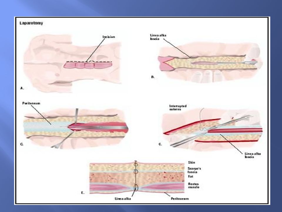
Definition A laparotomy is a large incision made into the abdomen. Exploratory laparotomy is used to visualize and examine the structures inside of the abdominal cavity.

45
PURPOSE Exploratory laparotomy is a method of abdominal exploration, a diagnostic tool that allows physicians to examine the abdominal organs. The procedure may be recommended for a patient who has abdominal pain of unknown origin or who has sustained an injury to the abdomen. Injuries may occur as a result of blunt trauma (e.g., road traffic accident) or penetrating trauma (e.g., stab or gunshot wound). Because of the nature of the abdominal organs, there is a high risk of infection if organs rupture or are perforated. In addition, bleeding into the abdominal cavity is considered a medical emergency. Exploratory laparotomy is used to determine the source of pain or the extent of injury and perform repairs if needed.
 or penetrating trauma (e.g., stab or gunshot wound). Because of the nature of the abdominal organs, there is a high risk of infection if organs rupture or are perforated. In addition, bleeding into the abdominal cavity is considered a medical emergency. Exploratory laparotomy is used to determine the source of pain or the extent of injury and perform repairs if needed.")
46
Laparotomy may be performed to determine the cause of a patient's symptoms or to establish the extent of a disease. For example, endometriosis is a disorder in which cells from the inner lining of the uterus grow elsewhere in the body, most commonly on the pelvic and abdominal organs. Endometrial growths, however, are difficult to visualize using standard imaging techniques such as x ray, ultrasound technology, or computed tomography (CT) scanning. Exploratory laparotomy may be used to examine the abdominal and pelvic organs (such as the ovaries, fallopian tubes, bladder, and rectum) for evidence of endometriosis. Any growths found may then be removed.
 scanning. Exploratory laparotomy may be used to examine the abdominal and pelvic organs (such as the ovaries, fallopian tubes, bladder, and rectum) for evidence of endometriosis. Any growths found may then be removed..")
47
Exploratory laparotomy plays an important role in the staging of certain cancers. Some other conditions that may be discovered or investigated during exploratory laparotomy include: cancer of the abdominal organs peritonitis (inflammation of the peritoneum, the lining of the abdominal cavity) appendicitis (inflammation of the appendix) pancreatitis (inflammation of the pancreas) abscesses (a localized area of infection) adhesions (bands of scar tissue that form after trauma or surgery) diverticulitis (inflammation of sac-like structures in the walls of the intestines) intestinal perforation ectopic pregnancy (pregnancy occurring outside of the uterus) foreign bodies (e.g., a bullet in a gunshot victims Internal bleeding.
 appendicitis (inflammation of the appendix) pancreatitis (inflammation of the pancreas) abscesses (a localized area of infection) adhesions (bands of scar tissue that form after trauma or surgery) diverticulitis (inflammation of sac-like structures in the walls of the intestines) intestinal perforation. ectopic pregnancy (pregnancy occurring outside of the uterus) foreign bodies (e.g., a bullet in a gunshot victims. Internal bleeding.")
48
INCISION Once an adequate level of anesthesia has been reached, the initial incision into the skin may be made. A scalpel is first used to cut into the superficial layers of the skin. The incision may be median (vertical down the patient's midline), paramedian (vertical elsewhere on the abdomen), transverse (horizontal), T-shaped, or curved, according to the needs of the surgery. The incision is then continued through the subcutaneous fat, the abdominal muscles, and finally, the peritoneum. Electrocautery is often used to cut through the subcutaneous tissue as it During a laparotomy, and an incision is made into the patient's abdomen (A). Skin and connective tissue called fascia is divided (B). The lining of the abdominal cavity, the peritoneum, is cut, and any exploratory procedures are undertaken (C). To close the incision, the peritoneum, fascia, and skin are stitched (E) has the ability to stop bleeding as it cuts. Instruments called retractors may be used to hold the incision open once the abdominal cavity has been exposed.
, paramedian (vertical elsewhere on the abdomen), transverse (horizontal), T-shaped, or curved, according to the needs of the surgery. The incision is then continued through the subcutaneous fat, the abdominal muscles, and finally, the peritoneum. Electrocautery is often used to cut through the subcutaneous tissue as it During a laparotomy, and an incision is made into the patient s abdomen (A). Skin and connective tissue called fascia is divided (B). The lining of the abdominal cavity, the peritoneum, is cut, and any exploratory procedures are undertaken (C). To close the incision, the peritoneum, fascia, and skin are stitched (E) has the ability to stop bleeding as it cuts. Instruments called retractors may be used to hold the incision open once the abdominal cavity has been exposed.")
50
ABDOMINAL EXPLORATION
The surgeon may then explore the abdominal cavity for disease or trauma. The abdominal organs in question will be examined for evidence of infection, inflammation, perforation, abnormal growths, or other conditions. Any fluid surrounding the abdominal organs will be inspected; the presence of blood, bile, or other fluids may indicate specific diseases or injuries. In some cases, an abnormal smell encountered upon entering the abdominal cavity may be evidence of infection or a perforated gastrointestinal organ

51
If an abnormality is found, the surgeon has the option of treating the patient before closing the wound or initiating treatment after exploratory surgery. Alternatively, samples of various tissues and/or fluids may be removed for further analysis. For example, if cancer is suspected, biopsies may be obtained so that the tissues can be examined microscopically for evidence of abnormal cells. If no abnormality is found, or if immediate treatment is not needed, the incision may be closed without performing any further surgical procedures. During exploratory laparotomy for cancer, a pelvic washing may be performed; sterile fluid is instilled into the abdominal cavity and washed around the abdominal organs, then withdrawn and analyzed for the presence of abnormal cells. This may indicate that a cancer has begun to spread.

52
CLOSURE Upon completion of any exploration or procedures, the organs and related structures are returned to their normal anatomical position. The incision may then be sutured (stitched closed). The layers of the abdominal wall are sutured in reverse order, and the skin incision closed with sutures or staples.
. The layers of the abdominal wall are sutured in reverse order, and the skin incision closed with sutures or staples.")
53
DIAGNOSIS Various diagnostic tests may be performed to determine if exploratory laparotomy is necessary. Blood tests or imaging techniques such as x ray, CT scan, and MRI are examples. The presence of intra peritoneal fluid (IF) may be an indication that exploratory laparotomy is necessary; one study indicated that IF was present in nearly three-quarters of patients with intra-abdominal injuries. Directly preceding the surgical procedure, an IV line will be placed so that fluids and/or medications may be administered to the patient during and after surgery. A Foley catheter will be inserted into the bladder to drain urine. The patient will also meet with the anesthesiologist to go over details of the method of anesthesia to be used.
 may be an indication that exploratory laparotomy is necessary; one study indicated that IF was present in nearly three-quarters of patients with intra-abdominal injuries. Directly preceding the surgical procedure, an IV line will be placed so that fluids and/or medications may be administered to the patient during and after surgery. A Foley catheter will be inserted into the bladder to drain urine. The patient will also meet with the anesthesiologist to go over details of the method of anesthesia to be used.")
54
AFTER CARE The patient will remain in the postoperative recovery roomfor several hours where his or her recovery can be closely monitored. Discharge from the hospital may occur in as little as one to two days after the procedure, but may be later if additional procedures were performed or complications were encountered. The patient will be instructed to watch for symptoms that may indicate infection, such as fever, redness or swelling around the incision, drainage, and worsening pain.

55
RISKS Risks inherent to the use of general anesthesia include nausea, vomiting, sore throat, fatigue, headache, and muscle soreness; more rarely, blood pressure problems, allergic reaction, heart attack, or stroke may occur. Additional risks include bleeding, infection, injury to the abdominal organs or structures, or formation of adhesions (bands of scar tissue between organs).
.")
56
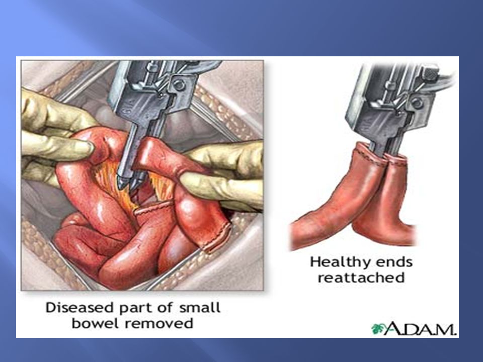
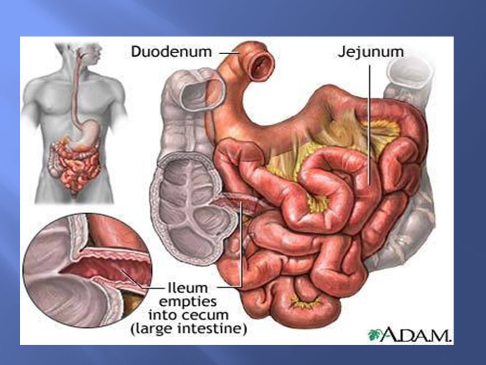
SMALL BOWEL RESECTION A small bowel resection is the surgical removal of one or more segments of the small intestine. Purpose The small intestine is the part of the digestive system that absorbs much of the liquid and nutrients from food. It consists of three segments: the duodenum, jejunum, and ileum; and is followed by the large intestine (colon).
.")
57
INTESTINAL OBSTRUCTION
This condition involves a partial or complete blockage of the bowel that results in the failure of the intestinal contents to pass through. Intestinal obstruction is usually treated by decompressing the intestine with suction, using a nasogastric tube inserted into the stomach or intestine. In cases where decompression does not relieve the symptoms, or if tissue death is suspected, bowel resection may be considered.

61
Injuries. Accidents may result in bowel injuries that require resection.
Precancerous polyps. A polyp is a growth that projects from the lining of the intestine. Polyps are usually benign and produce no symptoms, but they may cause rectal bleeding and develop into malignancies over time. When polyps have a high chance of becoming cancerous, bowel resection is usually indicated

62
DESCRIPTION The resection procedure can be performed using an open surgical approach or laparoscopically. There are three types of surgical small bowel resection procedures: Duodenectomy. Excision of all or part of the duodenum. Ileectomy. Excision of all or part of the ileum. Jejunectomy. Excision of all or a part of the jejunum.

63
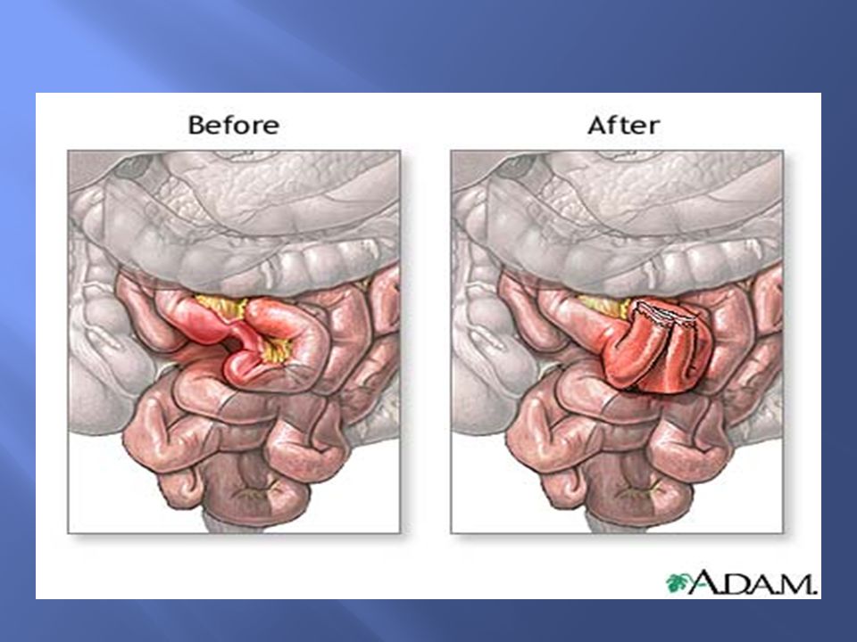
OPEN RESECTION Following adequate bowel preparation, the patient is placed under general anesthesia and positioned for the operation. The surgeon starts the procedure by making a midline incision in the abdomen. The diseased part of the small intestine (ileum or duodenum or jejunum) is removed. The two healthy ends are either stapled or sewn back together, and the incision is closed. If it is necessary to spare the intestine from its normal digestive work while it heals, a temporary opening (stoma) of the intestine into the abdomen ( ileostomy , duodenostomy, or jejunostomy) is made. The ostomy is later closed and repaired.
 is removed. The two healthy ends are either stapled or sewn back together, and the incision is closed. If it is necessary to spare the intestine from its normal digestive work while it heals, a temporary opening (stoma) of the intestine into the abdomen ( ileostomy , duodenostomy, or jejunostomy) is made. The ostomy is later closed and repaired.")
64
DIAGNOSIS and help prevent postoperative infection. A nasogastric tAs with any surgery, the patient is required to sign a consent form. Details of the procedure are discussed with the patient, including goals, technique, and risks. Blood and urine tests, along with various imaging tests and an electrocardiogram (EKG), may be ordered as required. To prepare for the procedure, the patient is asked to completely clean the bowel and is placed on a low residue diet for several days prior to surgery. A liquid diet may be ordered for at least the day before surgery, with nothing taken by mouth after midnight. Preoperative bowel preparation involving mechanical cleansing and administration of antibiotics before surgery is the standard practice. This involves the prescription of oral antibiotics (neomycin, erythromycin, or kanamycin sulfate) to decrease bacteria in the intestine ubeis inserted through the nose into the stomach on the day of surgery or during surgery. This removes the gastric secretions and prevents nausea and vomiting. A urinary catheter (thin tube inserted into the bladder) may also be inserted to keep the bladder empty during surgery, giving more space in the surgical field and decreasing chances of accidental injury
, may be ordered as required. To prepare for the procedure, the patient is asked to completely clean the bowel and is placed on a low residue diet for several days prior to surgery. A liquid diet may be ordered for at least the day before surgery, with nothing taken by mouth after midnight. Preoperative bowel preparation involving mechanical cleansing and administration of antibiotics before surgery is the standard practice. This involves the prescription of oral antibiotics (neomycin, erythromycin, or kanamycin sulfate) to decrease bacteria in the intestine ubeis inserted through the nose into the stomach on the day of surgery or during surgery. This removes the gastric secretions and prevents nausea and vomiting. A urinary catheter (thin tube inserted into the bladder) may also be inserted to keep the bladder empty during surgery, giving more space in the surgical field and decreasing chances of accidental injury.")
65
AFTERCARE Once the surgery is completed, the patient is taken to a postoperative or recovery unit where a nurse monitors recovery and ensures that bandages are kept clean and dry. Mild pain at the incision site is commonly experienced and the treating physician usually prescribes pain medication. Postoperative care also involves monitoring of blood pressure, pulse, respiration, and temperature. Breathing tends to be shallow because of the effect of anesthesia and the patient's reluctance to breathe deeply and experience pain that is caused by the abdominal incision. The patient is given instruction on the way to support the operative site during deep breathing and coughing. Fluid intake and output is measured, and the operative site is observed for color and amount of wound drainage. The nasogastric tube remains in place, attached to low intermittent suction until bowel activity resumes. Fluids and electrolytes are infused intravenously until the patient's diet can gradually be resumed, beginning with liquids and progressing to a regular diet as tolerated. The patient is generally out of bed approximately eight to 24 hours after surgery. Patients are usually scheduled for a follow-up examination within two weeks after surgery. During the first few days after surgery, physical activity is restricted.

66
RISKS Risks include all the risks associated with general anesthesia, namely, adverse reactions to medications and breathing problems. They also include the risks associated with any surgery, such as bleeding or infection. Additional risks associated specifically with bowel resection include: bulging through the incision (incisional hernia) narrowing (stricture) of the opening (stoma) blockage (obstruction) of the intestine from scar tissue.
 narrowing (stricture) of the opening (stoma) blockage (obstruction) of the intestine from scar tissue.")
68
PRIORITIZATION OF NURSING PROBLEMS
Acute pain related to surgical incision. Imbalanced Nutrition less than body requirement related to dietary modifications after surgery. Constipation related to surgery secondary to decreased mobilization. Impaired skin integrity related to surgical incision. Deficient fluid volume related to surgical procedure Risk for infection related to surgical incision.

69
NURSING CAREPLAN Subjective:
“ Im shivering and I feel weak” as verbalized by patient. Objective: Fever T- 38⁰C chills leakage from the wound of dressing increased pulse rate PR- 98bpm pain on the surgical site abdominal distention High risk for infection related to large surgical incision. Patient shows no evidence of infection as manifested by: Stable vital signs Afebrile Patient is stable and oriented No leakage from the wound dressing No abdominal distention Minimized the movement of the patient Done dressing daily with aseptic technique and check the dressing site for oozing Suction done to clear secretions and promote good ventilation Antibiotic therapy given like Metronidazole 500mg IV tid, Ciproxin 200mg IV bid Administered analgesics like Tramadol 50mg IM tid Immobilization reduces the risk of getting infection Will reduce the risk of infection To encourage adequate gas exchange It will reduce the chance of getting the infection To manage the post op pain After 12 hrs of nursing interventions the goals were met as evidenced by: Normal health person No signs of infection No oozing from the surgery site Active signs of wound healing Normal ROM Assessment: Planning: Implementation Evaluation Cues/Evidence Nursing Diagnosis Goals and desired outcome after 24 hours Nursing order/action Rationale for action

70
Assessment: Planning: Implementation Evaluation Cues/Evidence Nursing Diagnosis Goals and desired outcome Nursing order/action Rationale for action Subjective: “I cannot move properly and I’m having pain during motion” as verbalized by patient. Objective: Limited range of motion Inability to perform action as instructed Impaired physical mobility, acute pain secondary to exploratory and laparotomy and bowel resection with anastamosis. Patient will be able to perform his physical activity and free of complications as evidenced by: Participates in activites of daily living Performs physical activities independently Intact skin and absence of complications Normal bowel pattern Assisted patient for early ambulation. Encouraged adequate intake of fluids. Instructed or assisted patient with active and passive ROM exercises of affected and unaffected limbs. Determined presence of complications related to immobility such as pneumonia, elimination problem, decubitus ulcer. To maintain position and function and reduce the risk of pressure ulcers. To identify contributing factors of immobility To assess the presence of complications. Promote well being and maximized energy usage. Increases blood flow to muscles to improve muscle tone and maintain joint mobility. After 12 hrs of nursing intervention, the goals were met as evidenced by: Patient performs physical activities independently or with assisting devices as needed. Free of complications of immobility as normal bowel pattern.

71
PATIENT EDUCATON Review signs and symptoms of wound infection so early intervention may be instituted. Explain signs and symptoms of other post operations complications to report – elevated temperature , nausea, vomiting, abdominal distention changes in bowel function and stool consistency and color. Instruct the patient to report promptly blood in the stool or the coughing up of blood. Encourage the patient to turn , cough, deep breathe use of incentive spirometer and ambulation . discuss the importance of these functions during the recovery period. Review dietary changes such as increased fiber content and fluid intake and their importance in improving bowel function. Review actions and adverse effects of prescribed medications to encourage compliance and understanding of management. Assess the need for home health follow up , and initiate appropriate referrals if indicated.

72
CONCLUSION A case of post RTA polytrauma patient with peritonitis with bowel perforation and was with severe abdominal pain and vomiting. Initially seen by general surgeon. Surgical treatment exploratory laparotomy with bowel resection and anastomosis done. Patient is able to move. Health education given on home care. Patient was discharged. Patient was told to come for follow-up after 2 weeks.

73
BIBLIOGRAPHY Lippincott manual of Nursing Practice 9th edition
74
Thank you!!

Similar presentations


 The McGraw-Hill Companies, Inc. Permission required for reproduction or display. 23-1 Chapter 23 Abdominal and Gastrointestinal Disorders.>")


