Download presentation
Presentation is loading. Please wait.

1
بسم الله الرحمن الرحیم

2
Аnemia – pathologic state,accompanied by decrease in the level of hemoglobin and the quantity of erythrocytes per unit of volume of the blood.

3
Erythrocytes - less informative index of anemia than the level of hemoglobin therefore, in the general practice the basic criterion of severity is precisely Hb: Light degree of anemia - Hb 110-90 g / l, Light degree of anemia - Hb 110-90 g / l, The average degree of severity - Hb 90- 70 g / l, The average degree of severity - Hb 90- 70 g / l, Severe anemia - Hb below 70 g / liter Severe anemia - Hb below 70 g / liter

4
Laboratory Definition of Anemia Hgb: Hgb: Women: <12.0 Women: <12.0 Men: < 13.5 Men: < 13.5 Hct: Hct: Women: < 36 Women: < 36 Men: <41 Men: <41

5
RBC Life Cycle In the bone marrow, erythropoietin enhances the growth of differentiation of burst forming units- erythroid (BFU-E) and colony forming units-erythroid (CFU-E) into reticulocytes. In the bone marrow, erythropoietin enhances the growth of differentiation of burst forming units- erythroid (BFU-E) and colony forming units-erythroid (CFU-E) into reticulocytes. Reticulocyte spends three days maturing in the marrow, and then one day maturing in the peripheral blood. Reticulocyte spends three days maturing in the marrow, and then one day maturing in the peripheral blood. A mature Red Blood Cell circulates in the peripheral blood for 100 to 120 days. A mature Red Blood Cell circulates in the peripheral blood for 100 to 120 days. Under steady state conditions, the rate of RBC production equals the rate of RBC loss. Under steady state conditions, the rate of RBC production equals the rate of RBC loss. Erythropoetin role Erythropoetin role
 and colony forming units-erythroid (CFU-E) into reticulocytes. Reticulocyte spends three days maturing in the marrow, and then one day maturing in the peripheral blood. Reticulocyte spends three days maturing in the marrow, and then one day maturing in the peripheral blood. A mature Red Blood Cell circulates in the peripheral blood for 100 to 120 days. A mature Red Blood Cell circulates in the peripheral blood for 100 to 120 days. Under steady state conditions, the rate of RBC production equals the rate of RBC loss. Under steady state conditions, the rate of RBC production equals the rate of RBC loss. Erythropoetin role Erythropoetin role.")
7
Classification of Anemia I. Anemias resulting from acute blood loss I. Anemias resulting from acute blood loss II. Anemias resulting from a deficit of erythropoesis II. Anemias resulting from a deficit of erythropoesis 1) At the expense of maturation (mainly microcyte): violation of absorption and utilization of iron (iron) violation of transportation of iron (atransferrinemia) violation of recycling iron (thalassemia, sideroblastic anemia ) violation of reutilization of iron (anemia of chronic disease);
 At the expense of maturation (mainly microcyte): violation of absorption and utilization of iron (iron) violation of transportation of iron (atransferrinemia) violation of recycling iron (thalassemia, sideroblastic anemia ) violation of reutilization of iron (anemia of chronic disease);.")
8
Anemia (continued) 2) At the expense of differentiation (essentially normal): aplastic anemia (congenital and acquired) 3) At the expense of proliferation (mainly macrocytes) B12-DEFICIENCY anemia Folic-DEFICIENCY anemia.
 2) At the expense of differentiation (essentially normal): aplastic anemia (congenital and acquired) 3) At the expense of proliferation (mainly macrocytes) B12-DEFICIENCY anemia Folic-DEFICIENCY anemia.")
9
Anemia (continued) Anemias resulting from increased destruction of erythroid series cells - haemolytic: Anemias resulting from increased destruction of erythroid series cells - haemolytic: 1) caused by internal defects of erythrocytes membranopathy, enzimopathy, haemoglobinopathies; 1) caused by internal defects of erythrocytes membranopathy, enzimopathy, haemoglobinopathies; 2) the external (extracllular) effects: 2) the external (extracllular) effects: autoimmune, traumatic, etc. autoimmune, traumatic, etc. Classification D. Nathan, F. Oski, 2003, (book «Anemias in children», NA Finogenova et al, 2004.): Classification D. Nathan, F. Oski, 2003, (book «Anemias in children», NA Finogenova et al, 2004.):
: Classification D. Nathan, F. Oski, 2003, (book «Anemias in children», NA Finogenova et al, 2004.):.")
10
The clinical value of blood AutomaticalCountingUnits Measure- ment Normal Level Short Form HGB- Hemoglobin G/Liter120-160 Hb Hb RBC - erythrocyte 12 10 /L 3,9-5,9 Er Er HCT - Hematocryte % 36,0-48,0 Ht Ht

11
MCV- average volume of erythrocyte 3 1 micron = 1 - femtoliter (fl) 80 - 95 80 - 95 MCH – the average content of Hb in erythrocyte Pikogram 1 г =1012 pikograms 27,0-31,02 Colour Index (0,85-1,0) MCHC – average concentration of Hb in erythrocyte G/dl или g %, less g/l 32,0-36,0 RDW – width of the distribution curve of erythrocyte by volume % 11,5-14,5 anisocytosis
 MCH – the average content of Hb in erythrocyte Pikogram 1 г =1012 pikograms 27,0-31,02 Colour Index (0,85-1,0) MCHC – average concentration of Hb in erythrocyte G/dl или g %, less g/l 32,0-36,0 RDW – width of the distribution curve of erythrocyte by volume % 11,5-14,5 anisocytosis")
12
Measurements of Anemia Hemoglobin = grams of hemoglobin per 100 mL of whole blood (g/dL) Hemoglobin = grams of hemoglobin per 100 mL of whole blood (g/dL) Hematocrit = percent of a sample of whole blood occupied by intact red blood cells Hematocrit = percent of a sample of whole blood occupied by intact red blood cells RBC = millions of red blood cells per microL of whole blood RBC = millions of red blood cells per microL of whole blood MCV = Mean corpuscular volume MCV = Mean corpuscular volume If > 100 → Macrocytic anemia If > 100 → Macrocytic anemia If 80 – 100 → Normocytic anemia If 80 – 100 → Normocytic anemia If < 80 → Microcytic anemia If < 80 → Microcytic anemia RDW = Red blood cell distribution width RDW = Red blood cell distribution width = (Standard deviation of red cell volume ÷ mean cell volume) × 100 = (Standard deviation of red cell volume ÷ mean cell volume) × 100 Normal value is 11-15% Normal value is 11-15% If elevated, suggests large variability in sizes of RBCs If elevated, suggests large variability in sizes of RBCs
 Hemoglobin = grams of hemoglobin per 100 mL of whole blood (g/dL) Hematocrit = percent of a sample of whole blood occupied by intact red blood cells Hematocrit = percent of a sample of whole blood occupied by intact red blood cells RBC = millions of red blood cells per microL of whole blood RBC = millions of red blood cells per microL of whole blood MCV = Mean corpuscular volume MCV = Mean corpuscular volume If > 100 → Macrocytic anemia If > 100 → Macrocytic anemia If 80 – 100 → Normocytic anemia If 80 – 100 → Normocytic anemia If < 80 → Microcytic anemia If < 80 → Microcytic anemia RDW = Red blood cell distribution width RDW = Red blood cell distribution width = (Standard deviation of red cell volume ÷ mean cell volume) × 100 = (Standard deviation of red cell volume ÷ mean cell volume) × 100 Normal value is 11-15% Normal value is 11-15% If elevated, suggests large variability in sizes of RBCs If elevated, suggests large variability in sizes of RBCs")
13
Anemia due to Decreased Response to Erythropoietin Iron-Deficiency Iron-Deficiency Vitamin B12 Deficiency Vitamin B12 Deficiency Folate Deficiency Folate Deficiency Anemia of Chronic Disease Anemia of Chronic Disease

15
Iron Deficiency Iron Deficiency Can result from: Can result from: Pregnancy/lactation Pregnancy/lactation Normal growth Normal growth Blood loss Blood loss Intravascular hemolysis Intravascular hemolysis Gastric bypass Gastric bypass Malabsorption Malabsorption Iron is absorbed in proximal small bowel; decreased abosrption in celiac disease, inflammatory bowel disease Iron is absorbed in proximal small bowel; decreased abosrption in celiac disease, inflammatory bowel disease May manifest as PICA May manifest as PICA Tendency to eat ice, clay, starch, crunchy materials Tendency to eat ice, clay, starch, crunchy materials May have pallor, koilonychia of the nails, beeturia May have pallor, koilonychia of the nails, beeturia Peripheral smear shows microcytic, hypochromic red cells with marked anisopoikilocytosis. Peripheral smear shows microcytic, hypochromic red cells with marked anisopoikilocytosis.

16
Iron Deficiency Anemia - koilonychia

17
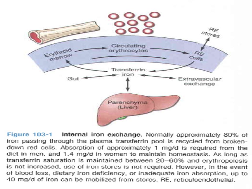
Iron in the Bod y

19
Serum Transferrin (Beta-globulin). Main function - transport of absorbed iron in the depot (liver, spleen), into the medullary erythroid predecessors and into the reticulocytes. Main function - transport of absorbed iron in the depot (liver, spleen), into the medullary erythroid predecessors and into the reticulocytes. Basic place of synthesis - liver. Basic place of synthesis - liver. An increase in the content of transferrin with lowering in the level of iron of serum is characteristic for the iron-deficiency state. An increase in the content of transferrin with lowering in the level of iron of serum is characteristic for the iron-deficiency state. A decrease in the level of transferrin can be with the damage of the liver (different genesis) and with the loss of protein (for example, in nephrotic syndrome). A decrease in the level of transferrin can be with the damage of the liver (different genesis) and with the loss of protein (for example, in nephrotic syndrome). The level of transferrin is increased in the last term of pregnancy. The level of transferrin is increased in the last term of pregnancy.
, into the medullary erythroid predecessors and into the reticulocytes. Main function - transport of absorbed iron in the depot (liver, spleen), into the medullary erythroid predecessors and into the reticulocytes. Basic place of synthesis - liver. Basic place of synthesis - liver. An increase in the content of transferrin with lowering in the level of iron of serum is characteristic for the iron-deficiency state. An increase in the content of transferrin with lowering in the level of iron of serum is characteristic for the iron-deficiency state. A decrease in the level of transferrin can be with the damage of the liver (different genesis) and with the loss of protein (for example, in nephrotic syndrome). A decrease in the level of transferrin can be with the damage of the liver (different genesis) and with the loss of protein (for example, in nephrotic syndrome). The level of transferrin is increased in the last term of pregnancy. The level of transferrin is increased in the last term of pregnancy..")
20
Transferrin LIMITATION The concentration of TF is subjected to the daily variations The concentration of TF is subjected to the daily variations Acute inflammation contributes to lowering the TF level Acute inflammation contributes to lowering the TF level CLINICAL SIGNIFICANCE Basic clinical index for the differentiation between the iron-deficiency ([TF] ↑ ) and hemolytic anemia ([TF] ↓ ) Basic clinical index for the differentiation between the iron-deficiency ([TF] ↑ ) and hemolytic anemia ([TF] ↓ ) More precise index than total iron binding capacity More precise index than total iron binding capacity After the liberation of iron from the complex, TF ion of Fe3+ must be restored into Fe2+ After the liberation of iron from the complex, TF ion of Fe3+ must be restored into Fe2+
![Transferrin LIMITATION The concentration of TF is subjected to the daily variations The concentration of TF is subjected to the daily variations Acute inflammation contributes to lowering the TF level Acute inflammation contributes to lowering the TF level CLINICAL SIGNIFICANCE Basic clinical index for the differentiation between the iron-deficiency ([TF] ↑ ) and hemolytic anemia ([TF] ↓ ) Basic clinical index for the differentiation between the iron-deficiency ([TF] ↑ ) and hemolytic anemia ([TF] ↓ ) More precise index than total iron binding capacity More precise index than total iron binding capacity After the liberation of iron from the complex, TF ion of Fe3+ must be restored into Fe2+ After the liberation of iron from the complex, TF ion of Fe3+ must be restored into Fe2+](http://images.slideplayer.com/20/6229516/slides/slide_20.jpg "Transferrin LIMITATION The concentration of TF is subjected to the daily variations The concentration of TF is subjected to the daily variations Acute inflammation contributes to lowering the TF level Acute inflammation contributes to lowering the TF level CLINICAL SIGNIFICANCE Basic clinical index for the differentiation between the iron-deficiency ([TF] ↑ ) and hemolytic anemia ([TF] ↓ ) Basic clinical index for the differentiation between the iron-deficiency ([TF] ↑ ) and hemolytic anemia ([TF] ↓ ) More precise index than total iron binding capacity More precise index than total iron binding capacity After the liberation of iron from the complex, TF ion of Fe3+ must be restored into Fe2+ After the liberation of iron from the complex, TF ion of Fe3+ must be restored into Fe2+")
21
Ferritin water-soluble complex of iron hydroxide with the protein apoferritin. water-soluble complex of iron hydroxide with the protein apoferritin. It is located in cells of the liver, spleen, bone marrow, in the reticulocytes. It is located in cells of the liver, spleen, bone marrow, in the reticulocytes. Ferritin is the basic protein in human which deposits iron and concentration of ferritin in the serum reflects the reserve of iron in the organism. Ferritin is the basic protein in human which deposits iron and concentration of ferritin in the serum reflects the reserve of iron in the organism.

22
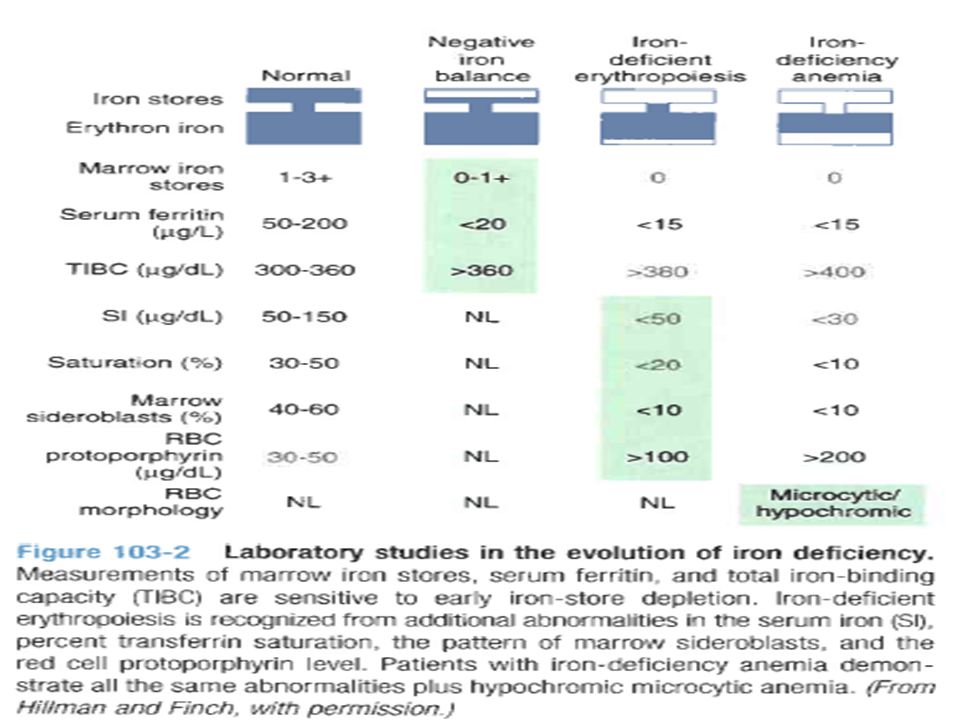
Iron Deficiency Anemia – Lab Findings Serum Iron Serum Iron LOW (< 50 micrograms/dL) LOW (< 50 micrograms/dL) Total Iron Binding Capacity (TIBC) Total Iron Binding Capacity (TIBC) HIGH ( > 360 micrograms/dL) HIGH ( > 360 micrograms/dL) Serum Ferritin Serum Ferritin LOW (< 20 nanograms/mL) LOW (< 20 nanograms/mL) Can be “falsely”normal in inflammatory states Can be “falsely”normal in inflammatory states
 LOW (< 50 micrograms/dL) Total Iron Binding Capacity (TIBC) Total Iron Binding Capacity (TIBC) HIGH ( > 360 micrograms/dL) HIGH ( > 360 micrograms/dL) Serum Ferritin Serum Ferritin LOW (< 20 nanograms/mL) LOW (< 20 nanograms/mL) Can be falsely normal in inflammatory states Can be falsely normal in inflammatory states")
24
Treatment of Iron Deficiency Anemia Oral iron salts Oral iron salts Ferrous sulfate – 325 mg(50 mg absorption) Ferrous sulfate – 325 mg(50 mg absorption) Side effects: constipation, black stools, positive hemmoccult test Side effects: constipation, black stools, positive hemmoccult test Vitamin C can facilitate iron absorption. Vitamin C can facilitate iron absorption.

25
Treatment of Iron Deficiency Anemia Diet: meat, liver, yeast, fish Diet: meat, liver, yeast, fish Oral preparations: recovery rate Hb does not differ from parenteral introduction, side effects are less, excessive introduction does not lead to hemosiderosis. Oral preparations: recovery rate Hb does not differ from parenteral introduction, side effects are less, excessive introduction does not lead to hemosiderosis. - Dosage : 1 hour prior to the meal - Dosage : 1 hour prior to the meal in the evening time (absorption increase in the second-half of a day) in the evening time (absorption increase in the second-half of a day)
 in the evening time (absorption increase in the second-half of a day).")
26
Possibilities : dark colour of stool and transitory dyspeptic disorders (nausea, diarrhea or watery stool) Possibilities : dark colour of stool and transitory dyspeptic disorders (nausea, diarrhea or watery stool) Check analysis of the blood: in 7-10 days – reticulocyte reaction; 4 weeks - increase Hb and Ht Check analysis of the blood: in 7-10 days – reticulocyte reaction; 4 weeks - increase Hb and Ht - Iron tolerance test(2 tablet-2 h-100 micro/dl During the normalization of the indices of the blood – reduce the dose of preparation
 Possibilities : dark colour of stool and transitory dyspeptic disorders (nausea, diarrhea or watery stool) Check analysis of the blood: in 7-10 days – reticulocyte reaction; 4 weeks - increase Hb and Ht Check analysis of the blood: in 7-10 days – reticulocyte reaction; 4 weeks - increase Hb and Ht - Iron tolerance test(2 tablet-2 h-100 micro/dl During the normalization of the indices of the blood – reduce the dose of preparation")
27
Parenteral Introduction of Iron in exceptional cases in exceptional cases in severe iron deficiency anemia in severe iron deficiency anemia intolerance of oral preparations (after repeated replacement and reduction in the dose) intolerance of oral preparations (after repeated replacement and reduction in the dose) diseases of gastro-intestinal tract diseases of gastro-intestinal tract syndrome of the disrupted intestinal absorbtion syndrome of the disrupted intestinal absorbtion after the extensive resection of the small intestine after the extensive resection of the small intestine continuous blood loss continuous blood loss
 intolerance of oral preparations (after repeated replacement and reduction in the dose) diseases of gastro-intestinal tract diseases of gastro-intestinal tract syndrome of the disrupted intestinal absorbtion syndrome of the disrupted intestinal absorbtion after the extensive resection of the small intestine after the extensive resection of the small intestine continuous blood loss continuous blood loss")
28
Complications of Parenteral Introduction Local reactions (pains, phlebitis) Local reactions (pains, phlebitis) General reactions (anaphylaxis, fever, head and articulate pains, vomiting, rash, bronchospasm). General reactions (anaphylaxis, fever, head and articulate pains, vomiting, rash, bronchospasm).Preparations: Venofer - for the intravenous introduction, Maltofer, Ferrum-Lek - intramuscular
.Preparations: Venofer - for the intravenous introduction, Maltofer, Ferrum-Lek - intramuscular.")
29
Overdose of Iron In the first 6-8 hours - epigastral pains, nausea, vomiting (including with the blood), diarrhea, pallor, sleepiness, acrocyanosis) For 12-24 hours - metabolic acidosis, leukocytosis, there can be spasms, coma, after 2-4 days - necroses of the liver and kidneys. For 12-24 hours - metabolic acidosis, leukocytosis, there can be spasms, coma, after 2-4 days - necroses of the liver and kidneys. Treatment: emetic means, stomach lavage, the method of milk with the egg white, Deferoksamin, Desferal, symptomatic therapy. Treatment: emetic means, stomach lavage, the method of milk with the egg white, Deferoksamin, Desferal, symptomatic therapy.

30
Iron Overload Syndrome Human does not have special mechanism of the excretion of iron! Its excessive introduction leads to hemosiderosis. Clinical manifestations: Gradual increase of the dimensions of the liver, spleen, cardiopathy, suprarenal insufficiency, diabetes mellitus Human does not have special mechanism of the excretion of iron! Its excessive introduction leads to hemosiderosis. Clinical manifestations: Gradual increase of the dimensions of the liver, spleen, cardiopathy, suprarenal insufficiency, diabetes mellitus Laboratory signs: Increase in serum iron (more than 30 mmol/liter), percentage of saturation transferrin by iron it is more than 50%, ferritin of serum it is more than 1000 ng/ml Increase in serum iron (more than 30 mmol/liter), percentage of saturation transferrin by iron it is more than 50%, ferritin of serum it is more than 1000 ng/ml
, percentage of saturation transferrin by iron it is more than 50%, ferritin of serum it is more than 1000 ng/ml Increase in serum iron (more than 30 mmol/liter), percentage of saturation transferrin by iron it is more than 50%, ferritin of serum it is more than 1000 ng/ml.")
31
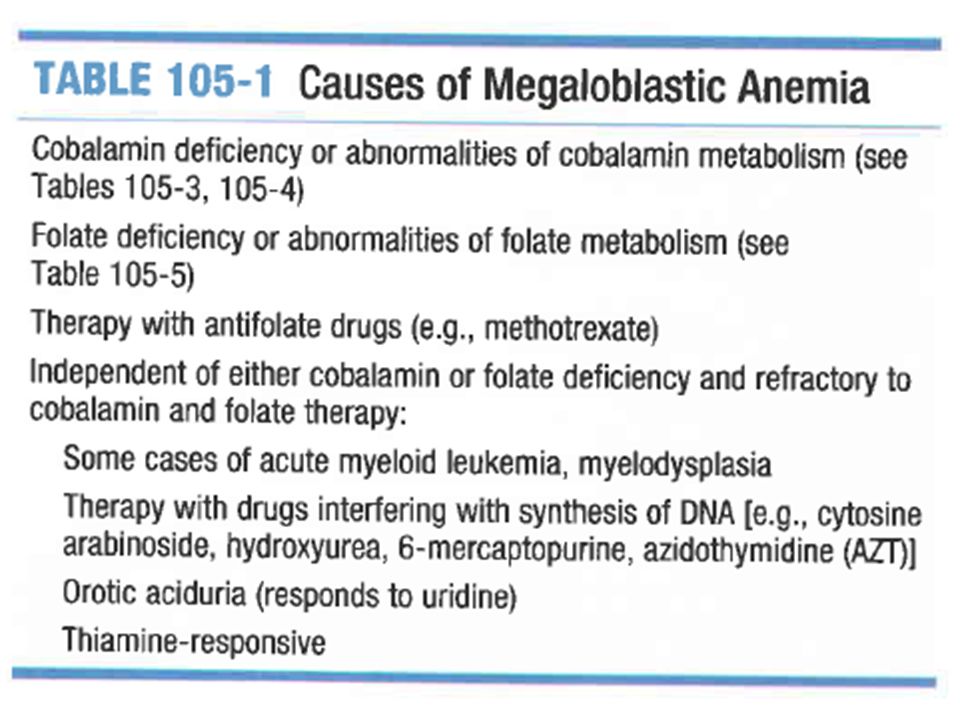
Megaloblastic Anemia A subclass of macrocytic anemia A subclass of macrocytic anemia (under morphologic classification) Or A subclass of anemias due to defective DNA synthesis A subclass of anemias due to defective DNA synthesis (pathogenetic classification)
 Or A subclass of anemias due to defective DNA synthesis A subclass of anemias due to defective DNA synthesis (pathogenetic classification)")
37
Vit.B12 Average diet contains 5 – 30 g Vit. B 12 daily Average diet contains 5 – 30 g Vit. B 12 daily The amount of Vit. B 12 in the body is about 2 – 5 mg. The amount of Vit. B 12 in the body is about 2 – 5 mg. Most of it is in the liver. Most of it is in the liver. The store is sufficient for 3-6 years in case of impaired absorbtion. The store is sufficient for 3-6 years in case of impaired absorbtion. The storage form is mainly adenosylcobalamin. The storage form is mainly adenosylcobalamin.

38
stomach Enterohepatic circulation Ileum cells Pancreas enzymes Parietal cell Duodenum and jejunum B 12 in diet R-Binder R - B 12 B 12 IF B 12 TC II B 12 İleum IF IF - B 12

41
Functions of Vit.B 12 2- Methyl FH 4 FH 4 Homocystein Methionin SAM B 1 2 B 1 2 Methionin synthase

42
Vit.B12 Food sources rich in Vit.B12 Food sources rich in Vit.B12 Liver Liver Kidney Kidney Muscle Muscle Egg Egg Milk,Cheese and other diary products Milk,Cheese and other diary products Seafood Seafood

43
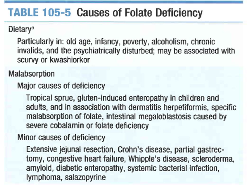
Folic acid Daily requirements Age 0 - 10 3.6 g /kg 0 - 10 3.6 g /kg > 10 3 g /kg > 10 3 g /kg Pregnants 500 g Pregnants 500 g Lactation +100 g Lactation +100 g Diet contains 100 - 500 g folate/day. Diet contains 100 - 500 g folate/day.

44
Folate absorbtion Mainly jejunum. Mainly jejunum. In the form of monoglutamate. In the form of monoglutamate. Methyltetrahydrofolate monoglutamate is the form it is found in serum. Methyltetrahydrofolate monoglutamate is the form it is found in serum.

45
Folate levels: Normal ranges Serum:6 – 21 g/L (RBC volume) Serum:6 – 21 g/L (RBC volume) Red cell: 160 – 640 g/L (RBC volume) Red cell: 160 – 640 g/L (RBC volume) Folate deficiency Serum folate : <4 g /L Serum folate : <4 g /L Red cell folate: <140 g /L Red cell folate: <140 g /L
 Serum:6 – 21 g/L (RBC volume) Red cell: 160 – 640 g/L (RBC volume) Red cell: 160 – 640 g/L (RBC volume) Folate deficiency Serum folate : <4 g /L Serum folate : <4 g /L Red cell folate: <140 g /L Red cell folate: <140 g /L")
46
Folate stores Total body folate: 5 – 20 mg Total body folate: 5 – 20 mg Storage place : Liver Storage place : Liver Storage form: Methyl- FH 4 polyglutamate Storage form: Methyl- FH 4 polyglutamate

47
Dihydrofolate THFA Methylene THFA Deoxyuridilate Thymidilate DNA- thymine Methyl THFA Homocystein Methyonine B 12 Dihydrofolate reductase serine glycine Thymidylate synthase

48
Tissues or organs other than bone marrow are also affected Skin,GIS, female genital system mucosal epithelium Skin,GIS, female genital system mucosal epithelium Congenital abn.(neural tube defects) Congenital abn.(neural tube defects) Neurologic changes (Vit.B 12 deficiency) Neurologic changes (Vit.B 12 deficiency) Peripheral neuropathy Peripheral neuropathy Subacute combined degeneration of spinal cord Subacute combined degeneration of spinal cord Cerebral -Mental changes Cerebral -Mental changes Hyperhomocysteinemia Hyperhomocysteinemia
 Congenital abn.(neural tube defects) Neurologic changes (Vit.B 12 deficiency) Neurologic changes (Vit.B 12 deficiency) Peripheral neuropathy Peripheral neuropathy Subacute combined degeneration of spinal cord Subacute combined degeneration of spinal cord Cerebral -Mental changes Cerebral -Mental changes Hyperhomocysteinemia Hyperhomocysteinemia")
49
Clinical findings(1) Clinical findings(1) Anemia: Anemia: Symptoms of anemia + palor+slight icterus Glossitis : Glossitis : Sore tongue, poor taste sensation, pain Papill. atrophy-beefy tongue

51
RBC Indexes: MCV MCV MCH MCH RDW RDW

52
Biochemical findings LDH( LDH -1> LDH - 2) LDH( LDH -1> LDH - 2) Bilirubin(indirect) Bilirubin(indirect) Ferritin and serum iron Ferritin and serum iron Haptoglobin Haptoglobin
 LDH( LDH -1> LDH - 2) Bilirubin(indirect) Bilirubin(indirect) Ferritin and serum iron Ferritin and serum iron Haptoglobin Haptoglobin")
54
Criteria of Effective Treatment Subjective improvement during the first days of treatment; Subjective improvement during the first days of treatment; Reticulocytosis, maximally expressed (to 20%) on 5-7 th day of treatment; Reticulocytosis, maximally expressed (to 20%) on 5-7 th day of treatment; Increase in hemoglobin and number of erythrocytes, beginning from the 2nd week of treatment; Increase in hemoglobin and number of erythrocytes, beginning from the 2nd week of treatment; The normalization of the blood index, number of leukocytes and thrombocytes in 3-4 weeks of treatment. The normalization of the blood index, number of leukocytes and thrombocytes in 3-4 weeks of treatment.

55
Anemia Case Study #1 A 72 year old male has the CBC findings shown. Peripheral RBCs are hypochromic & microcytic.

56
Anemia Case Study #2 A 48 year old male has become progressively more fatigued at the end of the day. This has been going on for months. In the past month he has noted paresthesias with numbness in his feet. A CBC demonstrates the findings shown.

57
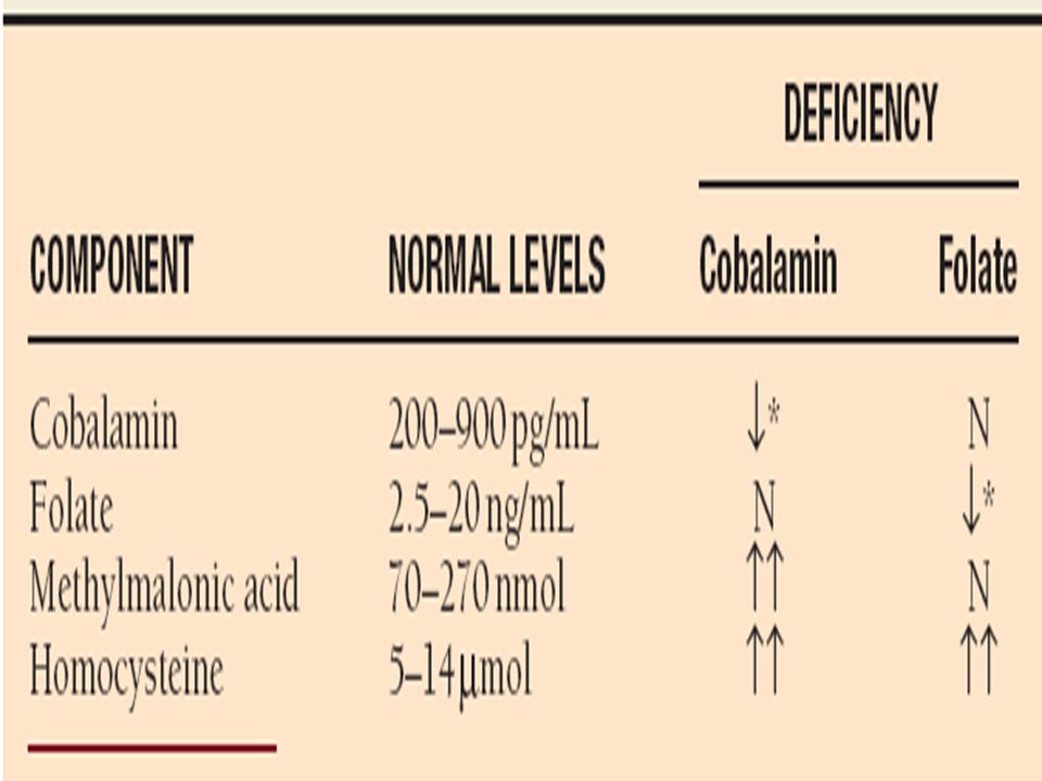
Case #3 Labs: Labs: Hgb: 5.1 g/dL Hgb: 5.1 g/dL MCV: 112 MCV: 112 RDW: 21% RDW: 21% Platelets: 109 Platelets: 109 WBC: 4.6 WBC: 4.6

58
Case #3 Which of the following blood levels are most likely in this patient? Which of the following blood levels are most likely in this patient? Vitamin B12 FolateMethylmalonic Acid Homocysteine (A)LowNormalHigh (B)LowNormal High (C)NormalLowHighNormal (D)NormalLowNormalHigh (D)Normal
LowNormalHigh (B)LowNormal High (C)NormalLowHighNormal (D)NormalLowNormalHigh (D)Normal.")
Similar presentations


, Moscow,>")
/ HYPOCHROMIC &/or (NORMO)/ MICROCYTIC ANEMIAS 1. Disorders of iron utilization a. iron deficiency b. anemia of.>")


. Complete Blood Count ( CBC)>")


i.>")