Download presentation
Presentation is loading. Please wait.

1
Introduction to Forensic Science
Forensic Pathology Greek: pathos – disease logos – study of Pathologist is a medical doctor (10-15 post secondary training), detective, public relations – much time spent communicating findings to law enforcement officers and/or jury
, detective, public relations – much time spent communicating findings to law enforcement officers and/or jury.")
2
The autopsy provides forensic evidence.
Forensic Pathology is the branch of medicine which analyses victims of crime scenes medically. They are the last physician for the deceased and their role is to discover and interpret the evidence left during the autopsy.

3
Forensic Pathology Pathology, the study of disease, is the broadest of the medical specialties. Pathologists don’t treat patients nor do surgery themselves- they consult with primary care and specialist physicians. Forensic Pathologist is a medical doctor with post secondary training.

4
Anatomic Pathology Diagnosis of disease and injury by the gross and microscopic examination of tissue specimens: Biopsies Organs pap smears bone marrow aspirates blood smears. The anatomic pathologist is also the one who performs autopsies.

5
Clinical Pathology Clinical pathology deals with the medical laboratory where the pathologist serves as medical director. The pathologist bears ultimate responsibility for medical laboratory test results.

6
Role of the Pathologist
Determine type of wound Measure the dimensions (length, width, depth) Position relative to anatomical landmarks Determine initial location if wound involves cutting, slashing, etc. Determine height of victim, other contributing factors like heart problems.
 Position relative to anatomical landmarks. Determine initial location if wound involves cutting, slashing, etc. Determine height of victim, other contributing factors like heart problems.")
7
Analysis of Wounds Not every crime victim is murdered.
Pathologists can contribute to proof of the severity of a crime or that a crime actually occurred in some cases for a living victim. Some victims are too young to testify and some are too severely injured to remember the crime. Wounds provide evidence of the crime.

8
Wound Categories Bruises (or contusions)
Abrasions (or grazes or scratches) Lacerations Incised wounds Puncture (or stab) wounds Gunshot wounds
 Lacerations. Incised wounds. Puncture (or stab) wounds. Gunshot wounds.")
9
This will be discussed with ballistics talk
Gunshot Wounds This will be discussed with ballistics talk

10
Bruises A bruise is "a hemorrhage into tissues produced by the escape of blood from blood vessels". Bruises may be found in the skin, muscles, and internal organs.

11
Bruises Bruises are typically produced by a blunt force impact, such as a blow or a fall. They may also be produced by squeezing or pinching, where the force is applied gradually and then maintained. Hickies or "love-bites" are superficial bruises.

12
Natural Bruises Bruises may occur in a variety of natural diseases in which there is an abnormality of the clotting mechanism of the blood, e.g. scurvy (vitamin C deficiency), leukemia, alcoholic liver disease. This bruising is "spontaneous" because the injury which produces it is so insignificant as to typically pass unnoticed. The presence of such natural disease will exaggerate the bruising effects of any trauma.
, leukemia, alcoholic liver disease. This bruising is spontaneous because the injury which produces it is so insignificant as to typically pass unnoticed. The presence of such natural disease will exaggerate the bruising effects of any trauma.")
13
Problems with Skin Bruises
Delayed appearance Ageing (relative) Site of Trauma Shape of object Degree of force Post-mortem bruises Post-mortem lividity
 Site of Trauma. Shape of object. Degree of force. Post-mortem bruises. Post-mortem lividity.")
14
Classic Causes of Bruises
Finger pad bruises: battered babies, manual strangulation Different ages: repeated assaults Shoulders and arms: forceful restraint Wrists and ankles: dragging Inner thighs: forceful intercourse Chest: resuscitation Bruising is uncommon in Suicides

15
Bruises The extent of bruising is inversely proportional to the sharpness of the impacting object. Bruises may be associated with other blunt force injuries such as abrasions and lacerations. As a general rule bruising is not associated with incised wounds or stab wounds where there is a free flow of blood from the cut blood vessels rather than leaking into the tissues.

16
Site of Trauma In contrast with abrasions, the location of a bruise does not necessarily reflect the precise point of injury. Leaking blood will follow the path of least resistance and gravity. For example, in the elderly, intense and widespread bruising of the lower thigh may follow a fracture of the hip; a bruise of the temple may move down to the cheek; a fractured jaw may result in bruising appearing on the neck.

17
Delayed Appearance Deep bruises may have delayed appearance at the skin surface. Deep bruises may require as long as 12 or 24 hours to become apparent, and some may never do so The more superficial the source of bleeding, the sooner the discoloration will be seen on the skin surface. In a living victim, a second examination in one or two days may show bruising. In the dead, a further examination one or two days after the original autopsy may show bruises which were not previously seen and reveal previously faint bruises. This may be particularly the case with "fingerpad bruises" produced by hands. Ultraviolet (UV) light may disclose bruises which are not otherwise identifiable at the time of examination.
 light may disclose bruises which are not otherwise identifiable at the time of examination.")
18
Autopsy and Bruising Bruising in Deep Tissue Documenting Bruising
1. Possibly life-threatening 2. Sometimes no external injury 3. Revealed in autopsy Documenting Bruising 1. Photography 2. Notes

19
Degree of Force The size of a bruise is an unreliable indicator of the degree of force causing it. However, a heavy impact is likely to produce a large bruise and a light impact to produce a small bruise. If bruising is slight, it is reasonable to assume that the degree of violence was slight.

20
Determining Degree of Force in Bruise Patterns
Location: Some areas of the body bruise more easily than others. The face bruises more readily than the hands. Bruising occurs more readily in loose tissues and where there is a large amount of subcutaneous fat Bruising is less apparent where the skin is strongly supported by fibrous tissue or if the muscle tone is good.

21
Determining Degree of Force in Bruise Patterns
Age Infants and the elderly tend to bruise more easily than young and middle aged adults. Infants have loose and delicate skin, and the abundant subcutaneous fat. Elderly have degenerative changes in the tissues which support the small blood vessels of the skin and subcutaneous tissues. Gender: Women bruise more easily than men because they have more subcutaneous fat and this is particularly true of obese women. Natural Disease Skin color

22
Causitive Object The shape of the bruise is most likely to reflect the shape of the causative object when the object is small and hard and death occurs soon after injury

23
Causitive Object A doughnut bruise is produced by an object with a rounded contour (e.g. baseball). Two parallel linear bruises result from a blow with a rod or stick Bruises can follow rounded contours if they are caused by a flexible object like a lash If the blow with the rod is struck against the buttocks, - a particularly pliable, curved, soft surface - the tissues are compressed and flattened under the impact; the resulting bruise will follow the curved contour of the buttocks. A pliable weapon such as a strap or electric flex may produce a similar appearance as it wraps around the body on impact.

24
Causitive Object Bruises produced by fingerpads as a result of gripping are usually larger than the fingerpads themselves. The pattern and location suggests the mechanism of causation: On the neck in throttling On the upper arms in restraint. Such bruises are referred to as patterned. A bruise which bears the imprint of the shape or contour of the impacting object is said to be patterned. A tracing of the pattern may be made to match to the causative object, or photographs of the injury and object may be superimposed. Patterned bruises of this type may be associated with patterned (imprint) abrasions. ligatures around the neck in strangulation the headlight or bumper of a vehicle in hit and run the muzzle or sight of a gun in contact gunshot wounds.
 abrasions. ligatures around the neck in strangulation. the headlight or bumper of a vehicle in hit and run. the muzzle or sight of a gun in contact gunshot wounds.")
25
Aging of Bruises Color changes a bruise goes through can give a rough estimate of time of injury Colors result from breakdown of hemoglobin from tissues Dark blue/purple (1-18 hours) Blue/brown (~1 to 2days) Green (~ 2 to 3 days) Yellow (~3 to 7 days) This rate assumes person is healthy, however. Contusions Bruise due to rupture of small blood vessels (skin or internal organs) Colors result from breakdown of hemoglobin from tissues
 Blue/brown (~1 to 2days) Green (~ 2 to 3 days) Yellow (~3 to 7 days) This rate assumes person is healthy, however. Contusions. Bruise due to rupture of small blood vessels (skin or internal organs) Colors result from breakdown of hemoglobin from tissues.")
26
Aging Bruises While accurate estimation of the age of a single bruise is not possible, a fresh bruise can be distinguished easily from one which is several days old. Establishing that bruises are of different ages may be of medical importance where there is an allegation of repeated assaults: Child abuse Wife beating Where pre-existing injuries need to be distinguished from those produced by a recent assault like a chronic alcoholic who was assaulted.

27
Post Mortem Bruises Bruising is a phenomenon of living tissue- since it usually requires circulating blood to push the blood from the veins. It isn’t possible to tell bruises that occurred causing death from those that occurred minutes earlier. You can only say they occurred at or about time of death.

28
Post Mortem Bruises It requires considerable violence to produce a bruise post mortem or after death. These bruises are smaller relative to the degree of force used. Post mortem bruises are most readily produced in areas of hypostasis (post mortem lividity, livor mortis) or where tissues can be forcibly compressed against bone. A bruise can develop on the head after the body is left lying on the back.
 or where tissues can be forcibly compressed against bone. A bruise can develop on the head after the body is left lying on the back.")
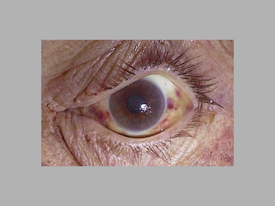
29
Post Mortem Lividity (hypostasis, livor mortis)
The settling, after death, of blood within the blood vessels under the influence of gravity. This results in a purplish discoloration of parts of the body that are lower while sparing areas of pressure contact - contact pallor. The pattern and distribution of lividity distinguishes it from bruising. A body found on its back has livor mortis on the dorsal (back) side with pale areas where the bone contacted the floor. In doubtful cases, an incision of the skin will cause blood to ooze from the cut ends of vessels in instances of lividity. In contrast, the blood within the tissues in bruises will not ooze. Washing the cut surface with running water will remove the blood from livid tissues but not the blood infiltrating the tissues in bruises. Confirmation of the distinction may be made by microscopic examination.
 side with pale areas where the bone contacted the floor. In doubtful cases, an incision of the skin will cause blood to ooze from the cut ends of vessels in instances of lividity. In contrast, the blood within the tissues in bruises will not ooze. Washing the cut surface with running water will remove the blood from livid tissues but not the blood infiltrating the tissues in bruises. Confirmation of the distinction may be made by microscopic examination.")
30
Decomposition Post mortem decomposition with its initial green discoloration of the anterior abdominal wall is readily distinguished from bruising. Putrefactive lysis of blood cells within the vessels and decompositional breakdown of the vessel walls results in diffusion of lysed blood into the adjacent tissues. Existing bruises are enlarged by this process. Later, putrefactive hemolytic staining of tissue may mask ante mortem bruising (e.g. in the neck muscles in case of choking).
.")
31
Patterns of Injury Bruises to the knuckles of the hands, together with bruises of the eyelids, bridge of the nose, cheeks and lips, suggest a fist fight. Bruising around the eyes (spectacle bruises) may be produced by direct blows, but also commonly result from a fracture of the base of the skull, e.g. in vehicle collisions or gunshot wounds to the head They may also follow blunt impact to the forehead producing jolting of the eyeballs in their sockets with tearing of small orbital blood vessels.
 may be produced by direct blows, but also commonly result from a fracture of the base of the skull, e.g. in vehicle collisions or gunshot wounds to the head. They may also follow blunt impact to the forehead producing jolting of the eyeballs in their sockets with tearing of small orbital blood vessels.")
32
Patterns of Injury Bruising of the genitalia and around the anus suggests sexual assault. Severe bruising of the genitalia, with or without laceration, can be produced by kicks. Counter-pressure bruising, with or without abrasion, to the back, (shoulder blades, sacrum and pelvis) suggests pressure against a firm surface as in forceful restraint on the ground. Similar bruising may be seen on boney prominences of the front of the pelvis.
 suggests pressure against a firm surface as in forceful restraint on the ground. Similar bruising may be seen on boney prominences of the front of the pelvis.")
33
Patterns of Injury In kicking assaults with the shod foot, bruises are invariably associated with multiple abrasions and lacerations. Gangs, individuals without weapons The bruises and abrasions may be patterned by the boot. Bruising is typically extensive and targeted on the face, neck, ears, groin, and kidney area. Internal bruising is usually severe.

34
Patterns of Injury Bruises are painful and therefore not commonly self-inflicted; extensive bruising creates a presumption of assault. Accidents generally are unforeseen and the injuries they produce tend not to follow a recognizable pattern. Some places bruise easily accidentally though: shins and hips.

35
Patterns of Injury Injuries in motor vehicle collisions almost invariably include abrasions and lacerations as well as bruises. Patterns of injury may allow reconstruction of incidents involving pedestrians or allow distinction between driver and front seat passenger.

36
Participation Question
Give me an example of forensic usefulness of analysis of bruises.

37
Abrasions Friction injury removing skin or tissue
An abrasion is "a portion of body surface from which the skin or mucous membrane has been removed by rubbing." Graze is synonymous with abrasion. A scratch is a linear abrasion produced by drawing a sharp point over the surface.

38
Abrasions Side impact produces a moving abrasion:
Indicates direction. Trace material (e.g. grit). Direct impact produces an imprint abrasion: Pattern of causative object. All abrasions reflect site of impact (in contrast with bruises). Assessment of age of abrasions is difficult. Post-mortem abrasions - Brown, leathery
. Direct impact produces an imprint abrasion: Pattern of causative object. All abrasions reflect site of impact (in contrast with bruises). Assessment of age of abrasions is difficult. Post-mortem abrasions - Brown, leathery.")
39
Incised Wounds (Cuts, Slashes, Stab)
Stab wounds or puncture wounds are penetrating injuries whose depth within the body is much greater than the dimensions of the wound on the body surface. Breach of the full thickness of the skin due to contact with a sharp edge.

40
Stab Wounds Forensic Importance Reflects sharp edge, not weapon type
No trace evidence Bleeds profusely Hemorrhage and air embolism They can be produced by any long thin object which impacts the body with sufficient force to penetrate. The typical instrument is a knife, but any sharp pointed, or keen-edged object will work. A wound which passes completely through a structure (such as the heart) is described as "perforating.“ A wound which enters a structure but does not exit it is described as “penetrating." Objects producing stab wounds fall into three general categories: Flat with a sharp point, e.g. a knife. Long and thin with a sharp point, e.g. a hat pin or needle. Long rigid objects which are blunt ended, e.g. a wooden stake.
 is described as perforating. A wound which enters a structure but does not exit it is described as penetrating. Objects producing stab wounds fall into three general categories: Flat with a sharp point, e.g. a knife. Long and thin with a sharp point, e.g. a hat pin or needle. Long rigid objects which are blunt ended, e.g. a wooden stake.")
41
Stab Wounds Should be Described at Autopsy:
Site relative to local anatomical landmarks as well as its distance from the midline and above the heel (or below the crown of the head). Shape and Size including the dimensions with the wound edges closed back. Direction (approximately) in three dimensions. Depth of the wound track at autopsy. Damage to tissues and organs along the wound track. Effects of damage described above. If a stabbing with a knife is "straight in and out" then the length of the stab wound on the skin surface is typically slightly less than the width of the blade, due to the elastic recoil of the skin following withdrawal of the weapon. This effect is exaggerated by the tendency of the wound to gape open so that the wound becomes wider but shorter; this amount of gaping reflects the alignment of the wound relative to natural lines of tension within the skin. The extent of wound gaping is also influenced by damage to underlying fascia and muscles. The length of a stab wound on the skin surface should be measured after the gaping edges have been approximated (using transparent tape), re-establishing the anatomical relationship of the skin edges that existed prior to injury. The post mortem examination of a body takes place with the body lying flat on its back, so that the position of the viscera will not necessarily be the same as when the person was standing up or sitting down during life. The depth of penetration of a stab wound should be interpreted with caution. If some fixed bone, e.g. a vertebra, is damaged at the limit of the wound track, then assessment of depth of penetration is easier. If the blade enters the skin obliquely then one edge will be beveled and the opposing edge will overhang (be undercut) This gives an indication of the direction of the underlying wound track. Withdrawal of the knife blade in a slightly different direction front the entry thrust may produce a notch along one edge of the wound; marked twisting of the blade (or twisting of the body) may exaggerate the notch effect, resulting in a cruciate (cross-shaped) wound.
. Shape and Size including the dimensions with the wound edges closed back. Direction (approximately) in three dimensions. Depth of the wound track at autopsy. Damage to tissues and organs along the wound track. Effects of damage described above. If a stabbing with a knife is straight in and out then the length of the stab wound on the skin surface is typically slightly less than the width of the blade, due to the elastic recoil of the skin following withdrawal of the weapon. This effect is exaggerated by the tendency of the wound to gape open so that the wound becomes wider but shorter; this amount of gaping reflects the alignment of the wound relative to natural lines of tension within the skin. The extent of wound gaping is also influenced by damage to underlying fascia and muscles. The length of a stab wound on the skin surface should be measured after the gaping edges have been approximated (using transparent tape), re-establishing the anatomical relationship of the skin edges that existed prior to injury. The post mortem examination of a body takes place with the body lying flat on its back, so that the position of the viscera will not necessarily be the same as when the person was standing up or sitting down during life. The depth of penetration of a stab wound should be interpreted with caution. If some fixed bone, e.g. a vertebra, is damaged at the limit of the wound track, then assessment of depth of penetration is easier. If the blade enters the skin obliquely then one edge will be beveled and the opposing edge will overhang (be undercut) This gives an indication of the direction of the underlying wound track. Withdrawal of the knife blade in a slightly different direction front the entry thrust may produce a notch along one edge of the wound; marked twisting of the blade (or twisting of the body) may exaggerate the notch effect, resulting in a cruciate (cross-shaped) wound.")
42
Stab Wounds: Shape of Weapon
A knife blade with a double edge will normally produce a symmetrical elliptical wound with both ends pointed, clean cut edges and without any associated bruising or marginal abrasion. A knife with a single-edged blade may show relative blunting ("fish-tailing") of one end of the entry slit. A single edged blade can produce a wound with two pointed ends, mimicking an injury from a double edged blade. A bayonet, which has a ridge along the back of the blade with a groove along each side, may produce a slit like an elongated letter "T". The cross-section of the blade may be accurately reproduced when the blade passes through bone, e.g. skull or sternum. Marks on bone may reflect if the instrument used is serrated of notched.
 of one end of the entry slit. A single edged blade can produce a wound with two pointed ends, mimicking an injury from a double edged blade. A bayonet, which has a ridge along the back of the blade with a groove along each side, may produce a slit like an elongated letter T . The cross-section of the blade may be accurately reproduced when the blade passes through bone, e.g. skull or sternum. Marks on bone may reflect if the instrument used is serrated of notched.")
43
Stab Wounds: Shape of Weapon
Stab wounds produced with relatively blunt instruments such as pokers, closed scissors and files, tend to bruise and scrape the wound margin. These blunter instruments also tend to lacerate, as well as cleanly penetrate, the skin; the blunter the point of the instrument and the thicker its shaft, the more likely is the entry hole to become a ragged, often cross shaped split. Forensic Pathologist sometimes practices wound type: The Body Farm.

44
Stab Wounds: Degree of Force
The most reliable estimate of blade width is made from the deepest wound with the shortest skin surface length. It is easy to over-estimate the amount of force required to produce a stab wound. The depth of a wound is not generally an indication of the degree of force used. The length of the wound on the skin surface may give a false impression of the true width of the weapon blade if: The blade has a marked taper and the entire length of the blade has not passed through the skin (i.e. a "shallow" stab). The blade has not passed "straight in and out" but instead there has been "rocking" of the blade, or it has been withdrawn at a different angle or plane to the original thrust.
. The blade has not passed straight in and out but instead there has been rocking of the blade, or it has been withdrawn at a different angle or plane to the original thrust.")
45
Stab Wounds: Degree of Force
The most critical factor is the sharpness of the point of the instrument; relatively little force is required to produce a stab wound provided a knife with a sharp point. After clothing, the skin offers the greatest resistance to penetration; once this is overcome, then the blade easily cuts into deeper tissue.

46
Stab Wounds: Degree of Force
The penetration of bone does imply a significant degree of force. The tip of the blade may break off when driven into bone and should be recovered for matching with the weapon. In estimating the force exerted by an assailant, consideration should be given to the possibility of counter pressure by the victim, e.g. running or falling forwards.

47
Stab Wounds: Length of Weapon
The depth of the wound (the length of the wound track, provides some indication of the length of the stabbing instrument). The wound track length may be less than the length of the instrument if the weapon was not thrust into the body to its full length. The wound track can be longer than the knife if there is force compressing tissues. If the thrust is forceful, the entire length of the blade enter the body and the tissues may be compressed. When the weapon is withdrawn, the resulting wound track in the decompressed tissues is longer than the blade of the weapon. A small pocket knife blade can produce an abdominal stab wound which penetrates the aorta, or a chest wound which penetrates the heart. Depth is a specific characteristic of stab wounds; although the injury to the skin may appear trivial, the great depth of these wounds, with severing of blood vessels and penetration of viscera, characteristically results in internal hemorrhage, often with little external bleeding. Compression of the body by a forceful stab is most pronounced in abdominal and chest wounds but occurs to a lesser extent with any soft tissue (muscle and fat).
. The wound track length may be less than the length of the instrument if the weapon was not thrust into the body to its full length. The wound track can be longer than the knife if there is force compressing tissues. If the thrust is forceful, the entire length of the blade enter the body and the tissues may be compressed. When the weapon is withdrawn, the resulting wound track in the decompressed tissues is longer than the blade of the weapon. A small pocket knife blade can produce an abdominal stab wound which penetrates the aorta, or a chest wound which penetrates the heart. Depth is a specific characteristic of stab wounds; although the injury to the skin may appear trivial, the great depth of these wounds, with severing of blood vessels and penetration of viscera, characteristically results in internal hemorrhage, often with little external bleeding. Compression of the body by a forceful stab is most pronounced in abdominal and chest wounds but occurs to a lesser extent with any soft tissue (muscle and fat).")
48
Stab Wounds: Clothing Cuts on the clothing should be noted and correlated with injuries to the body. More than one cut on the clothing may correspond with a single injury to the body as a result of folds in the clothing. Cuts to the clothing may not exactly overlie corresponding wounds to the body. There may be stab or slash marks on the clothing without corresponding injuries to the body, e.g. "defense"-type slashes to the arms.

49
Stab Wounds: Clothing Blood flow patterns on the clothing may indicate the position of the victim at the time of the stabbing. Blood drops on the tops of the shoes from a stab to the chest in a victim standing upright. Blood flow direction can change with movements of the body. Wound track can be indicated by undercutting and beveling of the external wound. Extrapolation from the direction of wound tracks to an opinion on the relative positions of an assailant and victim should be, since two potentially moving objects are involved. Wound track can be indicated by undercutting and beveling of the external wound. Extrapolation from the direction of wound tracks to an opinion on the relative positions of an assailant and victim should be always cautious, since two potentially moving objects are involved.

50
Stab Wounds: Cause of Death
Most deaths from stab wounds are homicides. Homicidal stab wounds are usually multiple, since most wounds leave the victim capable of some resistance for a measurable time during which the thrusts are repeated. Single homicidal stabbings are often associated with drugged, drunk, sleeping, or otherwise partially incapacitated victims and are almost always aimed at the heart. Wound directions and sites inaccessible to the victim also suggest homicide. Multiple scattered widely spaced wounds most, if not all, of which have penetrated deeply (suggesting relatively uniform force), with tracks in different directions and several lethal, also suggests homicide.
, with tracks in different directions and several lethal, also suggests homicide.")
51
Stab Wounds: Cause of Death
Homicidal stab wounds to the chest are all likely to be deep, penetrating the chest wall, and more than one may be lethal. Stabs in the back strongly suggest homicide. In cases of multiple scattered stabs, the larger the number the greater the certainty of murder. There is often a sexual motive to deaths with this type of "over-kill". In a stabbing action by an assailant, the weapon is usually held with the point downwards, so that most wounds in the victim are directed downwards. If the weapon is help with the point upwards, in a so-called "continental" manner, the wounds will be directed upwards.

52
Defense Stab Wounds "Defense wounds" are the result of the immediate and instinctive reaction of a victim to ward off anticipated injuries and may be seen in both homicidal and accidental deaths. Defense wounds result from raising the arm to ward off the attack or attempts to grasp the weapon. The resulting injuries may be stabs or slashes or both.

53
Defense Stab Wounds Attempts to grab the knife results in deep cuts to the palm of the hand and the palm side of fingers. With the hand in a gripping position the palm skin is loose and folded so that resultant cuts appear irregular and ragged. They may be duplicated by the thrust and withdrawal of the weapon. Penetration of the hand or arm is also a defense wound. The absence of defense wounds does not exclude homicide since the victim may be incapable of effective defense.

54
Suicidal Stab Wounds Suicide by stabbing is distinctive. The wounds, if multiple, have a location and direction accessible to the victim and are typically grouped in the "pit" of the stomach. Use of one hand is indicated by a consistent direction of penetration. Multiple wound tracks extending from the same slit in the skin reflects partial withdrawal of the weapon and further thrusts (possibly trial feelers), and suggests possible suicide.
, and suggests possible suicide.")
55
Suicidal Stab Wounds Typically a suicidal stabbing is to the bare skin and the clothing may be removed or pulled aside to effect this. Defense wounds do not occur in suicide, although the sharpness of a knife may be tested by running the blade across the tips of the fingers. Multiple scattered wounds weighs against suicide unless there was serious mental illness. The area of the heart is the preferred site for suicidal stabbing, but occasionally the abdomen or the neck is selected. Suicide may be attempted by penetration of the neck. Occasionally suicide is accomplished with a single stroke and the weapon may be left protruding from the wound. The hand of the decedent may be gripping the weapon in cadaveric spasm; this is proof that the victim was holding the weapon at the time of death and therefore creates a presumption, but not a proof, of suicide.

56
Suicidal Stab Wounds Fatalities from a single stab wound can be difficult and such a wound may be homicidal, suicidal, or accidental. Autopsy findings should always be interpreted in the light of information concerning the circumstances and scene of death. If the stab wound was inflicted during a fight then the usual defense is that it was accidental, the victim having ran or fallen on to the weapon. The position and direction of the wound may help resolve the issue.

57
Stab vs Slash Stab wounds are deep and not wide.
Slash wounds are wide and not deep.

58
Penetrating Wounds (Punctures)
Breach in full skin thickness and depth is greater than length Long, thin, sharp or blunt object. If sharp object then equals "stab wound".

59
Lacerations (Tears, Splits)
Splitting of the skin by the direct crushing of blunt trauma. Typically over bone, e.g. scalp, eyebrow, cheekbone.

60
Lacerations Distinguished from incised wounds by: Forensic Importance
Adjacent abrasion/bruise Ragged edge Tissue bridges in depth Forensic Importance Not related to object shape Trace evidence Relatively little blood loss (except scalp) Rarely suicidal
 Rarely suicidal.")
61
Lacerations Typical Examples Stellate pattern from poker end
Circles/crescents from hammer Y-shaped from metal rod Inside lips from blow to mouth. Stretching lacerations in vehicular accidents.

62
Bite Marks Double crescent of abrasions and bruises
Early Examination, loss of definition Swab for saliva, photograph Comparative value Child abuse, sexual assault.

63
Assault Any type of wound, combinations
Scattered, multiple directions, uniform force Defense injuries Several potentially lethal Clothing Secondary injuries

64
Order of Infliction Tentative or scattered first
Fatal and grouped last Distant shots before close shots.

65
Accidental Any area, single, clothing Defense injuries
Secondary injuries Check history (suicide attempts, assaults)
")
66
Blood Spatter Bruises and abrasions, none Lacerations, not much
Incised and stab wounds, often profuse.

67
Strangulation Strangulation implies pressure to the neck, and deaths due to strangulation are therefore of immense forensic importance. It can be defined as a circumferential squeezing of the neck that is independent of the gravitational weight or suspension of the head. Manual strangulation Ligature strangulation Choke holds

68
Signs of Strangulation
Obstruction of jugular veins with impaired venous return to the heart, leading to cyanosis (blue color), congestion (tissue swelling), and petechiae. Obstruction of carotid arteries. Stimulation of baroreceptors in the carotid sinuses and carotid sheaths. Elevation of larynx and tongue, closing the oropharynx.
, congestion (tissue swelling), and petechiae. Obstruction of carotid arteries. Stimulation of baroreceptors in the carotid sinuses and carotid sheaths. Elevation of larynx and tongue, closing the oropharynx.")
69
Petechia Very small hemorrhages (ranging in size from a pinpoint to a pinhead), which occur in tissues, may be described as petechia, or petechial hemorrhages (from the Italian petecchia, which has the Latinized plural petechiae). These hemorrhages may also be described as punctate (from the Latin punctum, a point).
, which occur in tissues, may be described as petechia, or petechial hemorrhages (from the Italian petecchia, which has the Latinized plural petechiae). These hemorrhages may also be described as punctate (from the Latin punctum, a point).")
71
Manual Strangulation Usually caused by men against women, and rarely against another man since a large disparity in physical strength between the assailant and victim is needed.

72
Signs of Manual Strangulation
Disc-like finger-tip bruises Abrasions Linear finger-nail scratches (from victim or assailant) Often limited signs of suffocation as fingers are more likely to probe deeper neck structures and cause reflex cardiac arrest
 Often limited signs of suffocation as fingers are more likely to probe deeper neck structures and cause reflex cardiac arrest.")
73
Signs of Manual Strangulation
Sustained pressure may cause congestion and blueness of the tongue, pharynx and larynx Hemorrhage under the skin of the neck and bruising of the strap muscles Damage to the larynx - particularly the superior horns of the thyroid cartilage, and the greater horns of the hyoid bone

74
Ligature Strangulation
Where a constricting band is tightened around the neck, there is usually gross congestion, cyanosis and petechiae in the face if the pressure is maintained for more than about 20 seconds. The ligature mark is a vital part of the evidence, as it often reproduces the pattern and dimensions of the ligature itself.

75
Ligature Strangulation
If the assailant has removed the ligature from the scene, and is subsequently arrested, possible ligatures found on the assailant or in his home can be compared with the mark on the victim's neck. Some modern techniques involving computer imaging are being developed to assist in this comparison process. A rising peak indicating a suspension point, is seen in cases of hanging or suspension. Victims may struggle less than manual strangulation. Ligatures that have been left on the neck after death leave a brown-colored dry leathery band, and there may be a red 'flare' of vital tissue reaction on either side of the ligature. The mark is usually horizontal, just above the 'Adam's apple'. It usually continues around the circumference of the neck, sometimes with a cross-over or knot. There is not be a rising peak indicating a suspension point, which is seen in cases of hanging or suspension. Ligature marks represent the nature of the ligature, i.e.. soft, fabric based ligatures may leave a diffuse mark, whilst wires or cords leave a deeper more defined mark. External skin markings may include scratches from the struggling victim, and the internal injuries may include those seen in manual strangulation, but are often less obvious or developed.

76
Choke Holds These include the so-called 'carotid sleeper' and 'bar arm' choke holds that are sometimes used in law-enforcement situations, although they are increasingly being outlawed in many jurisdictions. There is often little or no external neck injury visible, while hemorrhages in the strap muscles can be more extensive and broader in nature. If the bar arm hold has been of sufficient strength, the airway may have been obstructed, leading to 'air-hunger', and lead to violent struggling on the part of the restrained person.

77
Asphyxia Smothering - the covering of the mouth or nose (or external occlusion) e.g. by a plastic bag or in overlay deaths (may see abrasions etc in a homicidal smothering if the victim could put up a struggle) Gagging - the tongue is pushed backwards and upwards, and the gag becomes saturated with saliva and mucus causing further obstruction. Foreign body obstruction (those at risk being children/ infants, the intoxicated and those with neurological difficulties with swallowing etc) Swelling of the airway lining (anaphylactic hypersensitivity reactions, or thermal/ heat injury).
 e.g. by a plastic bag or in overlay deaths (may see abrasions etc in a homicidal smothering if the victim could put up a struggle) Gagging - the tongue is pushed backwards and upwards, and the gag becomes saturated with saliva and mucus causing further obstruction. Foreign body obstruction (those at risk being children/ infants, the intoxicated and those with neurological difficulties with swallowing etc) Swelling of the airway lining (anaphylactic hypersensitivity reactions, or thermal/ heat injury).")
78
Carbon Monoxide Carbon monoxide poisoning is a form of asphyxia that results when CO is breathed. Poorly ventilated houses with faulty heaters, housefires, and motor vehicle exhaust are the most common sources. Even small atmospheric concentrations of CO are dangerous, because CO binds to hemoglobin 200 times more avidly than oxygen. Drowsiness and headache occur at carboxyhemoglobin concentrations between 10 and 20%. Levels from 20 to 30% can be fatal to persons with pre-existing cardiac or respiratory disease. Levels above 30 to 40% can be fatal to anyone.

79
Note the bright "cherry red" or bright pink lividity to the hand.

80
Drowning Drowning may not produce extensive findings.
In 10 to 15% of cases, intense laryngospasm may even prevent water from entering the lungs. In some cases, some of the plant material in the water is aspirated into a bronchus, as seen through microscopic examination. A frothy fluid may exude from mouth and nose. Prolonged immersion may produce skin wrinkling and slippage.

81
Drowning Decomposition is some times held back by a phenomenon known as saponification: the process where certain soft tissues are said to saponify or literally to make soap. The process of saponification begins after decomposition has loosened and even partially removed a layer of skin. The underlying fatty layer is then exposed. This fat, in a warm, moist environment, undergoes a process called hydrolysis. These fatty acid tails from the fat layer combine with calcium and ammonium to form insoluble soaps.

82
Drowning Adipocere is made from the adipose layer of fat lying just under the skin. Adipocere appears as a grey-white waxy substance and its formation of adipocere inhibits further decomposition. Dry environments and the presence of oxygen inhibit adipocere formation. Adipocere usually indicates a postmortem interval of a least several months duration.

83
Role of the Forensic Pathologist in an Autopsy
Cause of Death medical diagnosis denoting disease or injury Proximate vs. immediate. Mechanism of Death altered physiology by which disease/injury produces death (arrhythmia, exsanguination, blood loss) Manner of Death Homicide Suicide: Not always easy to determine Accidental: may involve human negligence Natural Causes: disease or old age
 Manner of Death. Homicide. Suicide: Not always easy to determine. Accidental: may involve human negligence. Natural Causes: disease or old age.")
84
Participation Question
What is rigor mortis?

85
Normal Postmortem Changes
rigor mortis livor mortis desiccation putrefaction autolysis

86
Rigor Mortis Stiffening of muscles seconds or minutes after death
Rigor mortis results when [ATP] concentrations fall ATP = relaxed muscles No ATP = contracted muscles Rigor mortis stops when muscles begin to decompose ~ 36 hours after death Rigor mortis is used to estimate time of death (more discussion later)
")
87
Livor mortis Livor mortis – purplish discoloration of the body and organ surfaces Becomes visible 30 minutes to 2 hours after death Results from breakdown of hemoglobin – heme leaking into extravascular tissues Livor mortis is also used to estimate time of death.

88
Other Normal Postmortem Changes
Desiccation – mucous membranes (lips, eyes) shrivel and look darkly colored time depends on location of the body, environmental conditions Putrefaction – Greenish discoloration of skin Growth of bacteria unchecked by immune system causes gas production which may swell, rupture organs or make soft tissue appear swollen time again depends on environment of body (few days to weeks if colder)
 shrivel and look darkly colored. time depends on location of the body, environmental conditions. Putrefaction – Greenish discoloration of skin. Growth of bacteria unchecked by immune system causes gas production which may swell, rupture organs or make soft tissue appear swollen. time again depends on environment of body (few days to weeks if colder)")
89
Normal Postmortem Changes
Autolysis – cells begin to break open and ooze contents Liquefaction of soft tissues Proteins break down into amino acids which are further degraded by bacteria into “biogenic amines” this is what smells (putrescine, cadaverine)
")
Similar presentations


 Analyzes, compares, identifies, & interprets physical evidence at crime scenes.>")


