Download presentation
Presentation is loading. Please wait.

1
Organic Compounds AP Biology

2
The Chemistry of Carbon

3
The Uniqueness and Variety of Carbon

7
Don’t forget the structure and function relationship
Don’t forget the structure and function relationship. The shape of a molecule is important because structure often determines function (or, if you prefer, the shape probably evolved for a particular function). See page 41 in text.
. See page 41 in text.")
8
Chemical Groups

9
Functional Groups; take place in the chemical reactions.

10
Macromolecules Smaller organic molecules join together to form larger molecules (macromolecules) 4 major classes of macromolecules: Carbohydrates Lipids Proteins Nucleic acids

11
Polymers Long molecules built by linking a chain of repeating smaller units together polymers monomers = repeated small units Held together by covalent bonds (shared pairs of electrons) • great variety of polymers can be built from a small set of monomers • monomers can be connected in many combinations like the 26 letters in the alphabet can be used to create a great diversity of words • each cell has millions of different macromolecules
 • great variety of polymers can be built from a small set of monomers. • monomers can be connected in many combinations like the 26 letters in the alphabet can be used to create a great diversity of words. • each cell has millions of different macromolecules.")
12
How to build a polymer Condensation reaction Dehydration synthesis
Joins monomers by “taking” H2O out 1 monomer provides OH the other monomer provides H together these form H2O requires energy & enzymes

13
How to break down a polymer
Hydrolysis Use H2O to break apart monomers Reverse of condensation reaction H2O is split into H and OH H & OH group attach where the covalent bond used to be ex: Hydrolysis is used in digestion to break down large macromolecules Most macromolecules are polymers • build: condensation (dehydration) reaction • breakdown: hydrolysis An immense variety of polymers can be built from a small set of monomers
 reaction. • breakdown: hydrolysis. An immense variety of polymers can be built from a small set of monomers.")
14
Carbohydrates

15
Carbohydrates are composed of C, H, O
(CH2O)x C6H12O6 Function: energy u energy storage raw materials u structural materials Monomer: simple sugars (monosaccharides) ex: sugars & starches
x C6H12O6. Function: energy u energy storage. raw materials u structural materials. Monomer: simple sugars (monosaccharides) ex: sugars & starches.")
16
What functional groups?
carbonyl aldehyde ketone hydroxyl

17
Sugars Most names for sugars end in -ose
Classified by number of carbons 6C = hexose (glucose) 5C = pentose (fructose, ribose) 3C = triose (glyceraldehyde)
 5C = pentose (fructose, ribose) 3C = triose (glyceraldehyde)")
18
Sugar structure 5C & 6C sugars form rings in aqueous solutions (in cells). Notice carbons are numbered

19
Numbered carbons C 6' C O 5' C C 4' 1' C C 3' 2'

20
Simple & complex sugars
Monosaccharides simple 1 monomer sugars glucose Disaccharides 2 monomers sucrose Polysaccharides large polymers starch

21
Disaccharide formed by dehydration synthesis.
Two monosaccharides joined by a glycosidic linkage.

22
Building sugars Dehydration synthesis monosaccharides disaccharide |
maltose | glucose | glucose | maltose glycosidic linkage

23
Dehydration synthesis
monosaccharides disaccharide sucrose = table sugar | glucose | fructose | sucrose structural isomers glycosidic linkage

24
Polysaccharides Polymers of sugars costs little energy to build
easily reversible = release energy Function: energy storage starch (plants) glycogen (animals) building materials = structure cellulose (plants) chitin (arthropods & fungi) Polysaccharides are polymers of hundreds to thousands of monosaccharides
 glycogen (animals) building materials = structure. cellulose (plants) chitin (arthropods & fungi) Polysaccharides are polymers of hundreds to thousands of monosaccharides.")
25
Branched vs linear polysaccharides
Can you see the difference between starch & glycogen? Which is easier to digest? Glycogen = many branches = many ends Enzyme can digest at multiple ends. Animals use glycogen for energy storage == want rapid release. Form follows function.

26
Polysaccharide diversity
Molecular structure determines function isomers of glucose How does structure influence function???

27
Digesting starch vs. cellulose
Starch = all the glycosidic linkage are on same side = molecule lies flat Cellulose = cross linking between OH (H bonds) = rigid structure
 = rigid structure.")
28
Cellulose Most abundant organic compound on Earth
Cross-linking between polysaccharide chains: = rigid & hard to digest The digestion of cellulose governs the life strategy of herbivores. Either you do it really well and you’re a cow or an elephant (spend a long time digesting a lot of food with a little help from some microbes & have to walk around slowly for a long time carrying a lot of food in your stomach) Or you do it inefficiently and have to supplement your diet with simple sugars, like fruit and nectar, and you’re a gorilla.
 Or you do it inefficiently and have to supplement your diet with simple sugars, like fruit and nectar, and you’re a gorilla.")
29
Glycemic index Which food will get into your blood more quickly? apple
rice cakes corn flakes bagel peanut M&M

30
Glycemic index Ranking of carbohydrates based on their immediate effect on blood glucose (blood sugar) levels Carbohydrate foods that breakdown quickly during digestion have the highest glycemic indices. Their blood sugar response is fast & high.

31
Glycemic index Which food will get into your blood more quickly?
apple 36 rice cakes 82 corn flakes 84 bagel 72 peanut M&M 33 Why do we find sweet foods pleasurable to eat? Why are we attracted to sweet food?

32
Lipids

33
Lipids are composed of C, H, O
long hydrocarbon chain Diverse group fats phospholipids steroids Do not form polymers big molecules made of subunit smaller molecules not a continuing chain

34
dehydration synthesis
Fats Structure: glycerol (3C alcohol) + fatty acid fatty acid = long HC “tail” with COOH group at “head” Look at structure… What makes them hydrophobic? Note functional group = carboxyl dehydration synthesis
 + fatty acid. fatty acid = long HC tail with COOH group at head Look at structure… What makes them hydrophobic Note functional group = carboxyl. dehydration synthesis.")
35
Fat Triacylglycerol 3 fatty acids linked to glycerol
ester linkage = between OH & COOH BIG FAT molecule!!

36
Dehydration synthesis
Pulling the water out to free up the bond

37
Fats Long HC chain polar or non-polar? hydrophilic or hydrophobic?
Function: energy storage very rich 2x carbohydrates cushion organs insulates body think whale blubber! What happens when you add oil to water Why is there a lot of energy stored in fats? • big molecule • lots of bonds of stored energy So why are we attracted to eating fat? Think about our ancestors on the Serengeti Plain & during the Ice Age. Was eating fat an advantage?

38
Saturated fats All C bonded to H No C=C double bonds
long, straight chain most animal fats solid at room temp. contributes to cardiovascular disease (atherosclerosis) = plaque deposits Mostly animal fats
 = plaque deposits. Mostly animal fats.")
39
Unsaturated fats C=C double bonds in the fatty acids plant & fish fats
vegetable oils liquid at room temperature the kinks made by double bonded C prevent the molecules from packing tightly together Mostly plant lipids Think about “natural” peanut butter: Lots of unsaturated fats Oil separates out Companies want to make their product easier to use: Stop the oil from separating Keep oil solid at room temp. Hydrogenate it = chemically alter to saturate it Affect nutrition?

40
Phospholipids Structure: glycerol + 2 fatty acids + PO4
PO4 negatively charged other small molecules may also be attached adenine (ATP)
")
41
Phospholipids Hydrophobic or hydrophilic?
fatty acid tails = hydrophobic PO4 = hydrophilic head dual “personality” Phospholipids amphipathic = think interaction with H2O is complex & very important!

42
Phospholipids in water
Hydrophilic heads attracted to H2O Hydrophobic tails “hide” from H2O self-assemble into aggregates micelle liposome early evolutionary stage of cell?

43
Why is this important? Phospholipids define outside vs. inside
Where do we find phospholipids in cells? cell membranes

45
Phospholipids & cells Phospholipids of cell membrane
double layer = bilayer hydrophilic heads on outside in contact with aqueous solution hydrophobic tails on inside form core forms barrier between cell & external environment Phospholipid bilayer Note other molecules in membrane…

46
Steroids ex: cholesterol, sex hormones 4 fused C rings
different steroids created by attaching different functional groups to rings cholesterol

47
Diversity in steroids Same C skeleton, different functional groups

48
From Cholesterol Sex Hormones
What a big difference a little atom can make!

49
Cholesterol Important cell component animal cell membranes
precursor of all other steroids including vertebrate sex hormones high levels in blood may contribute to cardiovascular disease Cholesterol

50
Cholesterol helps keep cell membranes fluid & flexible

51
Proteins

52
Proteins Structure: monomer = amino acids 20 different amino acids
polymer = polypeptide protein can be 1 or more polypeptide chains folded & bonded together large & complex molecules complex 3-D shape

53
Amino acids O H | || —C— C—OH —N— R Structure: central carbon
amino group carboxyl group (acid) R group (side chain) variable group confers unique chemical properties of the amino acid —N— H | —C— C—OH || O R
 R group (side chain) variable group. confers unique chemical properties of the amino acid. —N— H. | —C— C—OH. || O. R.")
54
Nonpolar amino acids (Side Chains)
nonpolar & hydrophobic Why are these nonpolar & hydrophobic?

55
Polar amino acids (Side Chains)
polar or charged & hydrophilic Why are these polar & hydrophillic?

58
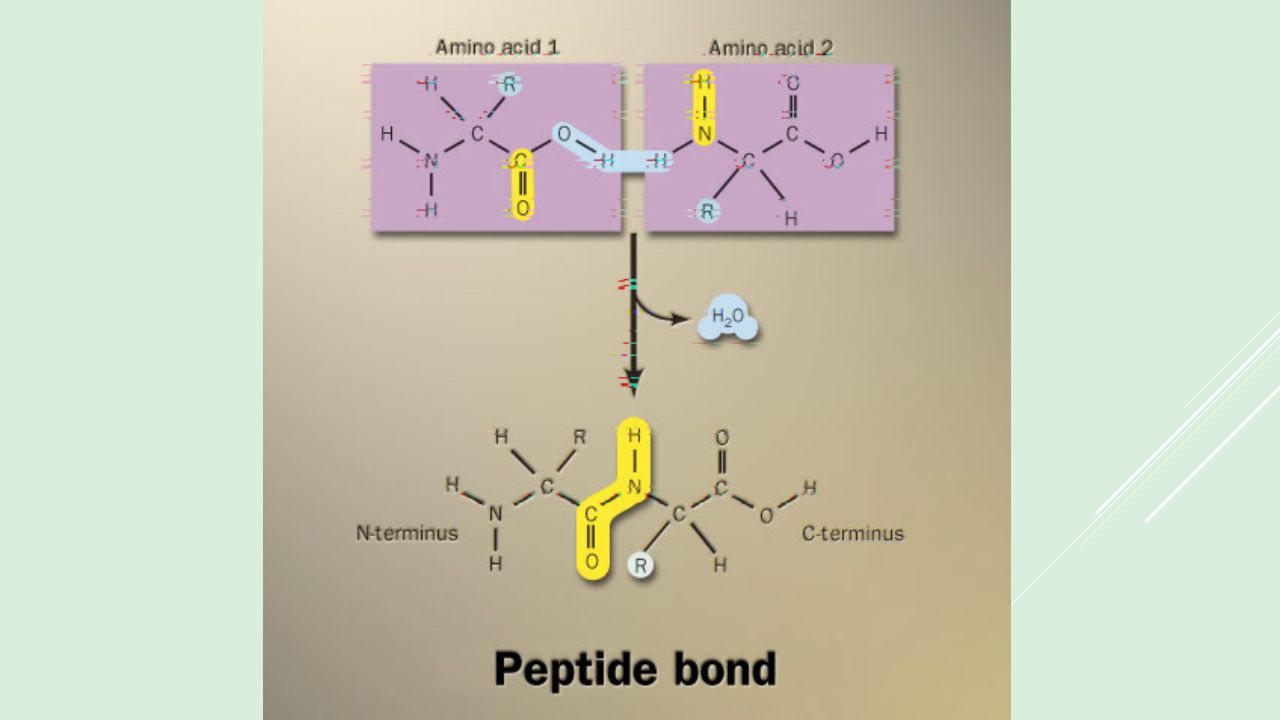
Building proteins Peptide bonds: dehydration synthesis
linking NH2 of 1 amino acid to COOH of another C–N bond free COOH group on one end is ready to form another peptide bond so they “grow” in one direction from N-terminal to C-terminal peptide bond

60
Building proteins Polypeptide chains N-terminal = NH2 end
C-terminal = COOH end repeated sequence (N-C-C) is the polypeptide backbone grow in one direction
 is the polypeptide backbone. grow in one direction.")
61
Protein structure & function
function depends on structure 3-D structure twisted, folded, coiled into unique shape Hemoglobin Hemoglobin is the protein that makes blood red. It is composed of four protein chains, two alpha chains and two beta chains, each with a ring-like heme group containing an iron atom. Oxygen binds reversibly to these iron atoms and is transported through blood. Pepsin Pepsin is the first in a series of enzymes in our digestive system that digest proteins. In the stomach, protein chains bind in the deep active site groove of pepsin, seen in the upper illustration (from PDB entry 5pep), and are broken into smaller pieces. Then, a variety of proteases and peptidases in the intestine finish the job. The small fragments--amino acids and dipeptides--are then absorbed by cells for use as metabolic fuel or construction of new proteins. Collagen– Your Most Plentiful Protein About one quarter of all of the protein in your body is collagen. Collagen is a major structural protein, forming molecular cables that strengthen the tendons and vast, resilient sheets that support the skin and internal organs. Bones and teeth are made by adding mineral crystals to collagen. Collagen provides structure to our bodies, protecting and supporting the softer tissues and connecting them with the skeleton. But, in spite of its critical function in the body, collagen is a relatively simple protein. pepsin hemoglobin collagen
, and are broken into smaller pieces. Then, a variety of proteases and peptidases in the intestine finish the job. The small fragments--amino acids and dipeptides--are then absorbed by cells for use as metabolic fuel or construction of new proteins. Collagen– Your Most Plentiful Protein. About one quarter of all of the protein in your body is collagen. Collagen is a major structural protein, forming molecular cables that strengthen the tendons and vast, resilient sheets that support the skin and internal organs. Bones and teeth are made by adding mineral crystals to collagen. Collagen provides structure to our bodies, protecting and supporting the softer tissues and connecting them with the skeleton. But, in spite of its critical function in the body, collagen is a relatively simple protein. pepsin. hemoglobin. collagen.")
62
Protein structure & function
function depends on structure all starts with the order of amino acids what determines that order of amino acids? Lysozyme Lysozyme protects us from the ever-present danger of bacterial infection. It is a small enzyme that attacks the protective cell walls of bacteria. Bacteria build a tough skin of carbohydrate chains, interlocked by short peptide strands, that braces their delicate membrane against the cell's high osmotic pressure. Lysozyme breaks these carbohydrate chains, destroying the structural integrity of the cell wall. The bacteria burst under their own internal pressure. Glycolytic enzymes Glucose powers cells throughout your body. Glucose is a convenient fuel molecule because it is stable and soluble, so it is easy to transport through the blood from places where it is stored to places where it is needed. Glucose is packed with chemical energy, ready for the taking. In a test tube, you can burn glucose, forming carbon dioxide and water and a lot of light and heat. Our cells also burn glucose, but they do it in many small, well-controlled steps, so that they can capture the energy in more useable forms, such as ATP (adenosine triphosphate). These ten enzymes control those small steps in the process of extracting energy from glucose. Glycolysis (sugar-breaking) is the first process in the cellular combustion of glucose. Glycolysis is endlessly fascinating. In these ten enzymes, you can find examples of many of the important molecular processes that animate our cells. They have been perfected by evolution to perform their diverse chemical tasks quickly and efficiently--adding, removing, and shifting atoms without making mistakes. The pathway is carefully regulated, so that glucose is only broken down when energy is needed Point out the active sites on each glycolytic enzym the 10 glycolytic enzymes used to breakdown glucose to make ATP lysozyme: enzyme in tears & mucus that kills bacteria
. These ten enzymes control those small steps in the process of extracting energy from glucose. Glycolysis (sugar-breaking) is the first process in the cellular combustion of glucose. Glycolysis is endlessly fascinating. In these ten enzymes, you can find examples of many of the important molecular processes that animate our cells. They have been perfected by evolution to perform their diverse chemical tasks quickly and efficiently--adding, removing, and shifting atoms without making mistakes. The pathway is carefully regulated, so that glucose is only broken down when energy is needed. Point out the active sites on each glycolytic enzym. the 10 glycolytic enzymes used to breakdown glucose to make ATP. lysozyme: enzyme in tears & mucus that kills bacteria.")
63
Primary (1°) structure Order of amino acids in chain
amino acid sequence determined by DNA slight change in amino acid sequence can affect protein’s structure & it’s function even just one amino acid change can make all the difference! Sickle cell anemia: 1 DNA letter changes 1 amino acid = serious disease Hemoglobin mutation: bends red blood cells out of shape & they clog your veins.

64
Sickle cell anemia glutamic acid is acidic & polar
valine is non-polar = tries to “hide” from water of cell by sticking to another hemoglobin molecules.

65
Secondary (2°) structure
“Local folding” Folding along short sections of polypeptide interaction between adjacent amino acids H bonds between R groups -helix -pleated sheet It’s a helix or B sheet within a single region. Can have both in one protein but a specific region is one or another

66
Secondary (2°) structure
 structure")
67
Tertiary (3°) structure
“Whole molecule folding” determined by interactions between R groups hydrophobic interactions effect of water in cell anchored by disulfide bridges (H & ionic bonds) How the whole thing holds together
 How the whole thing holds together.")
68
Quaternary (4°) structure
Joins together more than 1 polypeptide chain only then is it a functional protein collagen = skin & tendons hemoglobin Structure equals function wonderfully illustrated by proteins Collagen is just like rope -- enables your skin to be strong and flexible.

69
Protein structure (review)
R groups hydrophobic interactions, disulfide bridges 3° multiple polypeptides hydrophobic interactions sequence determines structure and… structure determines function. Change the sequence & that changes the structure which changes the function. 1° aa sequence peptide bonds 2° determined by DNA R groups H bonds 4°

70
Chaperonin proteins Guide protein folding
provide shelter for folding polypeptides keep the new protein segregated from cytoplasmic influences

71
Protein models Protein structure visualized by X-ray crystallography
extrapolating from amino acid sequence computer modelling lysozyme

72
Denature a protein Disrupt 3° structure pH salt temperature
unravel or denature protein disrupts H bonds, ionic bonds & disulfide bridges Some proteins can return to their functional shape after denaturation, many cannot Example: Eggs: Cooking an egg permanently denatures the proteins.

73
Nucleic Acids

74
Nucleic Acids Function: store & transmit hereditary information
Examples: RNA (ribonucleic acid) DNA (deoxyribonucleic acid) Structure: monomers = nucleotides
 DNA (deoxyribonucleic acid) Structure: monomers = nucleotides.")
75
Nucleotides 3 parts nitrogen base (C-N ring) pentose sugar (5C)
ribose in RNA deoxyribose in DNA PO4 group

76
Types of nucleotides 2 types of nucleotides different Nitrogen bases
purines double ring N base adenine (A) guanine (G) pyrimidines single ring N base cytosine (C) thymine (T) uracil (U)
 guanine (G) pyrimidines. single ring N base. cytosine (C) thymine (T) uracil (U)")
77
Building the polymer

78
Nucleic polymer Why is this important? Backbone sugar to PO4 bond
phosphodiester bond new base added to sugar of previous base polymer grows in one direction N bases hang off the sugar-phosphate backbone Why is this important?

79
RNA & DNA RNA single nucleotide chain DNA double nucleotide chain
N bases bond in pairs across chains spiraled in a double helix double helix 1st proposed as structure of DNA in 1953 by James Watson & Francis Crick (just celebrated 50th anniversary!)
")
80
Pairing of nucleotides
Nucleotides bond between DNA strands H bonds purine :: pyrimidine A :: T 2 H bonds G :: C 3 H bonds The 2 strands are complementary. One becomes the template of the other & each can be a template to recreate the whole molecule. Why is this important?

81
Information polymer Function series of bases encodes information
like the letters of a book stored information is passed from parent to offspring need to copy accurately stored information = genes genetic information All other biomolecules we spoke about served physical or chemical functions. DNA & RNA are information storage molecules. DNA well-suited for an information storage molecule: chemically stable stores information in the varying sequence of nucleotides (the genetic code) its coded sequence can be copied exactly by the synthesis of complementary strands; easily unzipped & re-zipped without damage (weak H bonds) damage to one strand can be repaired by addition of bases that match the complementary strand
 its coded sequence can be copied exactly by the synthesis of complementary strands; easily unzipped & re-zipped without damage (weak H bonds) damage to one strand can be repaired by addition of bases that match the complementary strand.")
83
Why is it important that the strands are bonded by H bonds?
DNA molecule Double helix H bonds between bases join the 2 strands A :: T C :: G Why is it important that the strands are bonded by H bonds? H bonds = biology’s weak bond • easy to unzip double helix for replication and then re-zip for storage • easy to unzip to “read” gene and then re-zip for storage

84
Copying DNA Replication 2 strands of DNA helix are complementary
have one, can build other have one, can rebuild the whole why is this a good system? when in the life of a cell does replication occur? when cells divide, they must duplicate DNA exactly for the new “daughter” cells Why is this a good system? mitosis meiosis

85
DNA replication “It has not escaped our notice that the specific pairing we have postulated immediately suggests a possible copying mechanism for the genetic material.” James Watson Francis Crick 1953 The greatest understatement in biology!

86
Watson and Crick … and others…
1953 | 1962 Discovered & published in 1953 Nobel Prize in 1962: Watson, Crick, Wilkins

87
Maurice Wilkins… and… 1953 | 1962

88
Rosalind Franklin ( ) A chemist by training, Franklin had made original and essential contributions to the understanding of the structure of graphite and other carbon compounds even before her appointment to King's College. Unfortunately, her reputation did not precede her. James Watson's unflattering portrayal of Franklin in his account of the discovery of DNA's structure, entitled "The Double Helix," depicts Franklin as an underling of Maurice Wilkins, when in fact Wilkins and Franklin were peers in the Randall laboratory. And it was Franklin alone whom Randall had given the task of elucidating DNA's structure. The technique with which Rosalind Franklin set out to do this is called X-ray crystallography. With this technique, the locations of atoms in any crystal can be precisely mapped by looking at the image of the crystal under an X-ray beam. By the early 1950s, scientists were just learning how to use this technique to study biological molecules. Rosalind Franklin applied her chemist's expertise to the unwieldy DNA molecule. After complicated analysis, she discovered (and was the first to state) that the sugar-phosphate backbone of DNA lies on the outside of the molecule. She also elucidated the basic helical structure of the molecule. After Randall presented Franklin's data and her unpublished conclusions at a routine seminar, her work was provided - without Randall's knowledge - to her competitors at Cambridge University, Watson and Crick. The scientists used her data and that of other scientists to build their ultimately correct and detailed description of DNA's structure in Franklin was not bitter, but pleased, and set out to publish a corroborating report of the Watson-Crick model. Her career was eventually cut short by illness. It is a tremendous shame that Franklin did not receive due credit for her essential role in this discovery, either during her lifetime or after her untimely death at age 37 due to cancer.
 that the sugar-phosphate backbone of DNA lies on the outside of the molecule. She also elucidated the basic helical structure of the molecule. After Randall presented Franklin s data and her unpublished conclusions at a routine seminar, her work was provided - without Randall s knowledge - to her competitors at Cambridge University, Watson and Crick. The scientists used her data and that of other scientists to build their ultimately correct and detailed description of DNA s structure in Franklin was not bitter, but pleased, and set out to publish a corroborating report of the Watson-Crick model. Her career was eventually cut short by illness. It is a tremendous shame that Franklin did not receive due credit for her essential role in this discovery, either during her lifetime or after her untimely death at age 37 due to cancer.")
89
Interesting note… Ratio of A-T::G-C affects stability of DNA molecule
2 H bonds vs. 3 H bonds biotech procedures more G-C = need higher T° to separate strands high T° organisms many G-C parasites many A-T (don’t know why) At the foundation of biology is chemistry!!
 At the foundation of biology is chemistry!!")
90
Another interesting note…
ATP Adenosine triphosphate modified nucleotide adenine ribose + Pi + Pi + Pi + +

91
Macromolecule Review

92
Carbohydrates Structure / monomer monosaccharide Function energy
raw materials energy storage structural compounds Examples glucose, starch, cellulose, glycogen glycosidic bond

93
Lipids Structure / building block
glycerol, fatty acid, cholesterol, H-C chains Function energy storage membranes hormones Examples fat, phospholipids, steroids ester bond (in a fat)
")
94
Proteins Structure / monomer amino acids levels of structure Function
enzymes u defense transport u structure signals u receptors Examples digestive enzymes, membrane channels, insulin hormone, actin peptide bond

95
Nucleic acids Structure / monomer nucleotide Function
information storage & transfer Examples DNA, RNA phosphodiester bond

Similar presentations


>")


