Download presentation
Presentation is loading. Please wait.

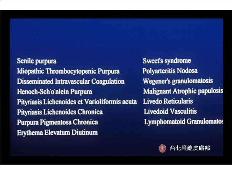
4
Senile purpura: Multiple purpuric macules

6
Idiopathic thrombocytopenic purpura: Multiple petechiae on the arm

8
Disseminated intravascular coagulation: Extensive geographic cutaneous infarction on the forearm

9
Disseminated intravascular coagulation: Extensive deep purple ecchymoses with hemorragic blister formation

11
Henoch-Schonlein purpura: Multiple palpable deep purple macules on the extensor side of lower extremities and buttocks

12
Henoch-Schonlein purpura: Small venules show fibrinoid necrosis with considerable red cell extravasation.

14
Henoch-Schonlein purpura: The DIF of IgA shows granular depositions at the papillary dermis.

16
Pityriasis lichenoides et varioliformis acuta : Multiple erythematous papules on the back and buttocks

17
Pityriasis lichenoides et varioliformis acuta : Multiple erythematous papules with necrotic and crusted lesions on the buttocks

18
Pityriasis lichenoides et varioliformis acuta : Multiple erythematous papules with necrotic and crusted lesions on the buttocks

19
Pityriasis lichenoides et varioliformis acuta : This view shows basal cell hydropic degeneration, moderate lymphohistiocytic infiltrate at the upper dermis and red cell extravasation.

20
Pityriasis lichenoides et varioliformis acuta : This view shows mild spongiosis, basal cell hydropic degeneration, moderate lymphohistiocytic infiltrate at the upper dermis and red cell extravasation.

22
Pityriasis lichenoides chronica : Multiple brownish-to-red scaly papules with and crusted lesions

24
Erythema elevatum diutinum : Multiple erythematous nodules present on the back of the hand.

25
Erythema elevatum diutinum : Multiple erythematous nodules present on the back of the hand.

26
Erythema elevatum diutinum : Multiple erythematous nodules present on the palm and fingers.

27
Erythema elevatum diutinum : Multiple erythematous nodules present on the palm and fingers.

29
Sweet’s syndrome : Erythematous, edematous plaques on the periorbital area with superficial tiny pustules.

30
Sweet’s syndrome : Erythematous, edematous plaques on the neck with pustules and crusted lesions.

31
Sweet’s syndrome : The epidermis shows subcorneal pustulation. There are diffuse mixed cell infiltrates in the dermis.

32
Sweet’s syndrome : The infiltrate consisted largely of neutrophils.

34
Polyarteritis nodosa: Several dermal and subcutaneous nodules on the back of the foot.

35
Polyarteritis nodosa: Several erythematous dermal and subcutaneous nodules with ulceration on the foot.

36
Polyarteritis nodosa: Necrotizing vasculitis is seen within the subcutaneous fat.

37
Polyarteritis nodosa: Necrotizing vasculitis is seen within the subcutaneous fat.

39
Wegener’s granulomatosis: Several erythematous-dark red ulcerating plaques and nodules on the foot.

40
Wegener’s granulomatosis: One erythematous-dark red ulcerating plaque with blister formation.

41
Wegener’s granulomatosis: Extensive necrotizing vasculitis is seen within the dermis with red cell extravasation.

42
Wegener’s granulomatosis: Close-up view shows necrotizing vasculitis with red cell extravasation.

44
Wegener’s granulomatosis: Nasal mucosa biopsy shows necrotizing vasculitis

46
Wegener’s granulomatosis: Transbronchial biopsy shows necrotizing vasculitis with extravascular granuloma formation.

47
Wegener’s granulomatosis: Chest films shows cavitation on the right upper lobs.

49
Dego’s syndrome: Small white papules with central depression and white fine scaling.

50
Dego’s syndrome: There is hyperkeratosis with epidermal atrophy

51
Dego’s syndrome: There is hyperkeratosis with epidermal atrophy with ectatic vessels.

52
Dego’s syndrome: There is hyperkeratosis with epidermal atrophy. Note the ectatic vessels.

54
Livedo reticularis: A mottled violaceous discoloration of the skin in a net-like pattern.

55
Livedo reticularis: A mottled erythematous discoloration of the skin in a net-like pattern.

57
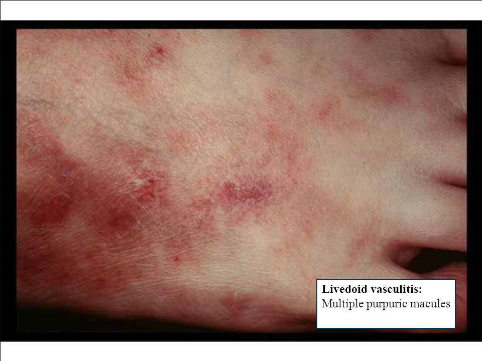
Livedoid vasculitis: Multiple purpuric macules

59
Livedoid vasculitis: Punched-out ulcers are seen on the ankle

61
Lymphomatoid granulomatosis: Multiple erythematous macules

62
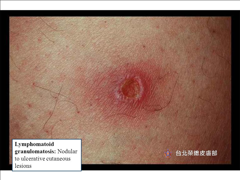
Lymphomatoid granulomatosis: Nodular to ulcerative cutaneous lesions

64
Lymphomatoid granulomatosis: Nodular lymphoid and/or granulomatous infiltration surrounding the vessels

65
Lymphomatoid granulomatosis: Nodular lymphoid infiltration surrounding the vessles

67
Purpura pigmentosa chronica: Multiple erythematous patches on the lower leg

68
Purpura pigmentosa chronica: Scaly petechial or purpuric macules

72
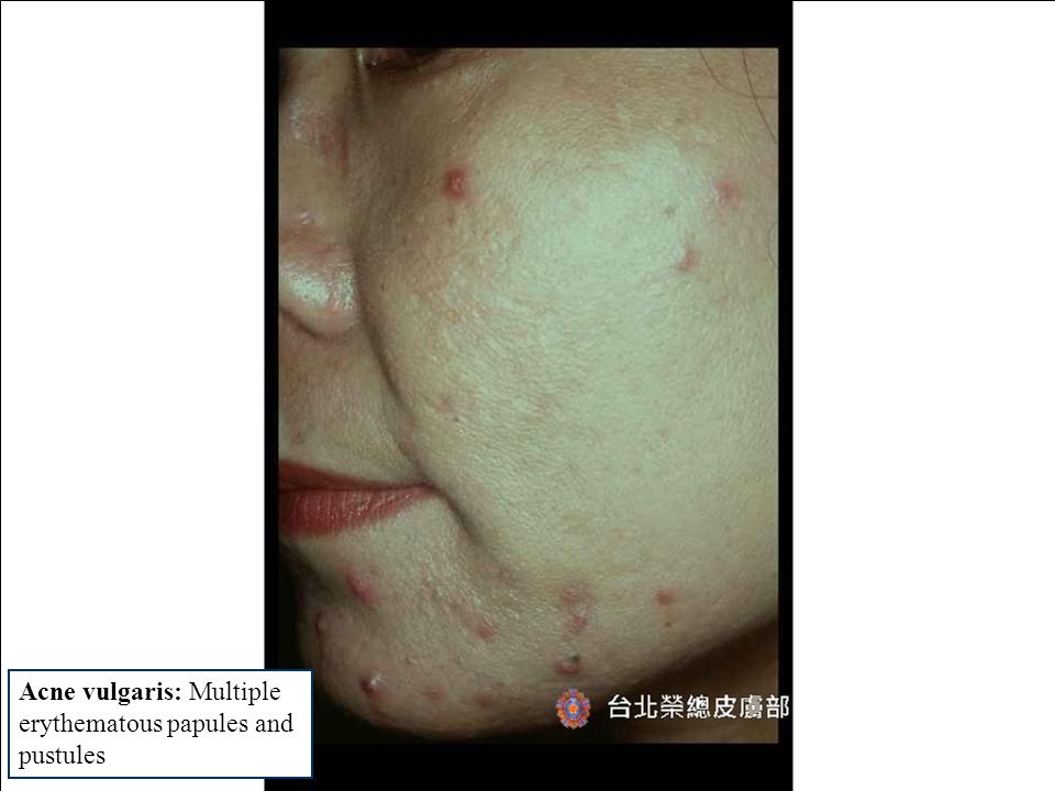
Acne vulgaris: Multiple erythematous papules and pustules

75
Steroid acne: Multiple erythematous papules and pustules

79
Neonatal: Multiple erythematous papules and pustules on a neonate

81
Rosacea: Swollen nose with sebaceous gland hyperplasia and fibrosis

82
Rosacea: Telangiectasia and erythema.

84
Androgenic alopecia: Alopecia with the typical M-shape pattern

87
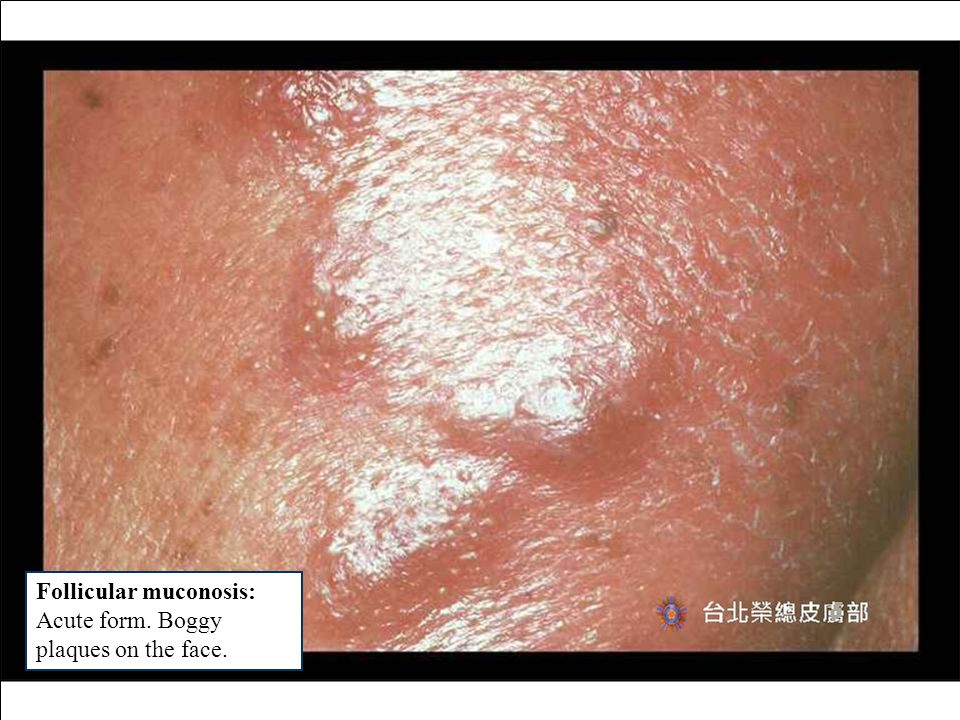
Follicular muconosis: Acute form. Boggy plaques on the face.

89
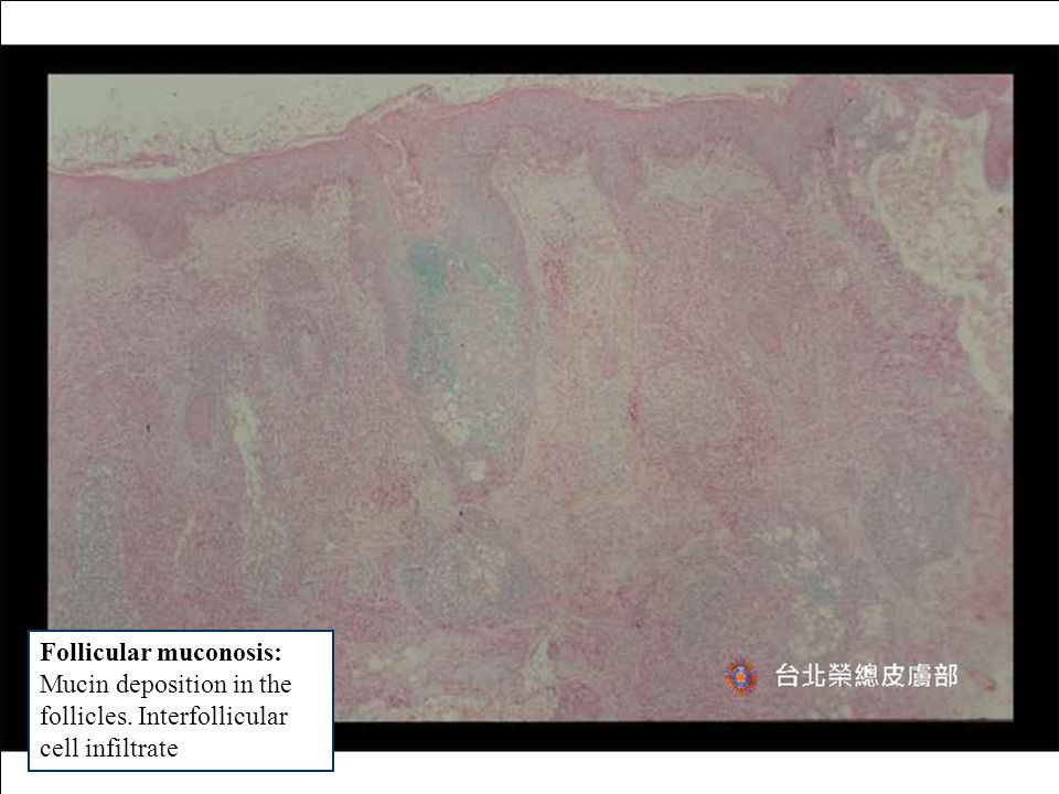
Follicular muconosis: Mucin deposition in the follicles. Interfollicular cell infiltrate

93
Relapsing polychondritis: Erythema, swelling, and pain of the cartilaginous portion of the ear

94
Relapsing polychondritis: Loss of basophilic staining and breakdown of lacunar structure of the cartilage

96
Relapsing polychondritis: Breakdown of lacunar structure of the cartilage

98
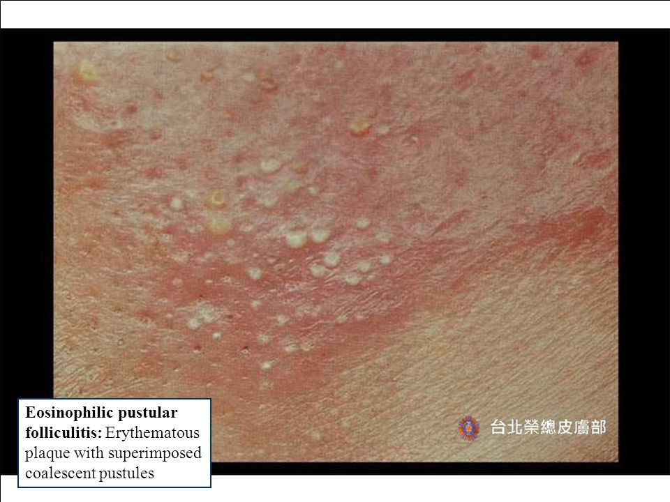
Eosinophilic pustular folliculitis: Erythematous plaque with superimposed coalescent pustules

100
Eosinophilic pustular folliculitis: Microabscess composed of eosinophils

102
Pityrosporum folliculitis: Pruritic eryhematous papules and pustules, primarily on the back, chest, and shoulders

105
Eosinophilic cellulitis: Recurrent painful or pruiritic plaques

106
Eosinophilic cellulitis: Recurrent painful or pruiritic plaques with bullae

108
Eosinophilic cellulitis: Diffuse eosinophilic infiltrate throughout the dermis

109
Eosinophilic cellulitis: Diffuse eosinophilic infiltrate throughout the dermis. Flame figures can be seen.

Similar presentations