Download presentation
Presentation is loading. Please wait.

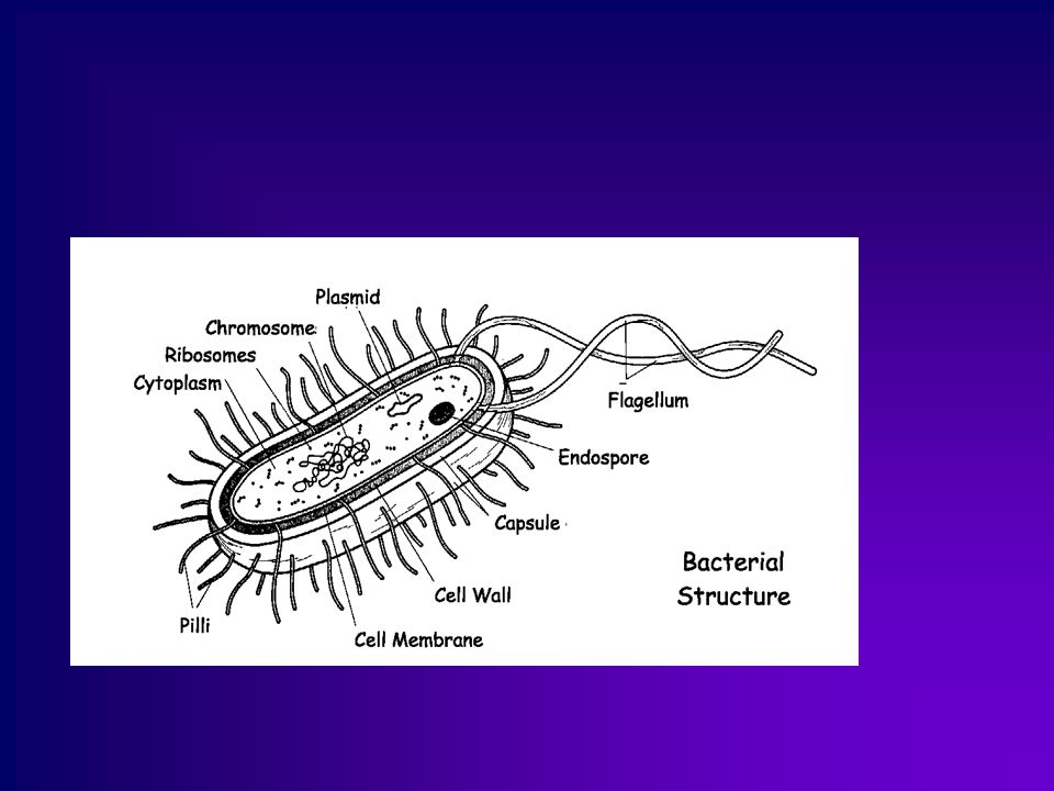
2
Acid-fast Stain (Ziehl-Neelsen stain )
")
3
Acid-fast stain (Ziehl-Neelsen stain )
It is a special bacteriological stain used to identify acid-fast organisms, mainly Mycobacteria. Mycobacterium tuberculosis is the most important of this group because it is responsible for tuberculosis (TB) and other important Mycobacterium species. Acid fast organisms like Mycobacterium contain large amounts of waxy lipid substances within their cell walls called mycolic acids. These acids resist staining by ordinary methods such as a Gram stain. It can also be used to stain a few other bacteria, such as Nocardia. The reagents used are Ziehl–Neelsen carbolfuchsin, acid alcohol, and methylene blue. Acid-fast bacilli will be bright red after staining.
 and other important Mycobacterium species. Acid fast organisms like Mycobacterium contain large amounts of waxy lipid substances within their cell walls called mycolic acids. These acids resist staining by ordinary methods such as a Gram stain. It can also be used to stain a few other bacteria, such as Nocardia. The reagents used are Ziehl–Neelsen carbolfuchsin, acid alcohol, and methylene blue. Acid-fast bacilli will be bright red after staining.")
4
Principle of acid fast stain
Cell wall of M.tuberculosis is impermeability to stains and dyes. But M.tuberculosis can be stained by acid-fast stain with long time heating, this mean that carbolfuchsin which is a phenolic stain is soluble in in the lipids of mycobacterial cell wall and the heating process o adding the targitol, increase the pentration of the carbolfuchsin. Bacteria except M.tuberculosis can be decolorized by 3% acid alcohol . So the color of M.tuberculosis is red and that of other Mycolic acid negtive bacteria is blue after Counterstain with methylene blue.

5
Acid-Fast Organisms Primary stain binds cell wall mycolic acids
Intense decolorization does not release primary stain from the cell wall of AFB, since the carbolfuchsin is more soluble in mycolic acid than in the decolrizer. Color of AFB-based on primary stain Counterstain provides contrasting background.

6
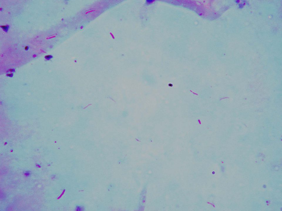
Acid fast bacteria

7
Preparation of AFB Smears
1. Make a smear of the sputum, dry and fix it. 2. Ziehl-Neelsen acid-fast stain: (1) Flood the slide with carbolfuchsin. Heat the slide to steaming for 5mins. Do not boil or allow the smear to dry. As stain evaporated from the slide, replenish with additional carbolfuchsin. Allow the slide to cool and rinse it thoroughly with water. (2) Decolorize the slide with acid alcohol until the red color no longer comes off in the decolorizer. It takes about 30secs. Rinse the slide with water. (3) Counterstain with methylene blue, allow the stain to react for 1min, rinse as above. (4) Bolt the slide carefully. Examine under microscope.
 Flood the slide with carbolfuchsin. Heat the slide to steaming for 5mins. Do not boil or allow the smear to dry. As stain evaporated from the slide, replenish with additional carbolfuchsin. Allow the slide to cool and rinse it thoroughly with water. (2) Decolorize the slide with acid alcohol until the red color no longer comes off in the decolorizer. It takes about 30secs. Rinse the slide with water. (3) Counterstain with methylene blue, allow the stain to react for 1min, rinse as above. (4) Bolt the slide carefully. Examine under microscope.")
8
Fixation of AFB Smears Fixation may kill some bacilli
makes smear stick to slide by heat or alcohol do not overheat safe smear ?

9
Primary Staining-ZN Carbol fuchsin staining
Uses higher fuchsin concentration Dissolve well !!! IUATLD/WHO : 0.3% references?? Heat well-apply long enough Cold staining prevents phenol’s toxicity!

10
Decolorization of Smears
must be complete not possible to de-stain too much repeat as needed use strong acids alcohol not absolutely needed

11
Counter-staining Provides good contrast for observation of AFB
background for focusing, not too strong methylene blue 0.3% ? diluted or < 1 min use of malachite green?

12
Microscopic Reading: Red slender rods on blue background
accept only typical shape, at least some depends condition of microscope! light! binocular, mechanical stage, good optics 100x oil immersion objective, 10x eyepieces Requires: patience, sincerity AFB microscopy is not difficult but tough.

15
Spore Staining

16
Endospore The name "endospore" is suggestive of a spore or seed-like form (endo means within). Endospore formation is usually triggered by a lack of nutrients, and usually occurs in Gram-positive bacteria. Endospores enable bacteria to lie dormant for extended periods, even centuries. Revival of spores millions of years old has been claimed. When the environment becomes more favorable, the endospore can reactivate itself to the vegetative state.

17
Endospore Most types of bacteria cannot change to the endospore form. Examples of bacteria that can form endospores include Bacillus and Clostridium. Endospore formation process called sporogenesis. An endospore is a dormant, tough, and non-reproductive structure produced by certain bacteria from the Firmicute phylum.

18
Endospore The endospore consists of the bacterium's DNA and part of its cytoplasm, surrounded by a very tough outer coating. Endospores can survive without nutrients. They are resistant to ultraviolet radiation, desiccation, high temperature, extreme freezing and chemical disinfectants. According to scientist Dr. Steinn Sigurdsson, "There are viable bacterial spores that have been found that are 40 million years old on Earth - and we know they're very hardened to radiation.“ Common anti-bacterial agents that work by destroying vegetative cell walls do not affect endospores. Endospores are commonly found in soil and water, where they may survive for long periods of time

19
Endospore Location The position of the endospore differs among bacterial species and is useful in identification. The main types within the cell are terminal, subterminal, and centrally placed endospores. Terminal endospores are seen at the poles of cells, whereas central endospores are more or less in the middle. Subterminal endospores are those between these two extremes, usually seen far enough towards the poles but close enough to the center so as not to be considered either terminal or central. Lateral endospores are seen occasionally.

20
Endospore staining (Schaeffer-fulton Method)
Prepare a smear of the bacteria Bacillus megatatium (a spore-producing organism) Flood the smear with malachite green Do not allow the stain to evaporate or completely evaporate. Remove from heat and allow slides to cool Once the slides are cool (important) rinse with water Flood the sample with safranin (30-60 seconds) Rinse the slide blot dry observe under microscopy. 10x, 40x, 100x (oil immersion).
 Flood the smear with malachite green. Do not allow the stain to evaporate or completely evaporate. Remove from heat and allow slides to cool. Once the slides are cool (important) rinse with water. Flood the sample with safranin (30-60 seconds) Rinse the slide blot dry observe under microscopy. 10x, 40x, 100x (oil immersion).")
21
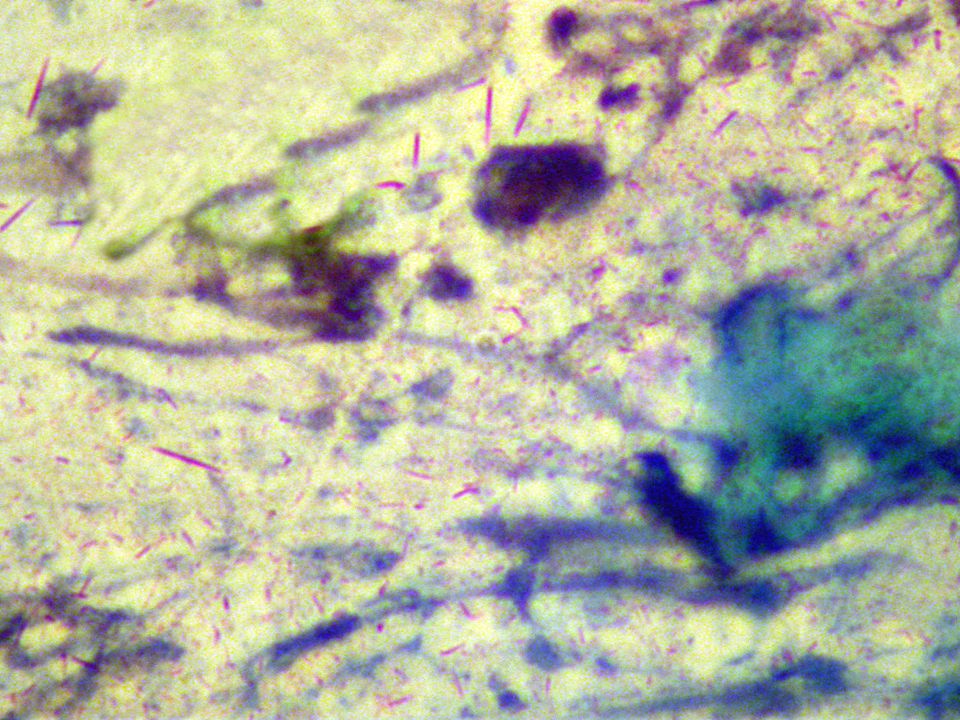
Endospore stain

22
Endospore stain

23
Capsule Stain

24
THE CAPSULE Capsules are structures that lay outside of an organism's cell wall and thus are in direct contact with the environment. Many, perhaps most, bacteria produce capsules under the right conditions Some capsules are composed of Carbohydrates or Glycoprotein.

26
Capsules functions Protect the cell from desiccation (drying))
Protect the cell from phagocytes (being engulfed by white blood cells) Provide a food reserve when certain organic compounds are in excess. A virulence determinant of pathogenic microbes They serve as binding or adhesion agents for sticking cells together and/or to a surface such as a rock in flowing stream or a tooth
 Provide a food reserve when certain organic compounds are in excess. A virulence determinant of pathogenic microbes. They serve as binding or adhesion agents for sticking cells together and/or to a surface such as a rock in flowing stream or a tooth.")
27
WHY WE USE CAPSULE STAIN ?
Bacterial Heat fixation cause capsule shrinkage Because most capsule materials are water soluble, simple stains will not adhere to them

28
Significant:- The capsule is a major virulence factor in the major disease-causing bacteria, such as:- Klebsiella pneumoniae or Enterobacter aerogenes (slant) Streptococcus lactis Escherichia coli
 Streptococcus lactis. Escherichia coli.")
29
Uses of Capsule Staining
Detection of dental plaque Light micrograph of S. mutans

30
A lesion on the 10the day of anthrax infection
2.Detection of Anthrax B. anthracis A lesion on the 10the day of anthrax infection

31
Dye use in capsule stain
. In this stain we use acidic and basic acidic dye as India Ink and Nigrosen use to stain the background of the slide but basic dye as methylene blue and crystal violet use to stain the cell

32
Note Older cultures are more likely to exhibit capsule production. When performing a capsule stain on your unknown, be sure the culture you take your sample from is at least five days old

33
MATERIALS Clean slid microscope India Ink and Methylene Wash bottle
Culture of bacteria .Sterile loop

34
Capsule Staining Procedure

35
Pictures of capsule stain

36
.

37
Capsule stain of Streptococcus lactis.

38
Capsule stain of Enterobacter aerogenes

Similar presentations


 Abdelraheem BA.>")
 Abdelraheem BA>")


