Download presentation
Presentation is loading. Please wait.

1
The Structure of the Heart Learning Objectives: Label the parts of the heart. Label the parts of the heart. How is the structure of the heart related to its function? How is the structure of the heart related to its function? Why is the heart made up of 2 adjacent pumps? Why is the heart made up of 2 adjacent pumps?

2
Homework P. 86-87. P. 86-87.

3
Inside the Heart Inside the Heart

4
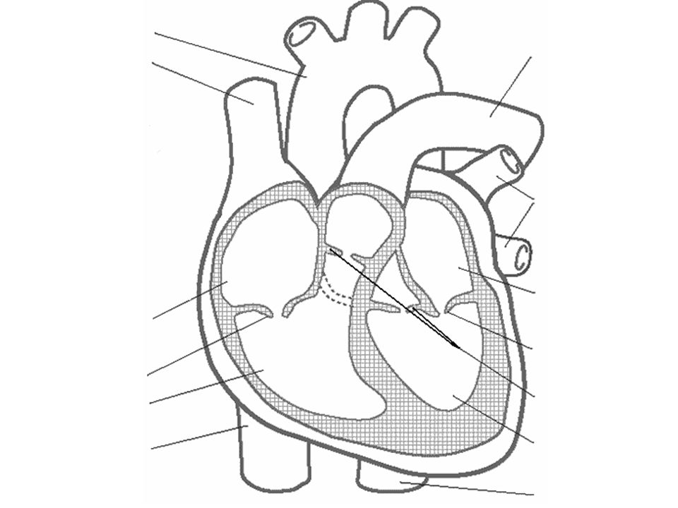
Chambers of the Heart Chambers of the Heart

6
Heart Structure text p.88-9 Label your heart diagram Label your heart diagram 1. Why does the heart have 2 separate pumps? 2. Why does the left ventricle have thicker cardiac muscle than the right ventricle? 3. Name the 4 main blood vessels serving the heart and describe what each one does. 4. Where are the coronary arteries found and what do they do?

8
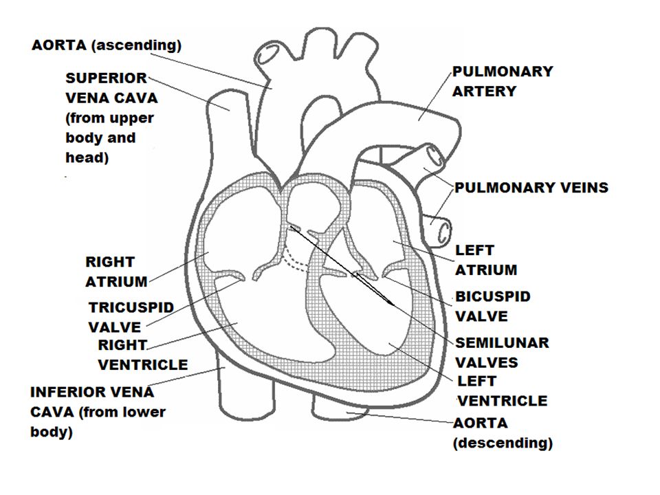
Mammalian Heart Structure Right ventricle Septum (dividing wall) Tricuspid valve Vena cavae Aorta Right atrium Semilunar valves Pulmonary artery Pulmonary veins Left atrium Bicuspid valve Left ventricle Cardiac muscle
 Tricuspid valve Vena cavae Aorta Right atrium Semilunar valves Pulmonary artery Pulmonary veins Left atrium Bicuspid valve Left ventricle Cardiac muscle")
9
Blood Vessels The Aorta is connected to the left ventricle and carries oxygenated blood to all parts of the body except the lungs. The Vena Cava is connected to the right atrium and brings deoxygenated blood back from the tissues of the body. The Pulmonary Artery is connected to the right ventricle and carries deoxygenated blood to the lungs. The Pulmonary Vein is connected to the left atrium and brings oxygenated blood back from the lungs.

10
The chambers of the heart are separated by valves which prevent blood from flowing in the wrong direction. Heart valves Heart valves There are valves between the atria and the ventricles… …and there are valves leading out of the ventricles. valve between right atrium and right ventricle valve between left atrium and left ventricle valve leading out of right ventricle valve leading out of left ventricle

11
How are valves held in place? How are valves held in place? The valves between the atria and ventricles are connected to the inner walls of the heart by tough tendons. valve open

12
How do valves work? How do valves work? A valve acts like a door that only opens in one direction. In the heart, the tendons holding the valve are like the arm holding the door. One end of each tendon is fixed to the wall of the heart and so the valve can only open in one direction.

13
How are valves held in place? How are valves held in place? The tendons allow the valves to close and hold the valve flaps in place. They prevent the valves from flipping up and turning inside out. Why is this important? valve open valve closed

14
The heart also needs its own blood vessels – coronary arteries and cardiac veins. They supply the heart muscle with oxygen so that the muscle cells can respire. And remove waste carbon dioxide. A blockage of these arteries leads to myocardial infarction (heart attack) because the heart muscle is deprived of oxygen and so dies.
 because the heart muscle is deprived of oxygen and so dies..")
18
The heart is the major organ of the circulatory system It is a fist-sized muscular pump consisting of four chambers The human heart recirculates the entire blood volume (5 dm 3 ) every minute when the body is at rest The ability of the heart to perform such work is due to the presence of specialised cardiac muscle in its walls The job of the heart is to pump blood around two separate circuits The left side of the heart pumps oxygenated blood out into the body’s arteries via the AORTA AORTA Deoxygenated blood returns to the right side of the heart via the VENA CAVA Deoxygenated blood is pumped to the lungs via the PULMONARY ARTERY Heart muscle receives its own supply of blood from the CORONARY ARTERIES PULMONARY ARTERY Superior VENA CAVA Inferior VENA CAVA The left side of the heart receives oxygenated blood from the lungs via the PULMONARY VEINS CORONARY ARTERIES Mammalian Heart Structure
 every minute when the body is at rest The ability of the heart to perform such work is due to the presence of specialised cardiac muscle in its walls The job of the heart is to pump blood around two separate circuits The left side of the heart pumps oxygenated blood out into the body’s arteries via the AORTA AORTA Deoxygenated blood returns to the right side of the heart via the VENA CAVA Deoxygenated blood is pumped to the lungs via the PULMONARY ARTERY Heart muscle receives its own supply of blood from the CORONARY ARTERIES PULMONARY ARTERY Superior VENA CAVA Inferior VENA CAVA The left side of the heart receives oxygenated blood from the lungs via the PULMONARY VEINS CORONARY ARTERIES Mammalian Heart Structure")
19
Questions List the correct sequence of the 4 main blood vessels and the 4 heart chambers that a red blood cell will pass through on its journey from the lungs through the heart and body and back again to the lungs. List the correct sequence of the 4 main blood vessels and the 4 heart chambers that a red blood cell will pass through on its journey from the lungs through the heart and body and back again to the lungs. Why does the left ventricle have a thicker wall than the right ventricle? Why does the left ventricle have a thicker wall than the right ventricle? Where would you find the following valves: Where would you find the following valves: Bicuspid? Bicuspid? Tricuspid? Tricuspid? Semi-lunar valves? Semi-lunar valves? What causes a heart attack? What causes a heart attack?

Similar presentations