Download presentation
Presentation is loading. Please wait.

1
Hemangioblastoma

2
Hemangioblastoma Epidemiology M:F is 2:1 Age: 30 - 50
Uncommon (1-3% of all intracranial neoplasms) Most common intraaxial, nonmetastatic posterior fossa tumor in adults Sometimes referred to as Lindau Tumors
 Most common intraaxial, nonmetastatic posterior fossa tumor in adults. Sometimes referred to as Lindau Tumors.")
3
Hemangioblastoma Arvid Lindau

4
Hemangioblastoma Sporadic (75%) Von Hippel Lindau Disease (25%)
VHL gene Tumor suppressor gene 3p25 Autosomal Dominant Inheritance Pattern Café au lait spots Tumors CNS Hemangioblastomas Pheochromocytoma Retinal Angiomas Clear Cell Renal Carcinoma Renal Cysts Pancreatic Islet Cell Tumors Pancreatic Cysts Extremely high probability of developing a second mutation in at least 1 cell in the body.

5
Hemangioblastoma Location Symptoms Cerebellum Spinal Cord Other
Brainstem Supratentorial Optic Nerves Peripheral Nerves Soft Tissues Symptoms Depend on location Brain / Posterior Fossa Ataxia, discoordination Headache Subarachnoid hemorrhage Spinal Cord Pain, weakness, numbness Eye Visual Changes Elevated Erythropoietin Secondary polycythemia

6
Hemangioblastoma Pathology Appearance Benign vascular neoplasm
WHO grade I Subtypes: Reticular and Cellular Arises from hemangioblasts Appearance Cherry Red Cystic filled with clear fluid Attached to the pia (rich vascular supply)
")
7
Hemangioblastoma No grading or staging system Workup
MRI of the brain and spine CT Abdomen - evaluate the kidneys, pancreas, adrenals Opthalmology consult Angiography (may help the surgeon plan their approach)
")
8
Hemangioblastoma

9
Hemangioblastoma A) Schematic representation of the distribution of CNS hemangioblastomas (red dots) in the 25 von Hippel-Lindau disease patients on MRI. Most (98%) of hemangioblastomas were found below the level of the tentorium in the cerebellum, brainstem, and spinal cord. (B–D) Contrast-enhanced MRI demonstrating representative locations of hemangioblastomas including the cerebellum (B), brainstem (C) and spinal cord (D). (B) Axial view through the cerebellum demonstrating a hyperintense enhancing hemangioblastoma (arrow) with surrounding edema (hypointense area surrounding the tumor) that frequently is associated with these lesions. (C) Sagittal view through the posterior fossa demonstrating a hyperintense enhancing brainstem (medullary) hemangioblastoma (arrow) with surrounding edema. (D) Sagittal view through the thoracic and lumbar spinal cord demonstrating two hyperintense enhancing hemangioblastomas (arrows). The superior tumor is associated with a large intraspinal cyst (syrinx) that is common with these neoplasms (arrowhead)
 Schematic representation of the distribution of CNS hemangioblastomas (red dots) in the 25 von Hippel-Lindau disease patients on MRI. Most (98%) of hemangioblastomas were found below the level of the tentorium in the cerebellum, brainstem, and spinal cord. (B–D) Contrast-enhanced MRI demonstrating representative locations of hemangioblastomas including the cerebellum (B), brainstem (C) and spinal cord (D). (B) Axial view through the cerebellum demonstrating a hyperintense enhancing hemangioblastoma (arrow) with surrounding edema (hypointense area surrounding the tumor) that frequently is associated with these lesions. (C) Sagittal view through the posterior fossa demonstrating a hyperintense enhancing brainstem (medullary) hemangioblastoma (arrow) with surrounding edema. (D) Sagittal view through the thoracic and lumbar spinal cord demonstrating two hyperintense enhancing hemangioblastomas (arrows). The superior tumor is associated with a large intraspinal cyst (syrinx) that is common with these neoplasms (arrowhead)")
10
Hemangioblastoma

11
Hemangioblastoma Other Treatment Options Endovascular Embolization
Antiangiogenic Therapy Advantage of SRS Alpha/Beta ratio for HB is thought to be close to that for normal responding tissue. Therefore, hypofractionated radiation is advantageous.

12
Hemangioblastoma Outcomes Generally curable with surgery
Local recurrence after surgery is higher with the following: VHL Syndrome Multiple Hemangioblastomas Younger Age Cellular Histology Cellular has a % recurrence rate Reticular has a % recurrence rate Subarachnoid dissemination is rare.

13
Retrospective Review Stanford University 1991 - 2007
Neurosurgery Vol. 65, No. 1, p. 79, 2009 Retrospective Review Stanford University 92 lesions in 31 pts 26 pts had VHL All treated with SRS Mean patient age: 41 Dose: Gy [Ave Gy] Ave. tumor volume: 1.8 cm3

14
Cerebellum 52 CPA 4 Thalamus 1 Brainstem 9 Cervical Cord 8 Thoracic Cord 7 Lumbar Cord 1

15
Median follow up of 69 months
16% progressed 22% regressed 62% stable Local control rates 36 months: % 60 months: % Lesion-associated symptoms improved in 36 / 41 tumors 5 patients developed radiation necrosis Authors concluded that SRS is safe and effective in the treatment of HBs and is an attractive alternative to surgery for patients, including those with VHL disease.

16
Neurosurgery Vol. 65, No. 1, p. 79, 2009

17
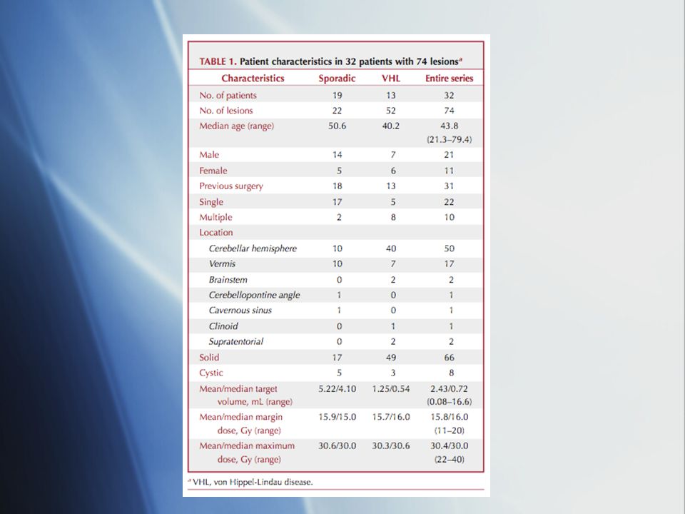
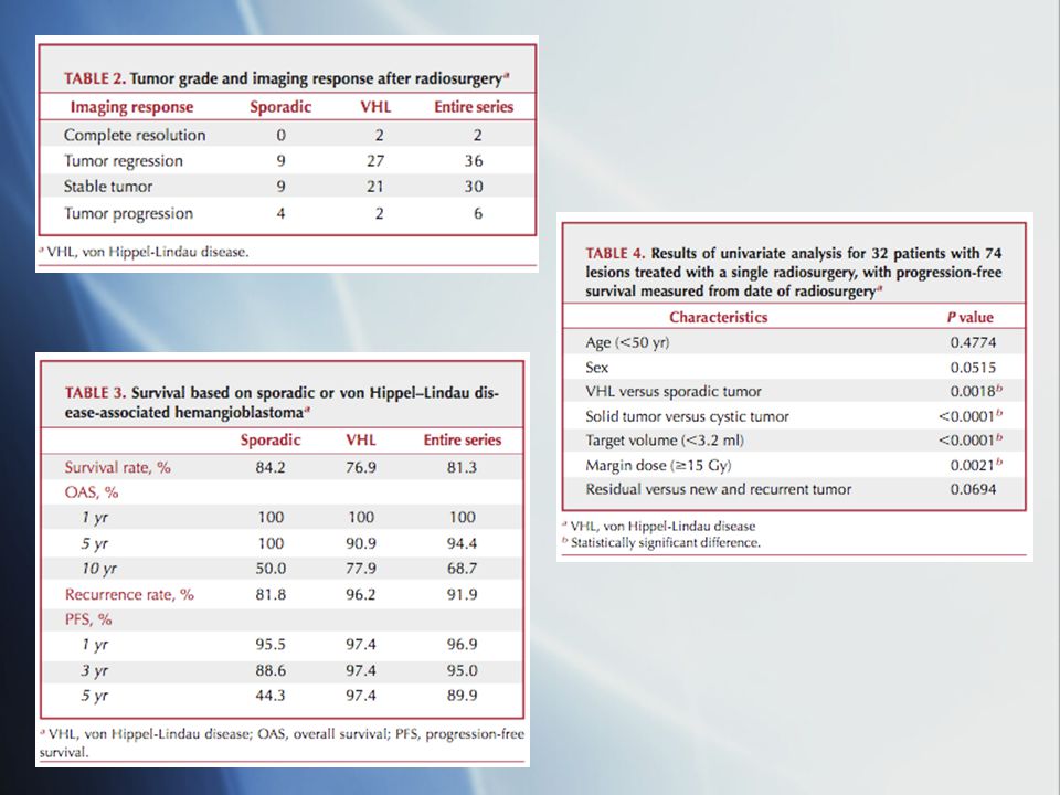
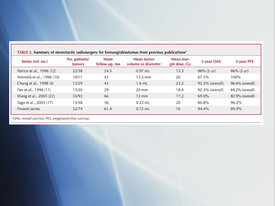
Neurosurgery 63: , 2008 Retrospective review University of Pittsburgh 32 patients; 74 tumors All received Gamma Knife SRS Median age: 44 13 pts had VHL (52 tumors) Median Dose: 16 Gy Median Volume: 0.72 ml Median follow up: 50 months
 Median Dose: 16 Gy. Median Volume: 0.72 ml. Median follow up: 50 months.")
23
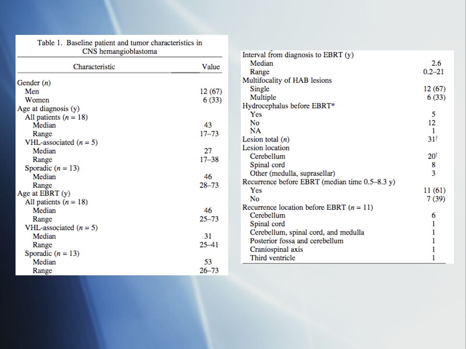
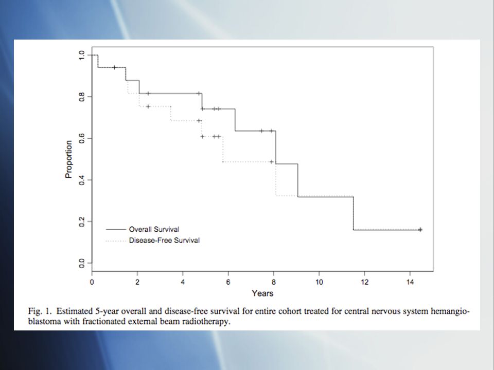
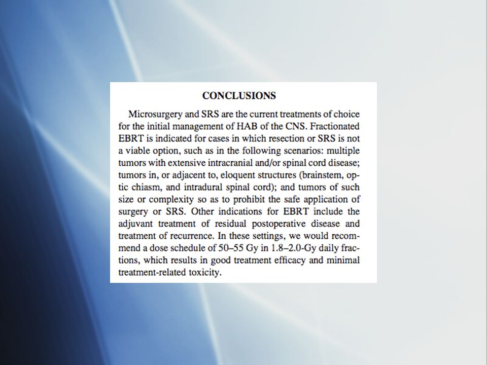
IJROBP, Vol. 69, No. 5, 2007 Retrospective Review, , PMH 18 patients (5 VHL; 13 sporadic); 31 lesions Doses ranged from Gy; Parallel opposed fields Median follow up: 5.1 years DFS at 5 and 10 years was 57% and 30%

Similar presentations


>")


![Case Report # [] Submitted by:Kandra Vogt, MSIV Faculty reviewer:Sandra A. A. Oldham, M.D. Date accepted:31 August 2007 Radiological Category:Principal.](/18/5671912/big_thumb.jpg "Case Report # [] Submitted by:Kandra Vogt, MSIV Faculty reviewer:Sandra A. A. Oldham, M.D. Date accepted:31 August 2007 Radiological Category:Principal.>")
 tumors arising from one of the many different cell types within.>")
 10.1 A 10.1 B 10.1 C Precontrast sagittal T1 wtd. MRI of.>")


 H. Louis Harkey Department of Neurosurgery University of Mississippi Jackson, MS.>")