Download presentation
Presentation is loading. Please wait.

1
IV.Bacterial Structure and Growth A.Bacterial Cells: An Overview B.Bacterial Cell Structures C.Factors that Influence Bacterial Growth

2
IV. A.Bacterial Cells: An Overview Shapes & Arrangements –Round Bacteria Coccus Staphylococcus Diplococcus Tetrad Streptococcus Sarcina –Rod-shaped Bacteria Bacillus Streptobacillus Diplobacillus Coryneform bacteria

3
IV. A.Bacterial Cells: An Overview Shapes & Arrangements (cont.) –Curved & Spiral Bacteria Vibrio Spirillum Spirochaete
 –Curved & Spiral Bacteria Vibrio Spirillum Spirochaete.")
4
IV. A.Bacterial Cells: An Overview Sizes –Typically ~ 0.1 - 20 m (with some exceptions) –Typical coccus: ~ 1 m (eg Staphylococcus) –Typical short rod: ~ 1 x 5 m (eg E. coli) –Barely within the best resolution of a good compound light microscope
 –Typical coccus: ~ 1 m (eg Staphylococcus) –Typical short rod: ~ 1 x 5 m (eg E. coli) –Barely within the best resolution of a good compound light microscope.")
5
IV. A.Bacterial Cells: An Overview

6
IV. B.Bacterial Cell Structures 1. Capsules 2. Cell Wall 3. Plasma Membrane 4. Cytoplasm & Cytoplasmic Inclusions 5. Ribosomes 6. Bacterial DNA 7. Pili 8. Flagella 9. Spores

7
IV. B. 1. Capsules Species and strain specific Structure –Polysaccharide or polypeptide layer outside cell wall –May be tightly or loosely bound –Detected by negative staining techniques

8
IV. B. 1. Capsules (cont.) Functions –Attachment –Resistance to desiccation –Nutrient Storage –Evasion of phagocytosis eg. in Streptococcus pneumoniae S strain is encapsulated & virulent R strain is nonencapsulated & nonvirulent
 Functions –Attachment –Resistance to desiccation –Nutrient Storage –Evasion of phagocytosis eg. in Streptococcus pneumoniae S strain is encapsulated & virulent R strain is nonencapsulated & nonvirulent.")
9
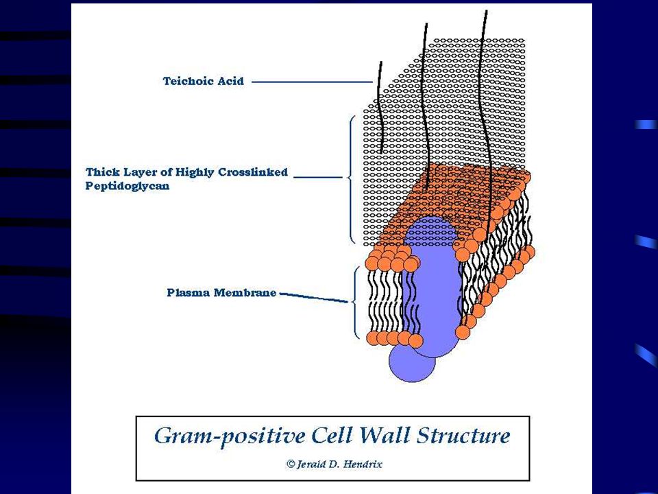
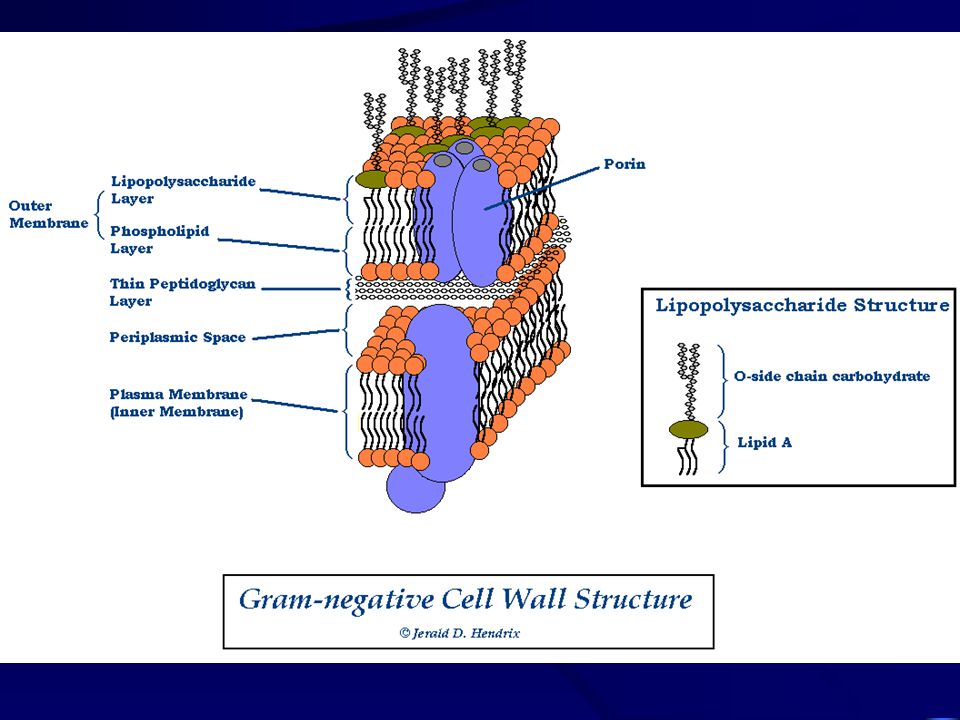
IV. B. 2. Cell Wall Gram Staining –Method developed by Gram in 1888 –Gram-positive cells stain purple Gram-negative cells stain pink –Later, it was discovered that the major factor determining Gram reactions is the bacterial cell wall structure –“Gram-positive” & “Gram-negative” These terms can mean either: Staining results, or Types of cell wall structure

11
IV. B. 2. Cell Wall Peptidoglycan Structure –Composition A Polysaccharide Composed of alternating units of N-acetylglucosamine (NAG) and N-acetylmuramic acid (NAM) –Peptide Crosslinking Between NAM units –Much thicker and more crosslinking in Gram-positive than in Gram-negative Bacteria
 and N-acetylmuramic acid (NAM) –Peptide Crosslinking Between NAM units –Much thicker and more crosslinking in Gram-positive than in Gram-negative Bacteria.")
12
IV. B. 2. Cell Wall Gram-positive Cell Wall –Thick Layer of Highly Crosslinked Peptidoglycan –Teichoic Acid Strands

14
IV. B. 2. Cell Wall Gram-negative Cell Walls –Outer Membrane Lipopolysaccharide Layer containing Lipid A Phospholipid Layer Outer Membrane Proteins –Thin Layer of Peptidoglycan with no teichoic acid –Periplasmic Space

16
IV. B. 2. Cell Wall Variations on Cell Wall Architecture –Acid-fast Cell Walls Similar to Gram-positive structure, but have Mycolic Acid: A waxy lipid Require special acid-fast staining technique Includes Mycobacterium and Nocardia

17
IV. B. 2. Cell Wall Variations on Cell Wall Architecture (cont.) –Mycoplasmas Bacteria that are naturally have no cell walls Includes Mycoplasma and Ureaplasma –Archaeobacteria Have unusual archaeobacterial cell walls with no peptidoglycan Have unusual metabolisms Share a more recent common ancestor with eukaryotes than with eubacteria (“true bacteria”)
 –Mycoplasmas Bacteria that are naturally have no cell walls Includes Mycoplasma and Ureaplasma –Archaeobacteria Have unusual archaeobacterial cell walls with no peptidoglycan Have unusual metabolisms Share a more recent common ancestor with eukaryotes than with eubacteria ( true bacteria ).")
18
IV. B. 3. Plasma Membrane Structure –Phospholipid Bilayer with Associated Proteins Functions –Maintain Cell Integrity –Regulate Transport –Specialized Functions in Bacteria

19
IV. B. 4.Cytoplasm & Cytoplasmic Inclusions Composition: –Viscous aqueous suspension of proteins, nucleic acid, dissolved organic compounds, mineral salts Cytoplasmic Inclusions: –Metachromatic Granules (Phosphate) –Starch Granules –Lipid Droplets –Sulfur Granules
 –Starch Granules –Lipid Droplets –Sulfur Granules.")
20
IV. B. 5. Ribosomes Suspended in Cytoplasm Sites of Protein Synthesis

21
IV. B. 6. Bacterial DNA Chromosomal DNA Plasmid DNA –R-Plasmids –F-Plasmids

22
IV. B. 7. Pili Hair-like structures on cell surface Functions –Attachment –Conjugation

23
IV. B. 8. Flagella Function –Motility Almost all motile bacteria are motile by means of flagella –Motile vs. nonmotile bacteria Structure –Filament Composed of the protein flagellin –Hook & Rotor Assembly Permits rotational "spinning" movement

25
IV. B. 8. Flagella Mechanism of Motility –“Run and Tumble” Movement controlled by the direction of the flagellar spin –Counterclockwise spin = Straight Run Clockwise spin = Random Tumble

26
IV. B. 8. Flagella Chemotaxis –Response to the concentration of chemical attractants and repellants –As a bacterium approaches an attractant: the lengths of the straight runs increase –As a bacterium approaches a repellant: the lengths of the straight runs decrease

27
IV. B. 9. Spores Function –To permit the organism to survive during conditions of desiccation, nutrient depletion, and waste buildup –Bacterial spores are NOT a reproductive structure, like plant or fungal spores Occurrence –Produced by very few genera of bacteria –Major examples Bacillus Clostridium

28
IV. B. 9. Spores Significance in Medicine & Industry –Spores are resistant to killing –Cannot be killed by 100°C (boiling) –Requires heating to 120°C for 15-20 min (autoclaving or pressure cooking)
 –Requires heating to 120°C for min (autoclaving or pressure cooking).")
29
IV. B. 9. Spores Sporulation –The process of spore formation –Governed by genetic mechanism –A copy of the bacterial chromosome is surrounded by a thick, durable spore coat –This forms an endospore within a vegetative cell –When the vegetative cell dies and ruptures, the free spore is released

30
IV. B. 9. Spores Spore Germination –When a spore encounters favorable growth conditions –The spore coat ruptures and a new vegetative cell is formed

31
IV. C.Factors that Influence Bacterial Growth Growth vs. Survival –Bacteria may tolerate or survive under more extreme conditions than their growth conditions

32
IV. C.Factors that Influence Bacterial Growth Nutrient Requirements –Energy Source Most bacteria are chemotrophs; a few are phototrophs –Carbon Source Most bacteria are heterotrophs; a few are autotrophs –Nitrogen, Phosphate, Sulfur, Trace Minerals

33
IV. C.Factors that Influence Bacterial Growth Nutrient Requirements (cont.) –Special Requirements examples: amino acids and enzyme cofactors (vitamins) Fastidious bacteria: Strains that are difficult or impossible to culture due to special growth requirements
 –Special Requirements examples: amino acids and enzyme cofactors (vitamins) Fastidious bacteria: Strains that are difficult or impossible to culture due to special growth requirements.")
34
IV. C.Factors that Influence Bacterial Growth Temperature –Psychrophiles Grow at ~0°C - 20°C –Mesophiles Grow at ~20°C - 45°C –Moderate Thermophiles Grow at ~45°C - 70°C –Extreme Thermophiles Grow at ~70°C - 100°C

35
IV. C.Factors that Influence Bacterial Growth pH –Acidophiles Grow at ~pH 1.0 - pH 6.0 –Neutrophiles Grow at ~pH 6.0 - pH 8.5 –Alkalophiles Grow above pH 8.5

36
IV. C.Factors that Influence Bacterial Growth Oxygen –Strict aerobes (Obligate aerobes) Use oxygen for respiration in their metabolism Require the presence of a normal oxygen concentration (~20%) for growth –Strict anaerobes (Obligate anaerobes) Oxygen is a poison for these microbes Cannot grow at all in the presence of oxygen
 Use oxygen for respiration in their metabolism Require the presence of a normal oxygen concentration (~20%) for growth –Strict anaerobes (Obligate anaerobes) Oxygen is a poison for these microbes Cannot grow at all in the presence of oxygen.")
37
IV. C.Factors that Influence Bacterial Growth Oxygen (cont.) –Aerotolerate anaerobes Do not use oxygen, but oxygen is not a poison for these Can grow equally well with or without oxygen –Facultative anaerobes Use oxygen for respiration, but can also grow without oxygen Grow better with oxygen that without oxygen
 –Aerotolerate anaerobes Do not use oxygen, but oxygen is not a poison for these Can grow equally well with or without oxygen –Facultative anaerobes Use oxygen for respiration, but can also grow without oxygen Grow better with oxygen that without oxygen.")
38
IV. C.Factors that Influence Bacterial Growth Oxygen (cont.) –Microaerophiles Require low concentrations (~5% - 10%) of oxygen for growth
 –Microaerophiles Require low concentrations (~5% - 10%) of oxygen for growth.")
Similar presentations


 Flagella 1) Functions in movement of the cell 2) 3 components.>")


 bacteria ◦ Live in oxygen free environment ◦ Produce.>")