Download presentation
Presentation is loading. Please wait.

1
JOURNAL CLUB SESSION Dr. SYED ABDUL QUADER(Ashraf)
Chairperson Dr. Sankar Narayan Dey Associate professor & Ex-Head of the department of Radiology Mymensingh Medical College Co-Chairperson Dr. MISBAH UDDIN AHMED Assistant professor & Head of the department of Radiology & Imaging Mymensingh Medical College PRESENTER Dr. SYED ABDUL QUADER(Ashraf) Student (MD-Final part) of Radiology & Imaging Mymensingh Medical college
 Student (MD-Final part) of Radiology & Imaging. Mymensingh Medical college.")
2
Title Computed Tomographic Evaluation of Traumatic Epidural and Subdural Haematoma with post operative correlation.

3
Author : Islam MS, Azad SA, Habib MA, Alam MR, Bhuiyan MSI. Source : Bangladesh Journal of Radiology and Imaging, 2009 ; Volume 17(2) : 40–45.
 : 40–45.")
4
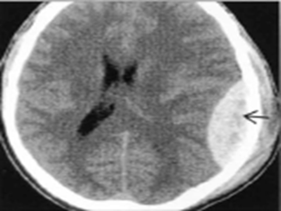
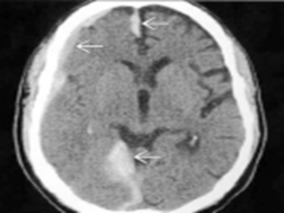
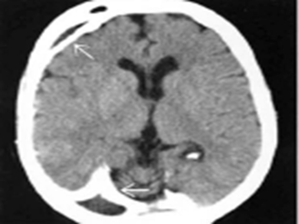
Introduction Head Injury is a serious health problem in all nations and responsible for approximately half of all deaths related to trauma. Intracranial lesions may be focal or diffuse, although these two forms frequently coexist. Focal lesions include epidural haematomas, subdural haematomas and contusions(or intracerebral haematomas). Epidural haematoma(EDH) lies in between the inner surface of the skull and stripes of the dural membrane. EDH are nearly always caused by, and located near a skull fracture.
. Epidural haematoma(EDH) lies in between the inner surface of the skull and stripes of the dural membrane. EDH are nearly always caused by, and located near a skull fracture.")
5
Introduction continues..
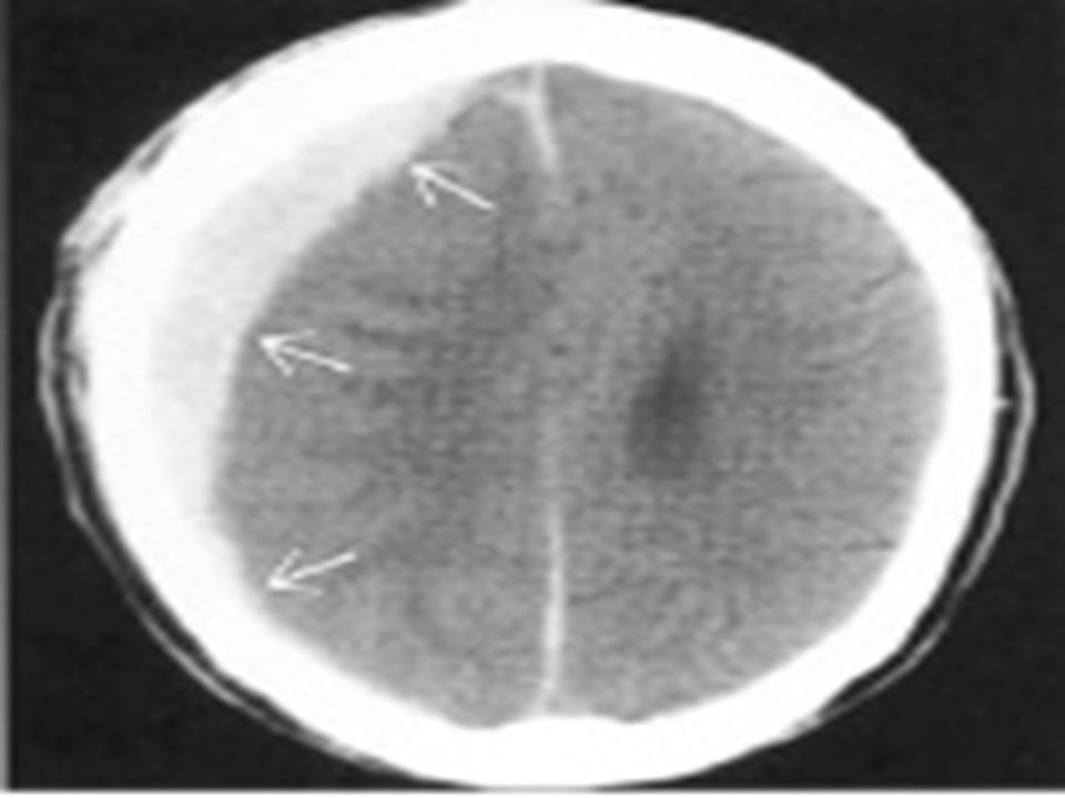
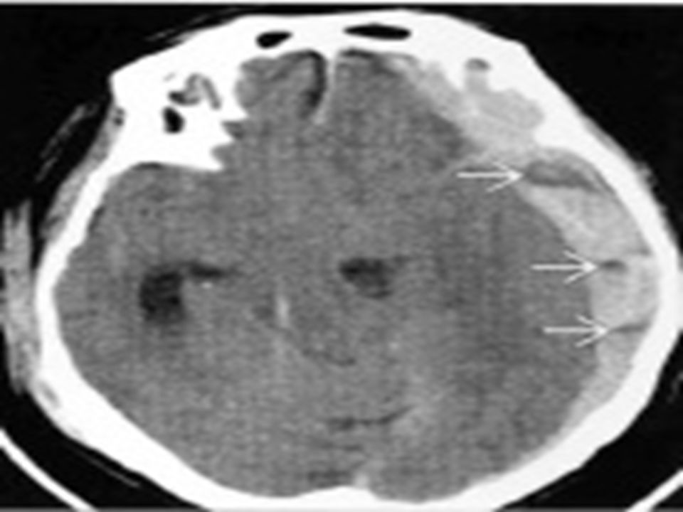
EDH, more common in temporal and temporoparietal areas(70% cases), usually forms within an hour from the time of injury but some time run a more chronic course. The classic CT appearance of EDH in 84% case is high density biconvex shape adjacent to the skull. Surgical evacuation is recommended for symptomatic lesions. Mortality is ~26%, is more related to growing speed of haematoma than to its location, being higher for the haematoma located in the temporal fossa.
, usually forms within an hour from the time of injury but some time run a more chronic course. The classic CT appearance of EDH in 84% case is high density biconvex shape adjacent to the skull. Surgical evacuation is recommended for symptomatic lesions. Mortality is ~26%, is more related to growing speed of haematoma than to its location, being higher for the haematoma located in the temporal fossa.")
6
Introduction continues..
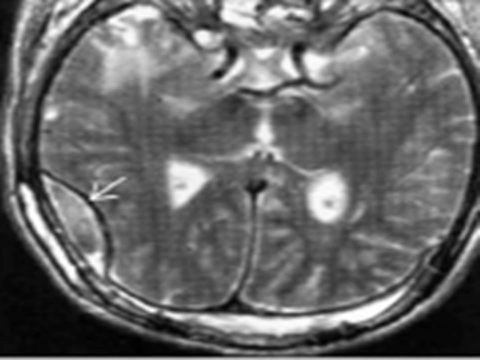
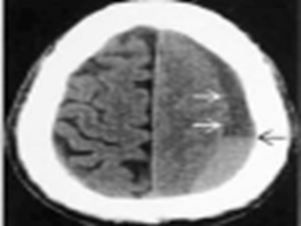
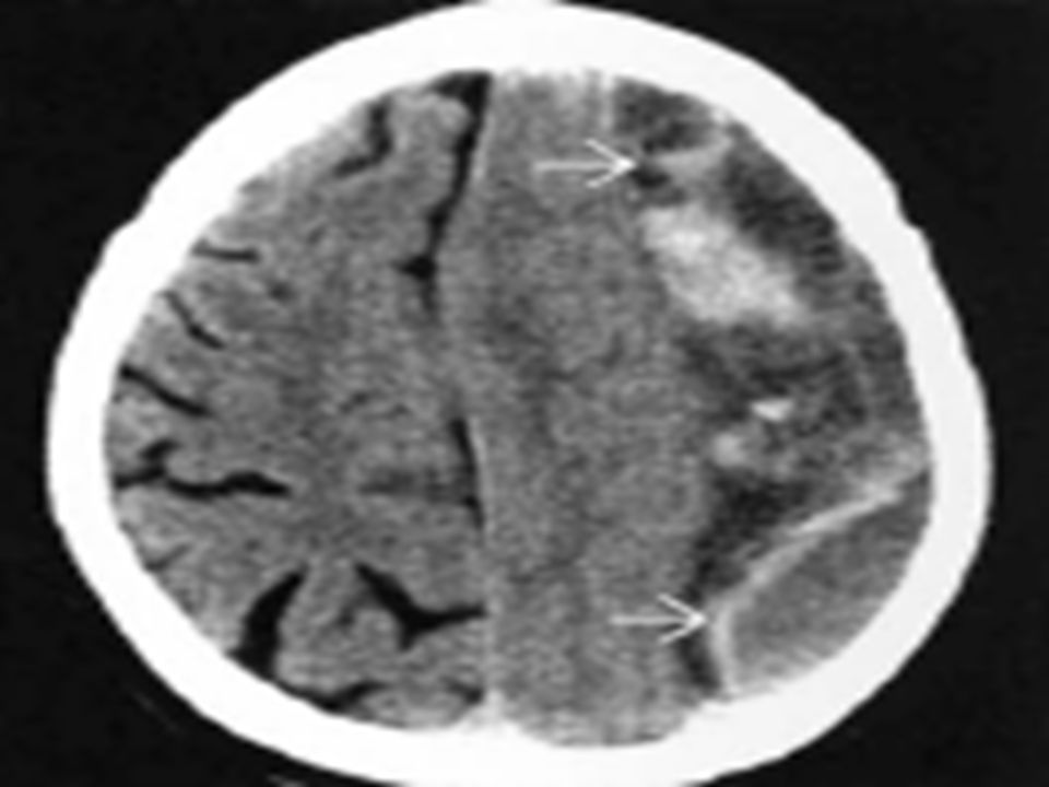
Subdural Haematoma(SDH) are much more common than EDH. They occur most frequently from a tearing of bridging veins between the cerebral cortex and draining sinuses. The injury in patients with SDH is usually much more severe, and prognosis is much worse than for EDH. Mortality in general is 60% but can be lowered by very rapid diagnosis & rapid surgical intervention and aggressive medical management. Computed Tomography(CT) scan of the head is currently the imaging modality of choice.
 are much more common than EDH. They occur most frequently from a tearing of bridging veins between the cerebral cortex and draining sinuses. The injury in patients with SDH is usually much more severe, and prognosis is much worse than for EDH. Mortality in general is 60% but can be lowered by very rapid diagnosis & rapid surgical intervention and aggressive medical management. Computed Tomography(CT) scan of the head is currently the imaging modality of choice.")
7
Introduction continues..
Early recovery of traumatic head injury patients largely depends upon the prompt diagnosis and meticulous management of these patients. Lots of lives can be spared by early and accurate diagnosis if CT scan can be established as a sensitive and specific modality for the diagnosis of EDH and SDH in patients having head injury.

8
Objectives To establish the CT scan as a good diagnostic modality for the diagnosis of EDH and SDH after head injury by considering surgical finding as a gold standard.

9
Materials and Methods Study Design : Cross sectional study.
Place of study: Department of Radiology and Imaging, Bangabandhu Sheikh Mujib Medical University & Dhaka Medical College Hospital, Dhaka in collaboration with department of Neurosurgery of the same hospitals. Study Period: From January 2010 to November 2010. Sample Size : 91 Sampling Technique : Purposive sampling

10
Materials and Methods continues..
Inclusion Criteria : Patients with head injury having epidural & / or subdural haematoma on CT and subsequently treated surgically. Exclusion Criteria : Non traumatic cases.

11
Ethical Consideration
Informed consent was required from all patients & / or from their guardians after the potential risks and benefits were explained.

12
Study Procedure Traumatic head injury patients who were admitted in the department of Neurosurgery BSMMU & DMCH, Dhaka with the clinical suspicion of having epidural & subdural haematoma and sent for CT scan in Radiology &Imaging department of the same hospital were initially enrolled for the study. 110 patients with traumatic intracranial haematoma(EDH/SDH) confirmed by CT scan who subsequently underwent surgical intervention were finally included in the study.
 confirmed by CT scan who subsequently underwent surgical intervention were finally included in the study.")
13
Study Procedure Continues…
CT scan was performed with a third generation CT, Siemens at DMCH & Hitachi CT scan Machine in BSMMU. Sample size was calculated for a power level of greater than 80%, an error of .05 and expected sensitivity of 94 to 96% based on previous report. 19 patients were withdrawn from the study as they were shifted to different hospitals other than BSMMU or DMCH for surgical procedure.

14
Study Procedure Continues…
For rest of the 91 patients diagnosis was established after surgical intervention. Surgical findings of all patients were correlated with CT scan findings. For the validity of study outcome, sensitivity, specificity, accuracy, positive predictive value and negative predictive value of CT scan in the diagnosis of EDH & SDH was calculated out after confirmation of the diagnosis by surgical intervention.

15
Data Analysis Data were processed and analysed by using soft-ware SPSS (Statistical Package for Social Sciences) version The test statistics used to analyzed the data were descriptive statistics, Kappa test. The level of significance was 0.05 and p <0.05 was considered significant.
 version The test statistics used to analyzed the data were descriptive statistics, Kappa test. The level of significance was 0.05 and p <0.05 was considered significant.")
16
Result and observations
110 patients with head injury were initially enrolled for the study. 19 patients were withdrawn from study. Mean age of the patients was 37.2(+/ ) years with maximum patients(36.2%) belonging to 16 to 30 years age group. Male and female ratio was 2.8: 1. 64.8% had history of road traffic accident as the cause of head injury.
 years with maximum patients(36.2%) belonging to 16 to 30 years age group. Male and female ratio was 2.8: % had history of road traffic accident as the cause of head injury.")
17
Result and observations Continues…
CT scan diagnosis showed that 13.2% had EDH, 54.9% had SDH and 31.9% had both EDH & SDH.

18
31.9 54.9 CT scan diagnosis of the patients with head injurey Pie diagram

19
Result and observations Continues…
Intraoperative diagnosis showed that 12.1% had EDH, 53.8% had SDH and 34.1% had both EDH & SDH.

20
53.8 Pie diagram

21
Result and observations Continues…
11 cases were diagnosed as EDH by CT scan and confirmed by intraoperative evaluation. One case was diagnosed as EDH by CT scan but could not be confirmed intraoperatively. Of 79 cases of other than EDH, which were diagnosed by CT scan, all were diagnosed other than EDH intraoperatively. Sensitvity of CT scan to diagnose EDH was 100%, specificity 98.8%, positive predictive value 91.7%, negative predictive value 100%, & accuracy 98.9%.

22
Result and observations Continues…
Table-1 Distribution of EDH by CT scan & intraoperative diagnosis(n=91) CT scan diagnosis Intra operative diagnosis EDH Total Others Kappa value 11(TP) 1(FP) 12 0(FN) 79(TN) 79 0.95 11 80 91
 CT scan diagnosis. Intra operative diagnosis. EDH. Total. Others. Kappa value. 11(TP) 1(FP) 12. 0(FN) 79(TN)")
23
Result and observations Continues…
48 cases were diagnosed as SDH by CT scan and confirmed by intraoperative evaluation. Two cases were diagnosed as SDH by CT scan but could not be confirmed intraoperatively. Of 41 cases of other than SDH, which were diagnosed by CT scan, one was diagnosed as having SDH & 40, other than SDH intraoperatively. Sensitvity of CT scan to diagnose SDH was 98.0%, specificity 95.2%, positive predictive value 96.0%, negative predictive value 97.6%, & accuracy 96.7%.

24
Result and observations Continues…
Table-2 Distribution of SDH by CT scan & intraoperative diagnosis(n=91) CT scan diagnosis Intra operative diagnosis SDH Total Others Kappa value 48(TP) 2(FP) 50 1(FN) 40(TN) 41 0.934 49 42 91
 CT scan diagnosis. Intra operative diagnosis. SDH. Total. Others. Kappa value. 48(TP) 2(FP) 50. 1(FN) 40(TN)")
25
Result and observations Continues…
28 cases were diagnosed as both EDH & SDH by CT scan and confirmed by intraoperative evaluation. One case was diagnosed as combined EDH & SDH by CT scan but could not be confirmed intraoperatively. Of 62 cases of not having both EDH & SDH, which was diagnosed by CT scan, three were diagnosed as having both EDH & SDH, 59 other than combined EDH & SDH intraoperatively. Sensitivity of CT scan to diagnose both EDH & SDH was 96.5%, specificity 98.3%, positive predictive value 96.6%, negative predictive value 95.2%, & accuracy 95.6%.

26
Result and observations Continues…
Table-3 Distribution of both EDH & SDH by CT scan & intraoperative diagnosis(n=91) CT scan diagnosis Intra operative diagnosis Both EDH & SDH Total Others Kappa value 28(TP) 1(FP) 29 3(FN) 59(TN) 62 0.901 31 60 91
 CT scan diagnosis. Intra operative diagnosis. Both EDH & SDH. Total. Others. Kappa value. 28(TP) 1(FP) 29. 3(FN) 59(TN)")
27
Discussion Mean age of patient was 37.02(+/ ) years and maximum 36.3% belonged to 16 to 30 years age group. Men receive more head injuries than women & it appears that their incidence is two to three times greater. RTA was the commonest mode of injury in this study(64.8%). Almost maximum(97.8%) patients had complaints of headache, 85.7% vomiting, 58.2% history of unconsciousness, 47.3% had clinical signs of skull fracture, 5.5% had post traumatic amnesia, 4.4% posttraumatic seizure & 3.3% neurological deficit.
. Almost maximum(97.8%) patients had complaints of headache, 85.7% vomiting, 58.2% history of unconsciousness, 47.3% had clinical signs of skull fracture, 5.5% had post traumatic amnesia, 4.4% posttraumatic seizure & 3.3% neurological deficit.")
28
Discussion Continues…
After CT evaluation 82.4% had skull fracture, 13.2% patients were diagnosed as EDH, 54.9% SDH, 31.9% both EDH & SDH. But intraoperatively 12.1% were evaluated as having EDH, 53.8% SDH, 34.1% both EDH & SDH. Sensitivity of CT scan to diagnose different type of haematoma ranged from 96.5% to 100%. Specificity 95.2% to 98.8%, positive predictive value 91.7% to 96.6%, negative predictive value 95.2% to 100% and accuracy 95.6% to 98.8%.

29
Limitation Sampling technique was purposive Small sample size
CT scan was not compared with any other imaging modality like MRI

30
Recommendation Large scale study is required including both traumatic and non-traumatic head injury evaluated by same CT &/or MRI machine and operated by same surgeon.

31
Conclusion CT scan is usually the 1st choice in the evaluation of EDH & SDH, primarily due to the rapidity of diagnosis. The present study also found its excellent efficacy. In Bangladesh this imaging modality is becoming available all over the country. Considering as a test of reference we can straight forwardly recommend this modality for early diagnosis of intracranial haematoma. So mortality & morbidity due to EDH & SDH can be reduced by early diagnosis & promt management.

32
Thank You All

Similar presentations