Download presentation
Presentation is loading. Please wait.

2
THE CLINICAL EFFICACY OF REPEAT BRAIN CT IN PATIENTS WITH TRAUMATIC INTRACRANIAL HAEMORRHAGE WITHIN 24 HRS AFTER BLUNT HEAD INJURY

3
INTRODUCTION Widespread availability of CT scanners in emergency &intensive care units have led to increase utilization of CT scanning in patients with traumatic brain injury(TBI) Repeat brain CT scans for all patients with TBI may facilitate early medical and surgical intervention and minimize secondary brain injury.
 Repeat brain CT scans for all patients with TBI may facilitate early medical and surgical intervention and minimize secondary brain injury.")
4
Contd. On the contrary repeat CT scan may increase
the unnecessary costs and risk of exposure to ionizing radiation as well as risk involved in the transfer of patients out of intensive care settings causing harm to critically ill patients.

5
The aim of this study was to study the efficacy
& variables associated with radiological deterioration from repeat brain CT scans possibly necessitating surgical interventions.

6
Patient selection &methods
It was retrospective review (Jan’03-Dec’06) of all the blunt head trauma patients with traumatic intracranial haemorrhage.
 of. all the blunt head trauma patients with. traumatic intracranial haemorrhage.")
7
INCLUSION CRITERIAN: Adult patients older than 16 yrs. of age.
Initial GCS score of 8 or greater as long as they have no planned immediate neurosurgical intervention after their initial CT scan. A repeat brain CT within 24 hrs. after trauma.

8
EXCLUSION CRITERIAN Patients with ventilatory support.
History of prior brain surgery. Chronic neurological conditions. Associate spinal cord injury. Patients with bleeding diathesis. Previous use of antiplatelets/anticoagulants. Patients undergone immediate craniotomy based on initial brain CT at admission.

9
In addition to above criterion other variables collected on admission included :
-age -gender -mechanism of injury -GCS score. Probability value less than 0.05 were accepted as statistically significant.

10
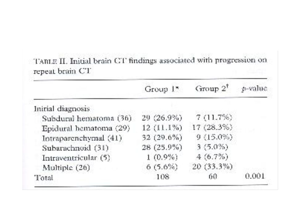
Findings of initial brain CT were categorized as:
-subdural haematoma(SDH) -epidural haematoma(EDH) -intraparenchymal haematoma(IPH) -subarachnoid haemorrhage(SAH) -intraventricular haemorrhage(IVH)
 -epidural haematoma(EDH) -intraparenchymal haematoma(IPH) -subarachnoid haemorrhage(SAH) -intraventricular haemorrhage(IVH)")
11
On repeat brain CT scans patient were categorized as:
-group 1(improved or unchanged conditions) -group 2(obvious increase in size of ICH ,amounting to 1 mm or more at least in one dimension or whose radiology reports declared an increase of one or more lesions)
 -group 2(obvious increase in size of ICH ,amounting to 1 mm or more at least in one dimension or whose radiology reports declared an increase of one or more lesions)")
13
Patient’s sex,initial GCS score and timing of the repeat CT scans were the strong predictors for the worsening of the lesions on repeat brain CT scans lesions. There were significantly more men in group 2(80%) than in group 1(61.6%) Mean GCS score was significantly higher in patients from group 1(14.3+_0.96)than in patients from group 2(11.9+_2.6)
 than in group 1(61.6%) Mean GCS score was significantly higher in patients from group 1(14.3+_0.96)than in patients from group 2(11.9+_2.6)")
14
The mean time between the initial and repeat
brain CT scan was significantly shorter for group 2(7.41+_5.98) than group 1(11.6+_7.52)
 than group 1(11.6+_7.52)")
16
Intraparenchymal haematoma, subdural haematoma, subarachnoid haemorrhage were common occurrence in group 1. Epidural haematoma and multiple lesions were more common in patients from group 2 as evident from radiological progression in same category.

17
After repeat brain CT scans, 28(47%)of the patients in group 2 ,comprising 17% of the entire population in this study group, underwent neurosurgical interventions. Of the 28 surgically treated patients of group 2 ,6(10%) exhibited neurological worsening and 22(37%) appear neurologically stable. No patient in group 1 underwent neurosurgical intervention.
 exhibited neurological worsening and 22(37%) appear neurologically stable. No patient in group 1 underwent neurosurgical intervention.")
18
22 out of 28 patients who underwent neurosurgical interventions were neurologically were stable at the time of repeat brain CT scans. Surgically treated lesions included: 1/36(3%) SDH’s 15/29(52%) EDH’s 1/41(2%) IPH’s 3/5(60%) IVH’s 8/26(31%) multiple lesions
 SDH’s. 15/29(52%) EDH’s. 1/41(2%) IPH’s. 3/5(60%) IVH’s. 8/26(31%) multiple lesions.")
19
Discussion Optimal management of patients with TBI includes neurosurgical intervention if needed, reduction of ICP, prevention of seizures and avoidance of hypoxia and hypotension. Patients with documented intracranial injuries often undergo frequent routine brain CT scans given that significant radiological changes may occur with minimal or no clinical and neurological changes.

20
Previous reports state that routine repeat brain CT scans are of little value in clinically observed patients with traumatic ICH,unless there is a corresponding deterioration of neurologic status. However; 22/60(37%) patients in group 2 who had undergone neurosurgical interventions had no neurological changes at the time of repeat CT scans.
 patients in group 2 who had undergone neurosurgical interventions had no neurological changes at the time of repeat CT scans.")
21
These results indicate that radiological deterioration on repeat brain CT scan might precede a significant neurological worsening in an affected patient. It will allow for the utilization of appropriate neurosurgical interventions to prevent the on-going neurological deterioration. The possibility exists that minor changes observed in patients from group 2 such as headache, nausea and drowsiness without a worsening GCS score, might be overlooked.

22
Predictors of radiological progression in current study were male sex, a short time interval between initial and repeat brain CT scans, a lower GCS at admission and subtypes of EDH or multiple lesions on the initial CT scans. It has also been supported by Oertel et al.

23
Certain subtypes of ICH’s were associated with radiological worsening (group 2):
-17(28%) EDH -20(33%) multiple injuries 3/5(60%) IVH These results demonstrate that presence of EDH,IVH,AND multiple lesions on the initial brain CT scan is a risk factor of neurosurgical intervention. -
 EDH. -20(33%) multiple injuries. 3/5(60%) IVH. These results demonstrate that presence of EDH,IVH,AND multiple lesions on the initial brain CT scan is a risk factor of neurosurgical intervention. -")
24
As a result of this study,it is suggested that repeat brain CT scans be performed in the 24 hrs. following blunt head trauma. It may minimise the potential neurological deterioration in patients with initial GCS score lower than 12 or with EDH or multiple lesions on their initial brain CT scan.

25
CONCLUSION Routine repeat brain CT scans within 24 hrs. in the blunt head trauma patients with traumatic ICH ,who were treated initially nonsurgically and remained neurologically stable,revealed radiological worsening in 34% of such patients.

26
Of the patients who showed radiological worsening on repeat brain CT scans,37% underwent neurosurgical interventions despite lack of significant neurological deterioration.

27
Based on these findings ,it is proposed that in those patients with an admission GCS score lower that 12 or with the EDH or multiple lesions on their initial brain CT scan, routine repeat scans should be performed within 24 hrs. of injury.

Similar presentations


 A David Mendelow, Department of Neurosurgery,>")


