Download presentation
Presentation is loading. Please wait.

1
Chapter 11 DNA & GENES

2
By the early 1950’s, most scientists were convinced
11-1 The Structure of DNA By the early 1950’s, most scientists were convinced That genes were made of DNA. The problem is that no one knew what it looked like. Then along came James Watson & Francis Crick.

3
James Watson and Francis Crick

4
A Winding Staircase Watson and Crick determined that a DNA molecule is a double helix—two strands twisted around each other, like a winding staircase. Nucleotides are the subunits that make up DNA. Each nucleotide is made of three parts: a phosphate group, a five-carbon sugar molecule, and a nitrogen-containing base

5
The five-carbon sugar in DNA nucleotides is called deoxyribose
Structure of a Nucleotide

6
DNA Double Helix

7
The nitrogen base in a nucleotide can be either a bulky, double-ring purine, or a smaller, single-ring pyrimidine.

8
Discovering DNA’s Structure
Chargaff’s Observations In 1949, Erwin Chargaff observed that for each organism he studied, the amount of adenine always equaled the amount of thymine (A=T) Likewise, the amount of guanine always equaled the amount of cytosine (G=C). However, the amount of adenine and thymine and of guanine and cytosine varied between different organisms
 Likewise, the amount of guanine always equaled the amount of cytosine (G=C). However, the amount of adenine and thymine and of guanine and cytosine varied between different organisms.")
9
X-Ray Diffraction

10
Watson and Crick’s DNA Model
In 1953, Watson and Crick built a model of DNA with the configuration of a double helix, a “spiral staircase” of two strands of nucleotides twisting around a central axis The double-helix model of DNA takes into account Chargaff’s observations and the patterns on Franklin’s X-ray diffraction photographs.

11
These base-pairing rules are supported by Chargaff’s observations
Pairing Between Bases An adenine on one strand always pairs with a thymine on the opposite strand, and a guanine on one strand always pairs with a cytosine on the opposite strand These base-pairing rules are supported by Chargaff’s observations The strictness of base-pairing results in two strands that contain complementary base pairs

12
In the diagram of DNA below, the helix makes it easier to visualize the base-pairing that occurs between DNA strands

13
The Replication of DNA When the double helix was discovered, scientists were very excited about the complimentary relationship between the sequences of nucleotides. Watson and Crick proposed that one DNA strand serves as a template on which the other strand is built.

14
Roles of Enzymes in DNA Replication
The complementary structure of DNA is used as a basis to make exact copies of the DNA each time a cell divided. The process of making a copy of DNA is called DNA replication DNA replication occurs during the synthesis (S) phase of the cell cycle, before a cell divides DNA replication occurs in three steps
 phase of the cell cycle, before a cell divides. DNA replication occurs in three steps.")
15
Step 1- DNA helicases open the double helix by breaking the hydrogen bonds that link the complementary nitrogen bases between the two strands. The areas where the double helix separates are called replication forks

16
Step 2 - At the replication fork, enzymes known as DNA polymerases move along each of the DNA strands. DNA polymerases add nucleotides to the exposed nitrogen bases, according to the base-pairing rules Step 3 - Two DNA molecules that form are identical to the original DNA molecule

18
Checking for Errors In the course of DNA replication, errors sometimes occur and the wrong nucleotide is added to the new strand. An important feature of DNA replication is that DNA polymerases have a “proofreading” role This proofreading reduces errors in DNA replication to about one error per 1 billion nucleotides

19
In eukaryotic cells, each chromosome contains a single, long strand of DNA
Each human chromosome is replicated in about 100 sections that are 100,000 nucleotides long, each section with its own starting point With multiple replication forks working in concert, an entire human chromosome can be replicated in about 8 hours

20
Replication Forks

21
Traits, such as eye color, are determined
11-2 RNA & Protein Traits, such as eye color, are determined By proteins that are built according to The instructions specified in the DNA.

22
Decoding the Information in DNA
Proteins, however, are not built directly from DNA. Ribonucleic acid is also involved Like DNA, ribonucleic acid (RNA) is a nucleic acid—a molecule made of nucleotides linked together
 is a nucleic acid—a molecule made of nucleotides linked together.")
23
RNA differs from DNA in three ways
1. RNA consists of a single strand of nucleotides instead of the two strands found in DNA 2. RNA nucleotides contain the five-carbon sugar ribose rather than the sugar deoxyribose, which is found in DNA nucleotides 3. In addition to the A, G, and C nitrogen bases found in DNA, RNA nucleotides can have a nitrogen base called uracil (U)
")
24
Comparing DNA and RNA The instructions for making a protein are transferred from a gene to an RNA molecule in a process called transcription Cells then use two different types of RNA to read the instructions on the RNA molecule and put together the amino acids that make up the protein in a process called translation

25
The entire process by which proteins are made based on the information encoded in DNA is called gene expression, or protein synthesis

26
Gene Expression

27
Transfer of Information
from DNA to RNA The first step in the making of a protein, transcription, takes the information found in a gene in the DNA and transfers it to a molecule of RNA RNA polymerase, an enzyme that adds and links complementary RNA nucleotides during transcription, is required

28
The three steps of transcription are
Step 1 RNA polymerase binds to the gene’s promoter Step 2 The two DNA strands unwind and separate Step 3 Complementary RNA nucleotides are added

30
Types of RNA

31
Genetic Code: Three-Nucleotide “Words”
Different types of RNA are made during transcription, depending on the gene being expressed When a cell needs a particular protein, it is messenger RNA that is made Messenger RNA (mRNA) is a form of RNA that carries the instructions for making a protein from a gene and delivers it to the site of translation
 is a form of RNA that carries the instructions for making a protein from a gene and delivers it to the site of translation.")
32
The information is translated from the language of RNA—nucleotides—to the language of proteins—amino acids The RNA instructions are written as a series of three-nucleotide sequences on the mRNA called codons The genetic code of mRNA is the amino acids and “start” and “stop” signals that are coded for by each of the possible 64 mRNA codons

34
RNA’s Roles in Translation
Translation takes place in the cytoplasm. Here transfer RNA molecules and ribosomes help in the synthesis of proteins Transfer RNA (tRNA) molecules are single strands of RNA that temporarily carry a specific amino acid on one end An anticodon is a three-nucleotide sequence on a tRNA that is complementary to an mRNA codon.
 molecules are single strands of RNA that temporarily carry a specific amino acid on one end. An anticodon is a three-nucleotide sequence on a tRNA that is complementary to an mRNA codon.")
35
Ribosomes are composed of both proteins and ribosomal RNA (rRNA)
Ribosomal RNA (rRNA) molecules are RNA molecules that are part of the structure of ribosomes Each ribosome temporarily holds one mRNA and two tRNA molecules
 molecules are RNA molecules that are part of the structure of ribosomes. Each ribosome temporarily holds one mRNA and two tRNA molecules.")
36
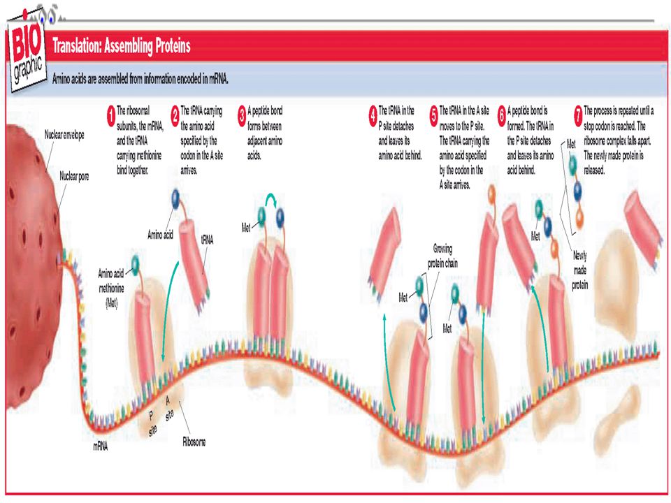
The seven steps of translation are:
Step 1 The ribosomal subunits, the mRNA, and the tRNA carrying methionine bind together Step 2 The tRNA carrying the amino acid specified by the codon in the A site arrives Step 3 A peptide bond forms between adjacent amino acids Step 4 The tRNA in the P site detaches and leaves its amino acid behind

37
Step 5 The tRNA in the A site moves to the P site
Step 5 The tRNA in the A site moves to the P site. The tRNA carrying the amino acid specified by the codon in the A site arrives Step 6 A peptide bond is formed. The tRNA in the P site detaches and leaves its amino acid behind Step 7 The process is repeated until a stop codon is reached. The ribosome complex falls apart. The newly made protein is released

39
THE END

Similar presentations