Download presentation
Presentation is loading. Please wait.

1
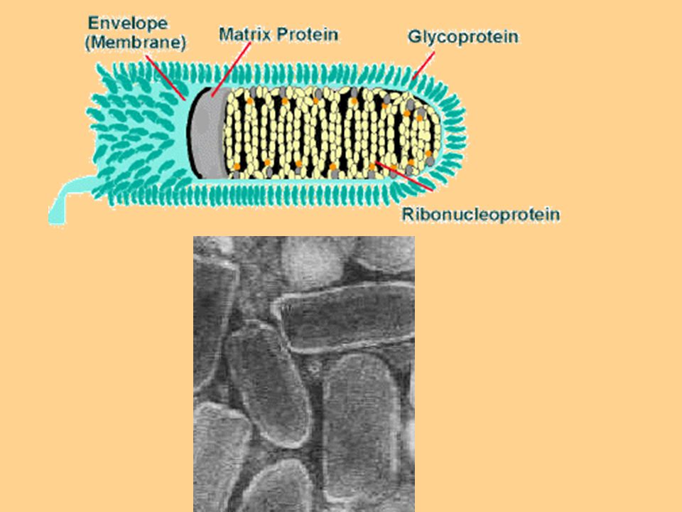
Rabdovirus The virion is from Rabdoviridea family.
Morphology: nucleocapside is like a bullet Genome: linear single stranded RNA, Negative sense, M protein, RNA-dependent RNA polymerase, Enveloped.

3
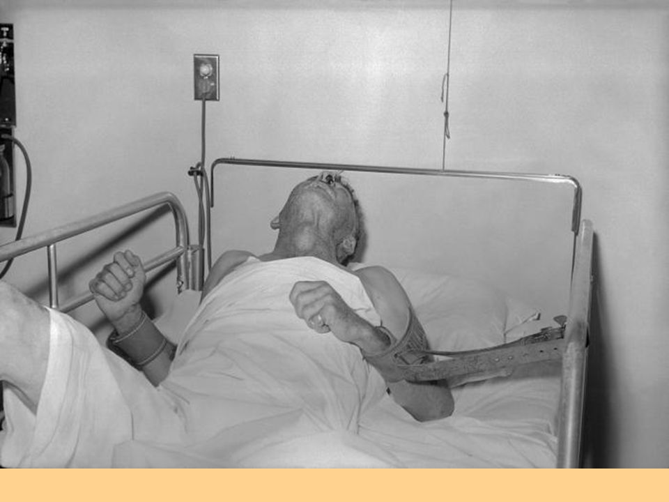
Disease: Rabies Several hosts including all warm-blooded living things, especially in dog, wolf, fox, bat. Human to human transmission is very rare. The virus can be found in nerve system, saliva, urine, lymph, milk. There is only one serotype of rabies virus. Glycoprotein G is a main cause of virus invading to nerve system.

4
Pathogenesis Virus amplification in muscles nerve - muscle synapses virus entering into peripheral nerves central nerve system Attachment to Asetylcholin entering to the brain and multiplication progressive encephalitis virus moving to saliva glands and other organs like pancreas, kidney, heart, retina, cornea, through peripheral nerves.

5
Clinical findings Incubation period: 20 days to 2 months (but seen between 1 week to 19 years) depends to the age, genetic background, race, the number of virus inoculated, the distance of virus to central nerve system. Rabies is an accidental viral infection in human.
 depends to the age, genetic background, race, the number of virus inoculated, the distance of virus to central nerve system. Rabies is an accidental viral infection in human.")
6
Clinical periods includes 3 stages:
I. Introductory short stage: 2-10 days: non-specific symptoms (headache, photophobia, nausea, vomiting, sore throat, fever, no appetite. II.2-7 days: partial paralysis, cerebral dysfunction, anxiety, sleeplessness, confusion, agitation, abnormal behavior, terror, hallucinations. Increased activity of simpatico system such as increased tear secretion, increased saliva, hydrophobia. III. Coma and death

8
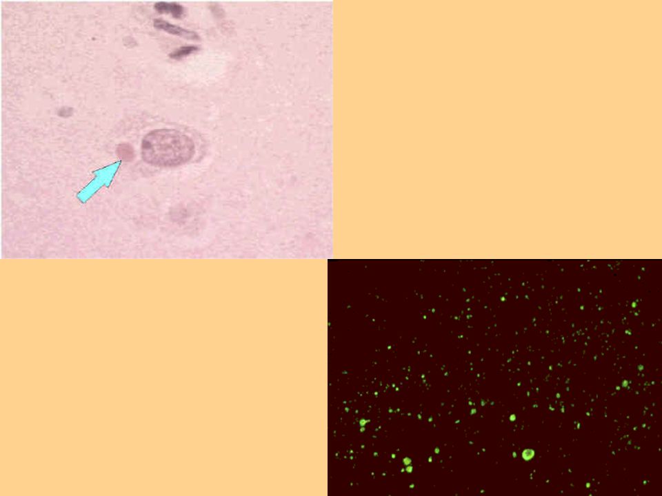
Lab diagnosis Negri bodies in brain or spinal cord (spherical, 2-10 microns granules. They contain rabies antigens and detectable by immunoflorosence (IF) methods. Virus isolation in humestesr or mice cells. PCR Serologic methods (Detecting antibodies)
 methods. Virus isolation in humestesr or mice cells. PCR. Serologic methods (Detecting antibodies)")
10
Prevention and treatment
All infected animals should be killed and their tissues should be tested in lab. Vaccination (inactivated virus) is used for people who have been bitted or their job is known as high risk carrier. Antibody (antiserum) prophylaxis. No treatment
 is used for people who have been bitted or their job is known as high risk carrier. Antibody (antiserum) prophylaxis. No treatment.")
11
Parainfluenza viruses
Common cold viruses Rinoviruses Adenoviruses Coronaviruses Enteroviruses Parainfluenza viruses Influenza viruses

12
Rinoviruses The main cause of the common cold From Picornaviruses
RNA single strand, + sense, The genome includes a small viral Pr at 5’ end. No envelope Ichozahedral 28-30 nm (as a small virus)
")
13
Important properties More than 100 serological types
Replicate better at 33C than 37C (affecting primarily nose and conjunctiva rather than the lower respiratory tract). Acid-labile, so killed by gastric acid when swallowed. The host range: humans and chimpanzees
. Acid-labile, so killed by gastric acid when swallowed. The host range: humans and chimpanzees.")
14
Pathogenesis Entering through the upper respiratory tract
There is a correlation between the concentration of viruses in nasal dischargse (or mucosa) and the severity of disease. The viruses are usually seen in 2-4 days after infection. Some times virus still is detectable by 3 weeks.
 and the severity of disease. The viruses are usually seen in 2-4 days after infection. Some times virus still is detectable by 3 weeks.")
15
The virus spread is limited to the epithelial surface of nasal mucosa.
Histopathology changes are limited to the surface of epithelium and includes: Edema and low penetration of cells. Cold weather does not cause common cold or even not increase the chance for it.

16
Clinical finding Incubation period: 2-4 days
Acute stage: sometimes to 7 days but dried cough can last by 2-3 weeks. Adults: 1-2 cold onset per year. Symptoms: sneeze. sore throat. chill, some times low fever, nasal congestion and loss of smell.

17
Clinical finding There is no way to distinguish common cold caused by rinoviruses from common colds by others viruses. Bacterial secondary infection: otitis, sinusitis, bronchitis, pneumonia.

18
Immunity Antibody appearance is late: 7-21 days after infection appearance in noise and serum at the same time. Antibody may clear virus from the body finally.

19
Epidemiolohy and Transmission
Worldwide More often is in early autumn and late spring and minimum in summer Transmission through fingers or shared things is more important than aerosol droplets. Family and school are two source of contamination for children.

20
Treatment and prevention
No spesific treatment No high chance for a vaccine production: The culture for high concentration of virus is difficult. 2. The immunity is short. 3. The serotypes are very high.

21
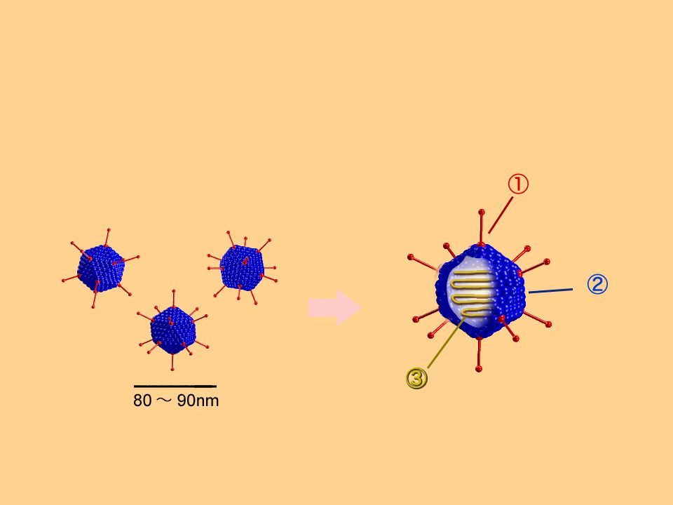
Adenoviruses Double-stranded linear DNA, icosahedral nucleocapsid (80-90 nm). The only viruses with a fiber protruding from each of the 12 vertices. The fiber is the organ of attachment and is a hemagglutinin. There are more than 40 known antigenic types and the fiber protein is the main type-specific antigen.

23
Diseases Upper respiratory tract infections: Pharyngitis, pharyngoconjunctivitis. Conjunctivitis Pneumon Keratoconjunctivitis Hemorrhagic cystitis Gastroenteritis Latent infections particularly in the adenoidal and tonsillar tissues.

24
Clinical findings of adenovirus infection
Varying degrees of fever, sore throat, coryza, and conjunctivitis. In the lower respiratory tract, atypical pneumonia is characterized primarily by fever and cough.

25
Lab diagnosis Isolation of the virus in cell culture
Detection of 4-fold rise in antibody titer. Serological tests: Hemagglutination test

26
Treatment and prevention
Most adenovirus infections resolve spontaneously. No antiviral therapy An enteric-coated capsule vaccine is used only in military recruits in USA but not available for civilian use.

27
The Influenza & Parainfluenza viruses
27

28
Orthomyxoviridae & Paramyxoviridae

29
Classification Family: Genus: Types: Type A Type B Influenza virus
Type C ORTHOMYXOVIRIDAE Genus: Types: 7 29

30
“myxo” refers to interaction with mucins (glycoproteins)
Different from paramyxoviruses : - segmented genome - smaller (average 110 nm in diameter against 150 nm).
.")
31
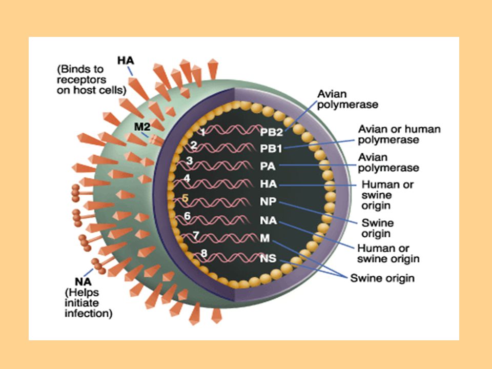
An enveloped viruse, helical symmetry capsid, segmented linear RNA genome
80 to 120 nm Surface antigens Internal Nucleocapsid: Nucleoprotein (7 or 8 RNA segments) Matrix protein (M) Lipid bilayer Haemaglutinin (HA) Neuraminidase (NA) 8 31
 Matrix protein (M) Lipid bilayer. Haemaglutinin (HA) Neuraminidase (NA)")
32
Surface glycoproteins
Haemagglutinin H or HA responsible for pathogenicity of the virus allows virus to adhere to endothelial cells in the respiratory tract main determinant of immunity Neuraminidase N or NA allows release of newly formed viruses within host determinant of disease severity 32

33
Antibody against the hemagglutinin neutralizes the infectivity of the virus and prevents disease. Ab against neuraminidase only reduces disease.

34
Influenza subgroups Influenza A Influenza B Influenza C
highly infective infects many species causes widespread epidemics Influenza B found only in humans capable of producing severe disease causes regional epidemics Influenza C causes mild disease humans are natural hosts, but isolates also found in pigs does not cause epidemics 34

35
Reassortment of segments of the genome RNA
Influenza viruses, especially type A show changes in the antigenicity of their hemagglutinin and neuraminidase proteins. epidemics. Influenza viruses antigenes Group-specific (internal ribonucleoprotein) antigenes. Type-specific (surface N and H) antigens.
 antigenes. Type-specific (surface N and H) antigens.")
36
Many species of animal (eg
Many species of animal (eg. Birds, swine, and hourses) have their own influenza A viruses. These animal viruses are probably the source of the new antigenic types. Antigenic shift: Major changes based on reassortment of genome pieces. Occurs every years Antigenic drift: Minor changes based on mutation occurs every year.
 have their own influenza A viruses. These animal viruses are probably the source of the new antigenic types. Antigenic shift: Major changes based on reassortment of genome pieces. Occurs every years. Antigenic drift: Minor changes based on mutation occurs every year.")
37
Antigenic shift appears to result from genetic recombination of human with animal or bird ,providing major antigenic change.This can cause a major epidemic or pandemic involving most or all age groups.

38
Epidemics and pandemics occur when the antigenicity of the virus has changed sufficiently that the preexisting immunity of many people is no longer effective.

39
39

40
Various combinations of RNA segments can result in a new subtype of virus (known as antigenic shift
It is even possible to include RNA strands from birds, swine, and human influenza viruses into one virus if a cell becomes infected with all three types of influenza.

42
Occurrence of influenza A viruses
Influenza A viruses 16 HA types 9 NA types Species affected humans, pigs, horses, birds, marine mammals In humans 3 HA types (H1, H2, H3) 3 NA types (N1, N2, N8) In birds all HA types all NA types 42
 3 NA types (N1, N2, N8) In birds all HA types. all NA types. 42.")
43
Influenza viruses nomenclature
For example: A / Beijing / 32 / 92 (H3N2) A virus type, here A Beijing place where the strain was isolated 32 strain number 92 year of first isolation H3N2 subtypes H3 and N2 virus sub type, here H3N2 43
 A virus type, here A. Beijing place where the strain was isolated. 32 strain number. 92 year of first isolation. H3N2 subtypes H3 and N2 virus sub type, here H3N")
44
ELECTRON MICROSOPE IMAGE OF H1N1 INFLUENZA VIRUS

45
Pathogenesis After the virus is inhaled, the neuraminidase degrades the protective mucus layer, allowing the virus to gain access to the cells of the upper and lower respiratory tract. Viremia rarely occurs, but there is necrosis of the superficial layers of respiratory epithelium.

46
Immunity Circulating IgG against the virus occurs after infection, but offers little protection. Secretory IgA in the respiratory tract is protective.

47
SYMPTOMS OF SWINE FLU IN HUMANS

48
Clinical findings Incubation period: 24-48 hours
Symptoms: fever, myalgias, headache, cough develop suddenly. The symptoms resolve spontaneously in 4-7 days but sometimes is complicated with secondary infections. Rey’s syndrome (Encephalopathy and liver degeneration life-threatening complication in children) following some viral infections, particularly influenza B and chikenpox, if they have been given Asprin to reduce the fever.
 following some viral infections, particularly influenza B and chikenpox, if they have been given Asprin to reduce the fever.")
49
Pneumonia Respiratory failure
COMPLICATIONS Pneumonia Respiratory failure Convulsion (muscles contract and relax rapidly and repeatedly, resulting in an uncontrolled shaking of the body)
")
50
When to Seek Emergency Medical Care
has difficulty breathing or chest pain has purple or blue discoloration of the lips is vomiting and unable to keep liquids down has signs of dehydration such as dizziness when standing, absence of urination, or in infants, a lack of tears when they cry has seizures (for example, uncontrolled convulsions) is less responsive than normal
 is less responsive than normal.")
51
RISK GROUPS Persons with certain chronic medical condition
School children Travelers to some high risk places Border workers Health care workers or public health workers

52
PREVENTION Prevention in swine or other animal hosts.
Prevention of transmission to humans. Prevention of its spread among humans.

53
Prevention of its spread among humans.
Frequent washing of hands with soap and water

54
Use of face masks

55
Use of towel while sneezing

56
Use of alcohol based sanitisers.

57
Lab diagnosis Virus isolation (by throat washing) with cell culture. Then flurescent-antibody staining of the infected cells by using antisera to influenza A and B. A rise in antibody titer of at least 4-fold in serum samples using hemmagglutination inhibition or complement fixation. PCR reactions
 with cell culture. Then flurescent-antibody staining of the infected cells by using antisera to influenza A and B. A rise in antibody titer of at least 4-fold in serum samples using hemmagglutination inhibition or complement fixation. PCR reactions.")
58
TREATMENT supportive care is required.
Antibiotics (to treat this disease, do help prevent bacterial pneumonia and other secondary infections.) Viral agent is used in severe infections. (Zanamivir is recommended by C.D.C.)
 Viral agent is used in severe infections. (Zanamivir is recommended by C.D.C.)")
59
SWINE INFLUENZA

60
WHAT IS SWINE INFLUENZA?
Swine flu is a respiratory disease caused by influenza viruses that infect the respiratory tract of pigs. Swine flu produces most of the same symptoms in pigs as human flu produces in people.

61
HISTORY OF SWINE INFLUENZA
The 1918 flu pandemic in humans was associated with H1N1 and influenza appearing in pigs, this may reflect a zoonosis either from swine to humans, or from humans to swine.

62
Since the isolation of virus in 1933 major antigenic change have occurred twice (1957-H2N2) and again in (1968-H3N3) Strains occuring between 1946 and 1957 have been called H1N1 strains.

63
VACCINE The current trivalent influenza vaccine is likely to provide protection against the new 2009 H1N1 strain.

64
TREATMENT supportive care is required.
Antibiotics (to treat this disease, do help prevent bacterial pneumonia and other secondary infections.) Zanamivir is recommended by C.D.C.
 Zanamivir is recommended by C.D.C.")
65
IN HUMANS Bed Rest Keep the sick person in a room separate from the common areas of the house. The U.S. Centers for Disease Controland Prevention recommends the use of Tamiflu (oseltamivir or Relenza (zanamivir) for the treatment and/or prevention of infection with swine influenza viruses.
 for the treatment and/or prevention of infection with swine influenza viruses.")
Similar presentations


 is a respiratory disease of pigs caused by type A influenza virus that regularly causes outbreaks of influenza in pigs. Swine.>")
 Virus & Pandemic Preparedness Generic H1N1 presentation developed for UN staff by Dr. Esther Tan, MSD, UNNY (Please customize.>")


 Virus>")


 Pharyngitis Tonsilitis Sinusitis & otitis media Croup (acute laryngotracheobronchitis)>")


>")

 Sneeze Heard Around The World: The Case Of The Bioengineered Bird Flu Case Study for AAC&U STIRS Project Jill M.>")
