Download presentation
Presentation is loading. Please wait.

3
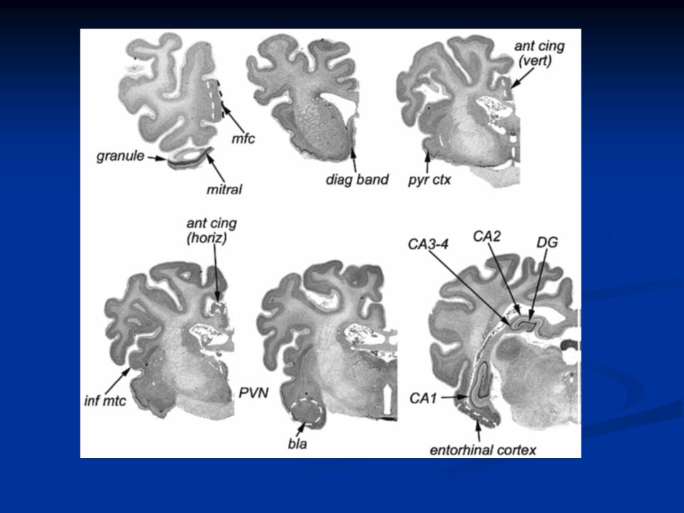
Results Animals with recognition displayed increased BDNF in the iTC, CA1 of the hippocampus, the diagonal band, basolateral amygdala and the anterior cingulate, medial frontal, entorhinal and pyriform cortices. Animals with recognition displayed increased BDNF in the iTC, CA1 of the hippocampus, the diagonal band, basolateral amygdala and the anterior cingulate, medial frontal, entorhinal and pyriform cortices. Activity in Visual AND Olfactory areas of cortex Activity in Visual AND Olfactory areas of cortex Suggests that reorganization of neural circuits underlying the visual recognition of lambs or the integration of olfactory/visual information is occurring even at this time (though accurate behavioral recognition at this stage can only be olfactory). Suggests that reorganization of neural circuits underlying the visual recognition of lambs or the integration of olfactory/visual information is occurring even at this time (though accurate behavioral recognition at this stage can only be olfactory).
. Suggests that reorganization of neural circuits underlying the visual recognition of lambs or the integration of olfactory/visual information is occurring even at this time (though accurate behavioral recognition at this stage can only be olfactory)..")
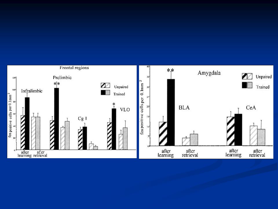
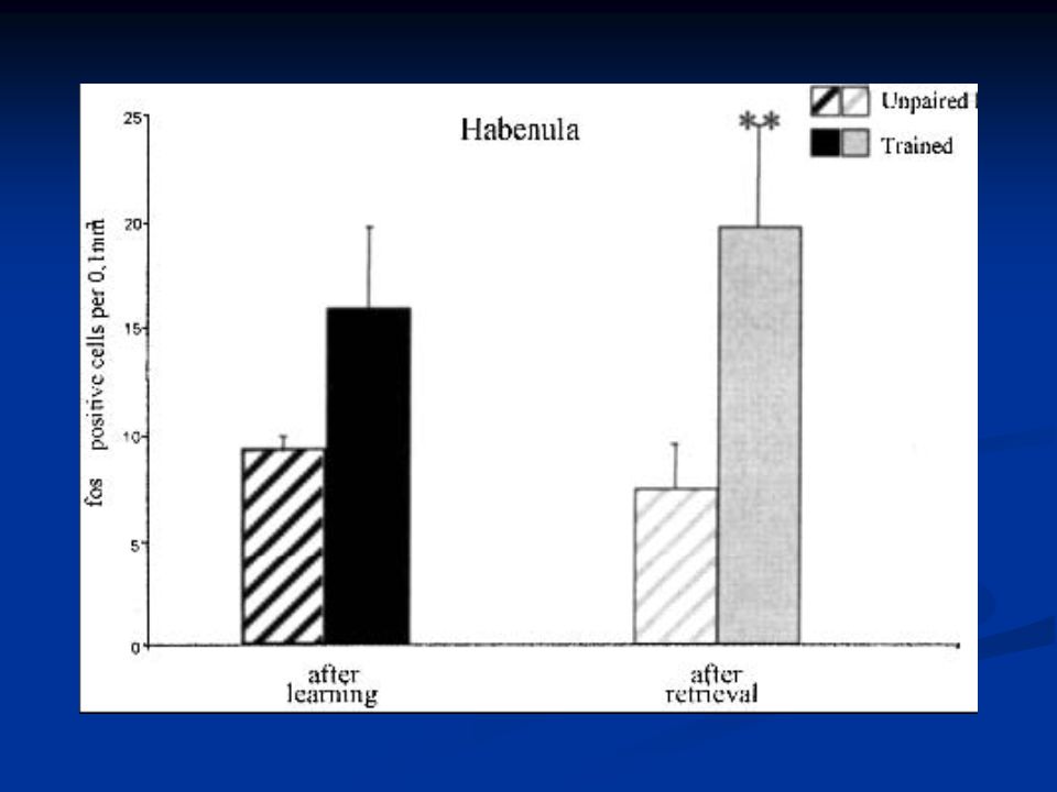
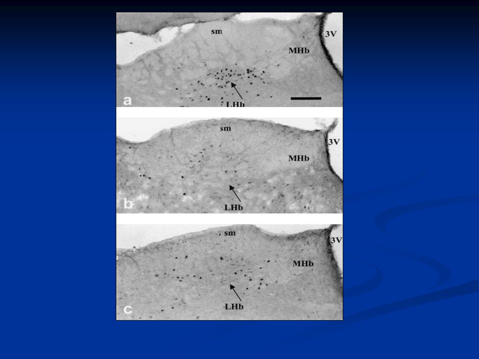
4
Solid Data? Experimental group engaged in 4.5 hours of maternal behavior not present in the control. Experimental group engaged in 4.5 hours of maternal behavior not present in the control. Stress in the control group? Stress in the control group?

5
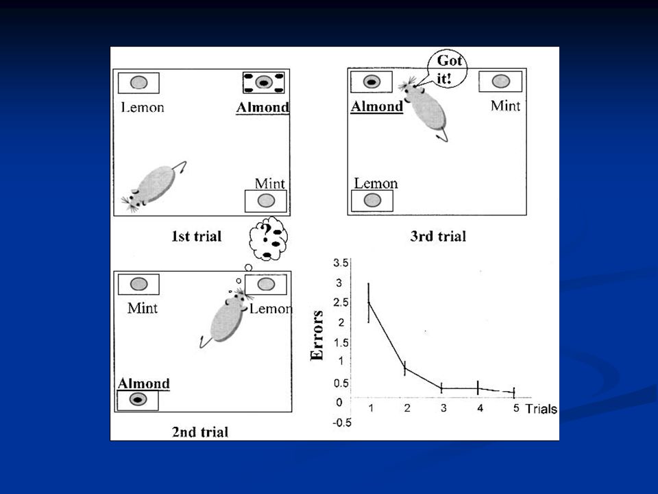
Mapping of Olfactory Memory Circuits: Region-Specific c-fos Activation After Odor- Reward Associative Learning of After its Retrieval Sophie Tronel and Susan J. Sara

6
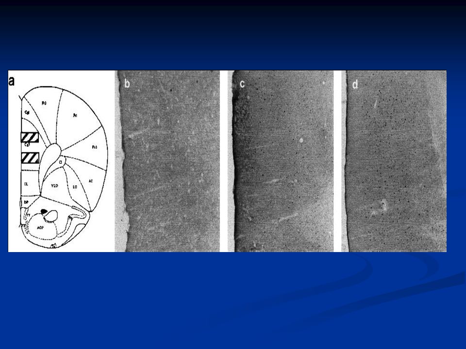
The Basics Looking for post-training activation of a network of closely related brain regions, particularly in the frontal cortex and the basolateral amygdala (BLA), that is specific to the learning of an odor reward sensation via immunoreaction to c-fos Looking for post-training activation of a network of closely related brain regions, particularly in the frontal cortex and the basolateral amygdala (BLA), that is specific to the learning of an odor reward sensation via immunoreaction to c-fos Retrieval does not differentially activate the same regions (amygdala is not active after retrieval whereas the lateral habenula shows activity). Retrieval does not differentially activate the same regions (amygdala is not active after retrieval whereas the lateral habenula shows activity).
..")
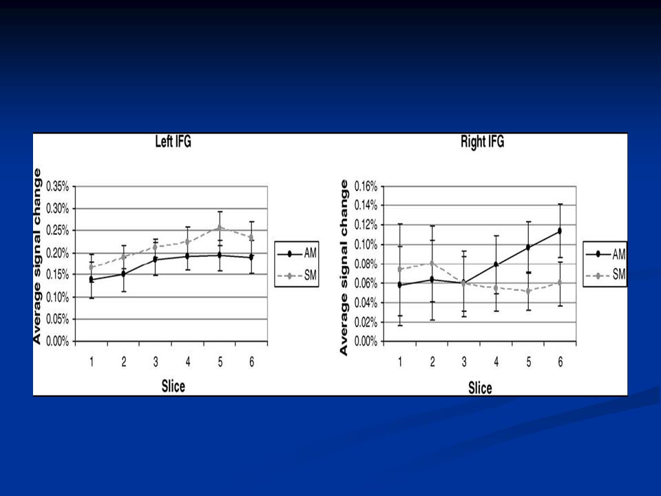
12
Co-activation of the amygdala, hippocampus and inferior frontal gyrus during autobiographical memory retrieval. Greenberg et al. Greenberg et al.

13
The Basics Functional MRI utilized to investigate the role of the medial temporal lobe and inferior frontal lobe regions in autobiographical recall. Functional MRI utilized to investigate the role of the medial temporal lobe and inferior frontal lobe regions in autobiographical recall. Autobiographical Memory: constructed of memories for personal life events. Autobiographical Memory: constructed of memories for personal life events. Tulving’s autonoetic conciousness Tulving’s autonoetic conciousness Dependent upon medial temporal and pre-frontal regions Dependent upon medial temporal and pre-frontal regions The three predictions... The three predictions...

14
fMRI Results Amygdala: Significant 3 way interaction of retrieval condition, hemisphere, and slice. Amygdala: Significant 3 way interaction of retrieval condition, hemisphere, and slice. Hippocampus: Greater activation during autobiographical retrieval and left hemisphere bias Hippocampus: Greater activation during autobiographical retrieval and left hemisphere bias IFG: same as amygdala plus a posterior gradient of activation in the right hemisphere during autobiograhical retrieval IFG: same as amygdala plus a posterior gradient of activation in the right hemisphere during autobiograhical retrieval

15
Group averaged hemodynamic responses

17
Schematic illustration of results from the correlational analysis for each retrieval condition.

18
Discussion Greater activity in left amygdala, hippocampus, and right IFG relative to semantic. Greater activity in left amygdala, hippocampus, and right IFG relative to semantic. Tighter coupling of activity across these three regions relative to semantic memory Tighter coupling of activity across these three regions relative to semantic memory Cohesion between medial temporal and ventral frontal lobes. Cohesion between medial temporal and ventral frontal lobes. IFG: contributes to both types of memory but activation prolonged in semantic IFG: contributes to both types of memory but activation prolonged in semantic

19
The neural origins of specific and general memory: the role of the fusiform cortex Rachel J. Garoff, Scott D. Slotnick, Daniel L. Schacter

20
The Basics Conducted an event related fMRI study to observe fusiform cortex activity Conducted an event related fMRI study to observe fusiform cortex activity Memory expression: specific/verbatim vs. non- specific recognition of the general sense Memory expression: specific/verbatim vs. non- specific recognition of the general sense Neural basis for non-specific recognition? Neural basis for non-specific recognition? The Fusiform Cortex: significance of activity. The Fusiform Cortex: significance of activity.

22
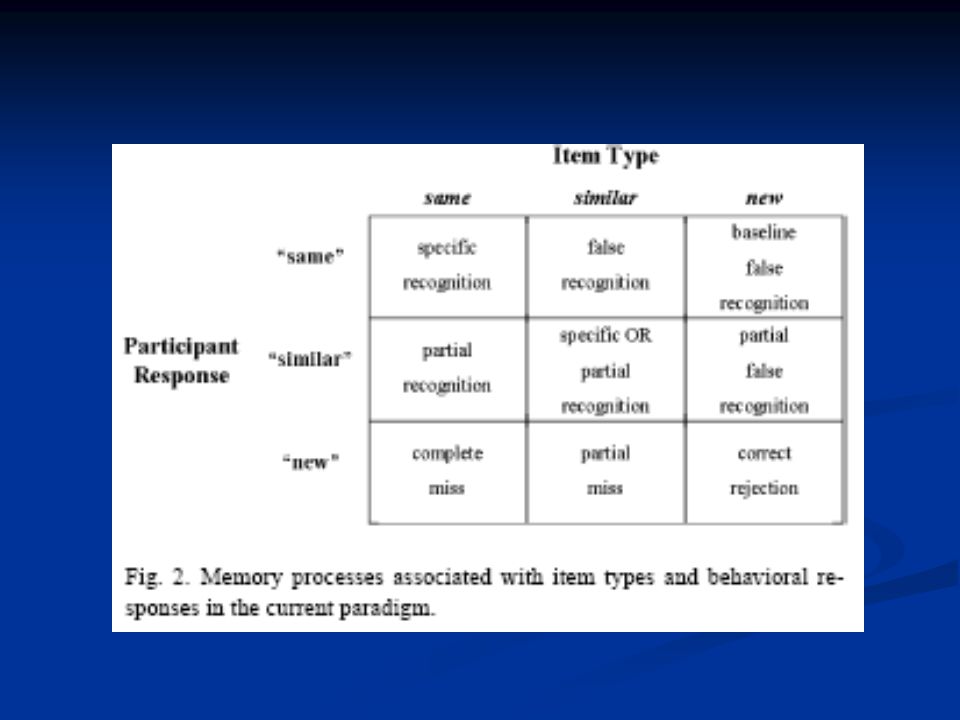
Behavioral responses associated with same similar and new items at recognition.

27
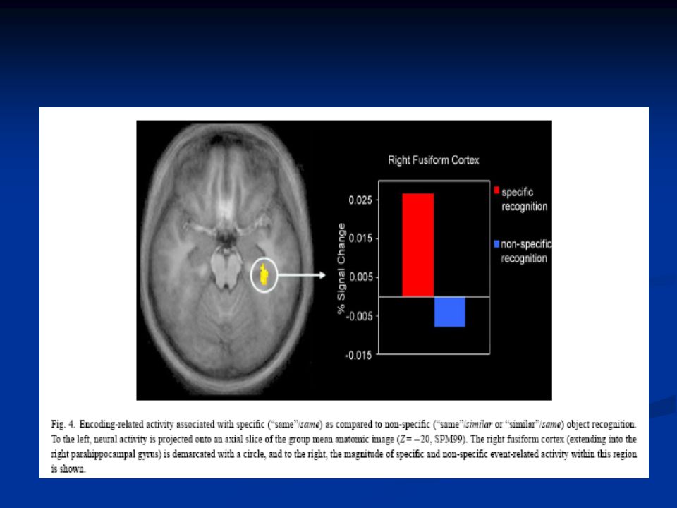
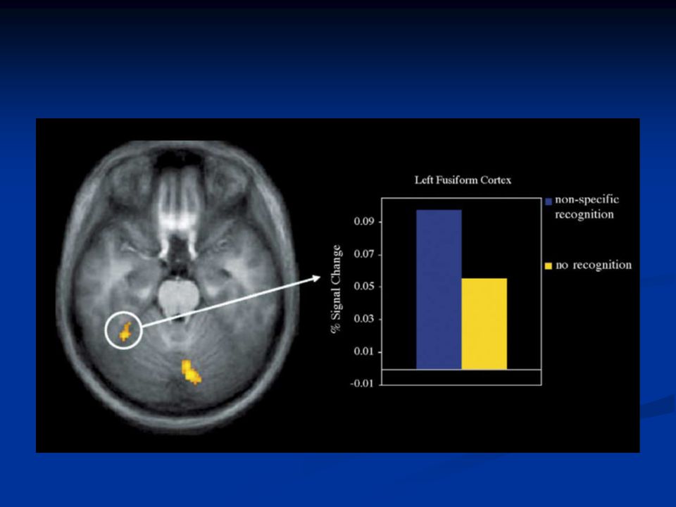
Results Activity in the right fusiform gyrus, extending into the parahippocampal gyrus, during encoding was preferentially associated with specific recognition. Activity in the right fusiform gyrus, extending into the parahippocampal gyrus, during encoding was preferentially associated with specific recognition. Significant difference between encoding related activity associated with non-specific recognition and baseline in left inferior frontal gyrus. Significant difference between encoding related activity associated with non-specific recognition and baseline in left inferior frontal gyrus.

Similar presentations


 Amir Shams Tabrizi.>")


 during Memory Encoding and Retrieval Investigators: C. Trott, D. Friedman, W. Ritter, M. Fabiani, J.G. Snodgrass.>")


. Visual imagery.>")
