Download presentation
Presentation is loading. Please wait.

1
Bilateral Clinical Anophthalmos Caleb Sawyer, MD Resident Jorge Corona, MD Faculty Advisor

2
Case Presentation 4 y/o Hispanic female with bilateral clinical anophthalmos 4 y/o Hispanic female with bilateral clinical anophthalmos Profound developmental delay Profound developmental delay Otherwise healthy Otherwise healthy Fitted with conformers 3 times since birth Fitted with conformers 3 times since birth

25
Definitions Anophthalmia = complete absence of an eye Anophthalmia = complete absence of an eye Clinical anophthalmia = small cystic remnant of globe is seen on pathologic examination Clinical anophthalmia = small cystic remnant of globe is seen on pathologic examination Microphthalmos = grossly visible, but small malformed globe Microphthalmos = grossly visible, but small malformed globe

26
Pathophysiology Pathophysiology Anophthalmia: Neuroectoderm of the primary optic vesicle fails to develop properly from the anterior neural plate of the neural tube during week 1-4 of embryological development. Anophthalmia: Neuroectoderm of the primary optic vesicle fails to develop properly from the anterior neural plate of the neural tube during week 1-4 of embryological development. Microphthalmia: development problem with optic vesicle in week 4 or later Microphthalmia: development problem with optic vesicle in week 4 or later

27
Morbidity Outgrowth of the globe drives growth and development of the bony orbit. Outgrowth of the globe drives growth and development of the bony orbit. Prevents fitting of prosthesis Prevents fitting of prosthesis Unilateral anophthalmos hemifacial hypoplasia Unilateral anophthalmos hemifacial hypoplasia Bilateral anophthalmos central hypoplasia Bilateral anophthalmos central hypoplasia

28
Associated Ocular Findings Orbital findings Orbital findings Small orbital rim and entrance Small orbital rim and entrance Reduced size of bony orbital cavity Reduced size of bony orbital cavity Extraocular muscles usually absent Extraocular muscles usually absent Lacrimal gland may be absent Lacrimal gland may be absent Small and maldeveloped optic foramen Small and maldeveloped optic foramen Eyelid findings Eyelid findings Foreshortening of the lids in all directions Foreshortening of the lids in all directions Absent or decreased levator function with decreased lid folds Absent or decreased levator function with decreased lid folds Contraction of orbicularis oculi muscle Contraction of orbicularis oculi muscle Shallow conjunctival fornix, especially inferiorly Shallow conjunctival fornix, especially inferiorly

29
Rare Condition U.S. congenital anophthalmos prevalence rate of 3 per 100,000. U.S. congenital anophthalmos prevalence rate of 3 per 100,000. Spanish Study of 1.1 million births: Spanish Study of 1.1 million births: 36/100,000 with eye malformations 36/100,000 with eye malformations 23/100,000 with anophthalmia/microphthalmia 23/100,000 with anophthalmia/microphthalmia No racial predilection No racial predilection No sex predilection No sex predilection

30
Causes Idiopathic/sporadic Idiopathic/sporadic Inherited as dominant, recessive, or sex-linked Inherited as dominant, recessive, or sex-linked Chromosome deletion in band 14q22-23 with associated polydactyly Chromosome deletion in band 14q22-23 with associated polydactyly Trisomy 13-15 Trisomy 13-15 Maternal infections or teratogenic exposure Maternal infections or teratogenic exposure 75% associated with syndromes 75% associated with syndromes

31
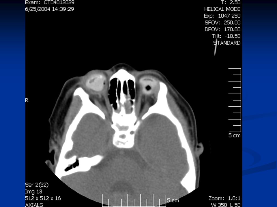
Role of head/orbit CT or MRI Look for extremely microphthalmic globe Look for extremely microphthalmic globe Bilateral anophthalmos Bilateral anophthalmos associated absence of the optic chiasm associated absence of the optic chiasm dysgenesis of the corpus callosum dysgenesis of the corpus callosum Unilateral anophthalmos may have severe craniofacial anomalies Unilateral anophthalmos may have severe craniofacial anomalies

38
3D CT Reconstruction

39
Treatment Options Progressive conformers Progressive conformers Easily extruded Easily extruded Balloon expanders Balloon expanders Easily extruded and require cooperation Easily extruded and require cooperation Progressive orbital implants Progressive orbital implants Require multiple surgeries Require multiple surgeries Hydrogel tissue expander implant Hydrogel tissue expander implant Good early results Good early results Late complications in scleral buckling Late complications in scleral buckling

42
Complications Significant cosmetic deformities if not treated early Significant cosmetic deformities if not treated early Fitted prostheses are completely immobile. Fitted prostheses are completely immobile. Shortened and immobile eyelids Shortened and immobile eyelids Even with treatment, results often are cosmetically disappointing. Even with treatment, results often are cosmetically disappointing.

43
Patient Education Treatment will be long and complicated Treatment will be long and complicated Multiple surgical treatments throughout a patient’s lifetime Multiple surgical treatments throughout a patient’s lifetime Consider genetic counseling in familial cases Consider genetic counseling in familial cases

46
References Bermejo E, Martinez-Frias ML. “Congenital eye malformations: clinical- epidemiological analysis of 1,124,654 consecutive births in Spain.”Am J Med Genet. 1998 Feb 17;75(5):497-504. Bermejo E, Martinez-Frias ML. “Congenital eye malformations: clinical- epidemiological analysis of 1,124,654 consecutive births in Spain.”Am J Med Genet. 1998 Feb 17;75(5):497-504. Chen D, Heher K. “Management of the anophthalmic socket in pediatric patients.” Curr Opin Ophthalmol. 2004 Oct;15(5):449-53. Review. Chen D, Heher K. “Management of the anophthalmic socket in pediatric patients.” Curr Opin Ophthalmol. 2004 Oct;15(5):449-53. Review. Dunaway DJ, David DJ. “Intraorbital tissue expansion in the management of congenital anophthalmos.” Br J Plast Surg. 1996 Dec;49(8):529-35. Dunaway DJ, David DJ. “Intraorbital tissue expansion in the management of congenital anophthalmos.” Br J Plast Surg. 1996 Dec;49(8):529-35. EMedicine http://www.emedicine.com/oph/topic572.htm EMedicine http://www.emedicine.com/oph/topic572.htmhttp://www.emedicine.com/oph/topic572.htm Mazzoli, Robert A; Raymond, William R IV; Ainbinder, Darryl J; Hansen, Elizabeth A. “Use of self-expanding, hydrophilic osmotic expanders (hydrogel) in the reconstruction of congenital clinical anophthalmos,” Current Opinion in Ophthalmology. 15(5):426-431, October 2004. Mazzoli, Robert A; Raymond, William R IV; Ainbinder, Darryl J; Hansen, Elizabeth A. “Use of self-expanding, hydrophilic osmotic expanders (hydrogel) in the reconstruction of congenital clinical anophthalmos,” Current Opinion in Ophthalmology. 15(5):426-431, October 2004. Yanoff: Ophthalmology, 2nd ed., 2004 Mosby, Inc. Yanoff: Ophthalmology, 2nd ed., 2004 Mosby, Inc. Young A, O'Keefe M. “Bilateral clinical anophthalmos.” Acta Ophthalmol Scand. 1997 Jun;75(3):308-10. Young A, O'Keefe M. “Bilateral clinical anophthalmos.” Acta Ophthalmol Scand. 1997 Jun;75(3):308-10.
: Bermejo E, Martinez-Frias ML. Congenital eye malformations: clinical- epidemiological analysis of 1,124,654 consecutive births in Spain. Am J Med Genet Feb 17;75(5): Chen D, Heher K. Management of the anophthalmic socket in pediatric patients. Curr Opin Ophthalmol Oct;15(5): Review. Chen D, Heher K. Management of the anophthalmic socket in pediatric patients. Curr Opin Ophthalmol Oct;15(5): Review. Dunaway DJ, David DJ. Intraorbital tissue expansion in the management of congenital anophthalmos. Br J Plast Surg Dec;49(8): Dunaway DJ, David DJ. Intraorbital tissue expansion in the management of congenital anophthalmos. Br J Plast Surg Dec;49(8): EMedicine EMedicine Mazzoli, Robert A; Raymond, William R IV; Ainbinder, Darryl J; Hansen, Elizabeth A. Use of self-expanding, hydrophilic osmotic expanders (hydrogel) in the reconstruction of congenital clinical anophthalmos, Current Opinion in Ophthalmology. 15(5): , October Mazzoli, Robert A; Raymond, William R IV; Ainbinder, Darryl J; Hansen, Elizabeth A. Use of self-expanding, hydrophilic osmotic expanders (hydrogel) in the reconstruction of congenital clinical anophthalmos, Current Opinion in Ophthalmology. 15(5): , October Yanoff: Ophthalmology, 2nd ed., 2004 Mosby, Inc. Yanoff: Ophthalmology, 2nd ed., 2004 Mosby, Inc. Young A, O Keefe M. Bilateral clinical anophthalmos. Acta Ophthalmol Scand Jun;75(3): Young A, O Keefe M. Bilateral clinical anophthalmos. Acta Ophthalmol Scand Jun;75(3):")
Similar presentations