Download presentation
Presentation is loading. Please wait.

1
Prepared by : Khansa’ Mohd Rashid Norhana Rahmat
BUPHTHALMOS Prepared by : Khansa’ Mohd Rashid Norhana Rahmat

2
Definition Increase in IOP
restricted to developmental abnormalities in trabecular meshwork in the angle of the eye in absence of other secondary causes or related systemic congenital anomalies (e.g: Sturge-Weber syndrome, Neurofibromatosis, Lowe syndrome etc.)
")
3
Incidence Only 1% from all glaucoma 75% Bilateral 65% Male
80% diagnoses in the first year of life Most cases appear to be sporadic However, an autosomal recessive pattern of transmission of the disease is evidenced

4
Etiology In all developmental glaucoma, there is maldevelopment of iridocorneal angle (goniodysgenesis) which includes:- Trabeculodysgenesis: maldevelopment of the trabecular meshwork Corneodysgenesis: maldevelopment of the cornea Iridodysgenesis: maldevelopment of the iris. The hallmark for developmental glaucoma is isolated trabeculodysgenesis.

5
Underdeveloped trabecular meshwork (Trabeculodysgenesis)
Normal trabecular meshwork Underdeveloped trabecular meshwork (Trabeculodysgenesis)
")
6
Symptoms Early: Late: Triad of photophobia, epiphora and blepharospasm
Due to corneal edema Late: Hazy cornea Large eye “ox eye” Defective vision

7
Signs Cornea Sclera Large & hazy Increased corneal diameter
Normal = 10.5 mm Buphthalmos = > 12 mm Haab’s striae: transverse, tears in Descemet’s membrane Sclera Bluish discoloration due to thinning of sclera

8
Anterior chamber Iris Pupil Lens
Deep: due to bulging of cornea & flattening of lens Iris Iridodenesis (tremulous) May have stromal hypoplasia Pupil Sluggish reaction Lens Phacodenesis (tremulous) Relatively small and displaced posteriorly Flattened due to stretching of the suspensory ligaments
 May have stromal hypoplasia. Pupil. Sluggish reaction. Lens. Phacodenesis (tremulous) Relatively small and displaced posteriorly. Flattened due to stretching of the suspensory ligaments.")
9
Fundus Refraction Glaucomatous optic cupping (late)
Axial myopia, but less than expected, due to:- Flat cornea Flat lens Deep anterior chamber and the lens is relatively become more posterior in position

10
Increased tear lake in the left eye
Increased corneal diameter of the left eye in comparison with the right eye

11
Haab's striae as seen on retroillumination
Hazy cornea Haab’s striae Haab’s striae Haab's striae as seen on retroillumination

12
Dense opacification of cornea in advanced cases
Stromal edema superiorly (arrow)
")
13
Diagnosis Positive family history Corneal assessment Tonometry
Corneal diameter more than 12 mm Haab’s striae Hazy cornea Tonometry Perkins handheld or Tono Pen is preferable in children IOP more than 21 mmHg IOP measured under general anaesthesia is less than real IOP due to:- Effect of general anaesthesia Low scleral rigidity Flat cornea

14
Gonioscopy Ophthalmoscopy Abnormal angle structures or membrane
Iris is directly inserted into trabecular meshwork (either flat insertion or concave insertion) Ophthalmoscopy Glaucomatous cupping Cup to disc ratio is more than 0.3
 Ophthalmoscopy. Glaucomatous cupping. Cup to disc ratio is more than 0.3.")
15
Perkins handheld tonometer
Tono Pen Examination of a 3 months infant with buphthalmos under general anesthesia using Schiotz tonometer. Perkins handheld tonometer

16
Treatment Treatment of buphthalmos is only surgical : Goniotomy
Trabeculotomy Trabeculectomy Drainage devices

17
Goniotomy Indication : How? Clear cornea with diameter less than 13mm.
Goniotomy knife introduced in the A.C under gonioscopic control to cut the abnormal mesodermal membrane at the angle in 2/5 of the circumference.

18
Result Complication Goniotomy can successfully treat congenital
glaucoma 80% to 90% of the time in cases the symptoms start when the child is 1 month to 2 years old. Complication The greatest complication after goniotomy is a return of high pressure in the eyes. If the pressure in the eye increases, the procedure may need to be repeated.

19
Goniotomy

20
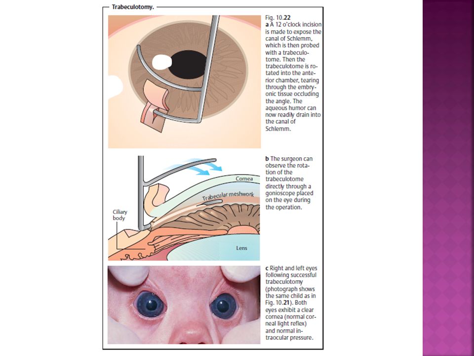
Trabeculotomy How? Indication
Corneal diameter more than 13mm or less than 13mm with hazy cornea. How? Trabeculotomy is performed from the scleral side at the limbus under the conjunctival flap. A fine wire-like instrument (trabeculotome) is inserted into Schlemm’s canal from an external incision and the trabecular meshwork is torn by rotating the instrument into the anterior chamber.
 is. inserted into Schlemm’s canal from an external. incision and the trabecular meshwork is torn. by rotating the instrument into the anterior. chamber.")
22
Result Trabeculotomy can successfully treat
congenital glaucoma 80% to 90% of the time in cases the symptoms start at the age of one month to two years old. Trabeculotomy is not as successful in children whose glaucoma was present at birth or began late in childhood.

23
Complication The most common problem after trabeculotomy is scarring of the new opening in the eye which prevents fluid from draining out of the eye. Other complications : Blurring of vision Bleeding in the eye Sudden, permanent loss of central vision Infection in the eye Malignant glaucoma is rare

24
Trabeculotome Trabeculotomy

25
Trabeculectomy Indication How ?
After failure of trabeculotomy or in advanced cases with corneal diameter more than 13mm. How ? Trabeculectomy is a surgical procedure involves removal of part of the trabeculum in the eye to relieve pressure caused by glaucoma.

26
Procedures of trabeculectomy

27
Trabeculectomy

28
Drainage devices such as Molteno- seton implant or Ahmad’s valve are indicated in difficult recurrent cases where conventional procedure above failed. Molteno implant

29
Thank you

Similar presentations


. Optic nerve head damage. Corresponding loss.>")


