Download presentation
Presentation is loading. Please wait.

1
Embryology of the eye The eye is formed from both ectoderm and mesenchyme. The neuroectoderm that is derived from the neural tube gives rise to (the retina, the fibers of the optic nerve, and the smooth muscle of the iris). The surface ectoderm on the side of the head forms( the corneal and conjunctival epithelium, the lens, and the lacrimal and tarsal glands). The mesenchyme forms( the corneal stroma, the sclera, the choroid, the iris, the ciliary musculature, part of the vitreous body, and the cells lining the anterior chamber).
. The surface ectoderm on the side of the head forms( the corneal and conjunctival epithelium, the lens, and the lacrimal and tarsal glands). The mesenchyme forms( the corneal stroma, the sclera, the choroid, the iris, the ciliary musculature, part of the vitreous body, and the cells lining the anterior chamber)..")
2
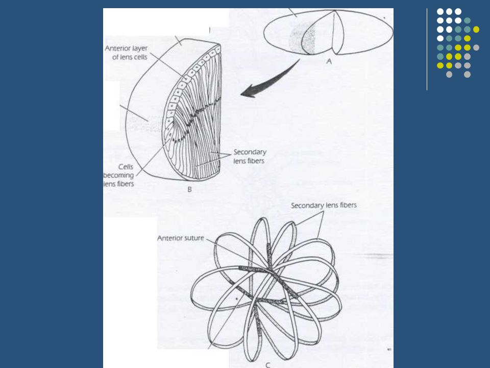
The eyeball The rudimentary eyeball develops as an ectodermal diverticulum from the lateral aspect of the forebrain. The diverticulum grows out laterally toward the side of the head, and the end becomes slightly dilated to form the optic vesicle, while the proximal portion becomes constricted to form the optic stalk. At the same time, a small area of surface ectoderm overlying the optic vesicle thickens to form the lens placode. The lens placode invaginates and sinks below the surface ectoderm to become the lens vesicle, the optic vesicle becomes invaginated to form the double-layered optic cup.

8
Orbit and Ocular Adnexa During infancy and childhood, the orbital volume increases, the shape of the orbital opening becomes less circular and more like a horizontal oval, the lacrimal fossa becomes more superficial, and the angle formed by the axes of the 2 orbits assumes a less divergent position. The palpebral fissure measures approximately 18 mm horizontall y and 8 mm vertically at birth and changes very little during the first year of life, but a rapid increase in palpebral fissure length occurs during the first decade. causing the round infant eye to acquire an elliptical adult shape.

9
Iris, Pupil, and Anterior Chamber Most iris color changes occur over the first 6 to 12 months of life. as pigment accumulates in the iris stroma and melanocytes, but iris pigmentation may continue.

10
The optic nerve The ganglion cells of the retina develop axons that converge to a point where the optic stalk leaves the posterior surface of the optic cup. This site will later become the optic disc. The axons now pass among the cells that form the inner layer of the stalk. Gradually, the inner layer encroaches on the cavity of the stalk until the inner and outer layers fuse.

14
Developmental Malformations and Anomalies Eyelids Epicanthic folds Signs- bilateral vertical folds of skin that extend from the upper or lower Lids towards the medial canthi;

15
Coloboma 1. Definition- partial or full-thickness Eyelid defect occurring when eyelid Development is incomplete. 2. Pathogenesis failure of migration of lid ectoderm to fuse the lid folds, or mechanical forces such as Amniotic bands. 3. Upper lid - at junction of the middle and inner thirds. 4. Lower lid - at junction of the middle and outer thirds

16
Globe Microphthalmos 1. Definition- total axial length at least2 standard deviations below age-similar controls usually unilateral. 2. Pathogenesis non-specific growth Failure in response to a variety of Prenatal insults. 3. Classification a. Simple- isolated. b. Complex(colobomatous\

17
Anophthalmos 1. Pathogenesis either acomplete Failure of budding of the optic vesicle or early developmental arrest. 2. Simple- associated with absence of Extraocular muscles,short conjunctival sac. and microbleoharon. 3. Anophthalmos with cyst (congenital cystic eyeball) - globe is replaced by a cyst
 - globe is replaced by a cyst.")
18
Iris, Pupil, and Anterior Chamber Most iris color changes occur over the first 6 to 12 months of li fe. as pigment accumulates in the iris stroma and melanocytes, but iris pigmentation may continue. Compared with

22
References 1. Lecture notes in ophthalmology 2. Parson’s disease of the eye

Similar presentations


: This is a cross section through the forebrain of a human embryo. Identify the DIENCEPHALON, OPTIC STALK,>")


>")


 separated by the palpebral fissue Eyelashes Tarsal glands Lacrimal apparatus Vision Accessory structures.>")


