Download presentation
Presentation is loading. Please wait.

1
RSNA 2010 Talking Points NCI had two booths: The caBIG exhibit which comprised of seven workstations showcasing the NBIA, AIM, Virtual PACS, AVT, XIP, caIntegrator and Clinical Trials Suite – this was in the Learning Center (research, teaching and continuing ed focused). NCI Office of Communications had a “standard” booth in one of the main exhibit halls providing NCI (including caBIG) communications material. We crossed advertised each others’ booths – NCI OC wants to coordinate with us for next year’s RSNA We also got advertised in the main NCI calendar Eliot being one of the founders and leaders of RSNA helps us get good spot in both the Learning Center as well as in the RSNA paper and electronic Meeting Program. We had several people express a serious interest in wanting to get more information about how they could use one or more of the caBIG® products in their research or clinical trials, both in the US and internationally. Kelly and I are following up with each one and adding to our Imaging deployment tracking spreadsheet. There were also about 20 caBIG related presentations from members of the caBIG® Imaging community, including Dr. Rubin’s feature presentation on AIM and commercial PACS vendors in the Reading Room of the Future exhibit as well as Dr. Siegel’s RSNA Keynote on caBIG® Imaging (in this deck).
 communications material. We crossed advertised each others’ booths – NCI OC wants to coordinate with us for next year’s RSNA We also got advertised in the main NCI calendar Eliot being one of the founders and leaders of RSNA helps us get good spot in both the Learning Center as well as in the RSNA paper and electronic Meeting Program. We had several people express a serious interest in wanting to get more information about how they could use one or more of the caBIG® products in their research or clinical trials, both in the US and internationally. Kelly and I are following up with each one and adding to our Imaging deployment tracking spreadsheet. There were also about 20 caBIG related presentations from members of the caBIG® Imaging community, including Dr. Rubin’s feature presentation on AIM and commercial PACS vendors in the Reading Room of the Future exhibit as well as Dr. Siegel’s RSNA Keynote on caBIG® Imaging (in this deck)..")
2
Eliot’s RSNA Keynote on caBIG® Imaging

3
RSNA 2010 NCI caBIG® Update Eliot Siegel, M.D. University of Maryland School of Medicine Department of Diagnostic Radiology

4
Introduction One of the major original goals of caBIG® was to determine out how to create a system that would enable extraction of data for research or clinical decision support that would: Allow access to a variety of types and sources of data including genomic, proteomic, clinical, lab, demographic, and diagnostic imaging Take advantage of analytic potential of grid computing to combine and cross-reference these for analysis for research and clinical care

5
The caBIG® Imaging workspace has worked to build basic tools toward this goal and the TCGA Imaging workspace project represents an example of the potential for caBIG® to have a major impact on the way in which data are shared, research conducted, and patient care is provided

6
Introduction to the caBIG® in Vivo Imaging Workspace caBIG® in vivo Imaging workspace established April 2005 a little more than a year after the establishment of the other caBIG® workspaces NCI funded effort by far the biggest and most productive effort in imaging informatics today Subject matter experts from around country with representation from major Universities, informatics experts, industry, NCI

7
Review of Relevant Workspace Projects XIP, AIM, Middleware, NBIA

8
Rapid application development environment for diagnostic imaging tasks that researchers and others use to create targeted workflows customized for specific projects

10
Annotations and Image Markup (AIM) Being Adopted by Increasing Number of Research and Commercial Systems Represents a “standard” means of adding information/knowledge to an image in a research or clinical environment to allow easy and automated search for image “content”
 Being Adopted by Increasing Number of Research and Commercial Systems Represents a standard means of adding information/knowledge to an image in a research or clinical environment to allow easy and automated search for image content")
11
Imaging Middleware (including GridCAD and Virtual PACS) Grid computing has received surprisingly little attention. One application has been to allow multiple computers to work in parallel on a single task such as CAD detection of lung nodules or to give multiple opinions using multiple algorithms Middleware software is used to create interoperability between DICOM devices and the caGRID which uses a service oriented architecture

12
NBIA: National Cancer Imaging Archive Initially designed as repository for LIDC and RIDER CT lung nodule studies Expanded to include multiple additional types of image collections with role based security to share with public or a selected group or to support ongoing clinical trials or other reader studies Open source and free Meant to be “federated” to create virtual database across multiple instances of NCIA software

13
NBIA Demo: Home Page

14
NBIA Demo: Using the Search Criteria

15
NBIA PC DICOM Viewer: Cedara i- Response

16
The Cancer Genome Atlas (TCGA) In Vivo Imaging Project Initial Phase
 In Vivo Imaging Project Initial Phase")
17
TCGA The Cancer Genome Atlas Collaboration between National Human Genome Research Institute and NCI The Cancer Genome Atlas (TCGA) is a comprehensive and coordinated effort to accelerate our understanding of the genetics of cancer using innovative genome analysis technologies.
 is a comprehensive and coordinated effort to accelerate our understanding of the genetics of cancer using innovative genome analysis technologies.")
18
The Cancer Genome Atlas TCGA researchers have identified four distinct molecular subtypes of glioblastoma multiforme (GBM), and demonstrated that response to aggressive chemotherapy and radiation differed by subtype These findings, reported in the January 19 issue of Cancer Cell, may result in more personalized approaches to treating groups of GBM patients based on their genetic alterations
, and demonstrated that response to aggressive chemotherapy and radiation differed by subtype These findings, reported in the January 19 issue of Cancer Cell, may result in more personalized approaches to treating groups of GBM patients based on their genetic alterations")
19
TCGA Second Study in Cancer Cell Another study published in April by The Cancer Genome Atlas Research Network also in Cancer Cell used epigenomic profiling Maps specific chemical changes or 'marks' to different areas of the genome, to reveal a new subtype of Glioblastoma Multiforme (GBM) Most patients with GBM survive only 12-15 months after their initial diagnosis However, patients with this specific subtype, called Glioma CpG Island Methylator Phenotype (G-CIMP), have a median survival of three years
 Most patients with GBM survive only months after their initial diagnosis However, patients with this specific subtype, called Glioma CpG Island Methylator Phenotype (G-CIMP), have a median survival of three years")
20
Goals of TCGA Imaging Workspace Project Investigate the added value of highly structured interpretation and quantification of MRI images of the TCGA dataset using AIM Determine the correlation between MRI imaging and genotypic information and response to therapy and prognosis Revise Cell article to include impact of MRI data Determine the potential for these tools in routine clinical practice

21
Feature Set – Controlled Vocabulary 20 features clustered by categories. Lesion Location Morphology of Lesion Substance Morphology of Lesion Margin Alterations in Vicinity of Lesion Extent of Resection Goal is to capture imaging features of entire tumor and imaging features of resection specimen.

22
Examples Non-standardized Features May correspond to Angiogenesis, Oxygenation, Apoptosis, Cellularity Infiltration Margination Edema Non-enhancing tumor. Enhancement Irregular Nodular Indistinct Infiltrative Necrosis Physiologic Diffusion Perfusion

23
Well marginated Non-enhancing

24
Infiltrative & Necrotic Type

25
Nodular Predominantly Non-enhancing

26
Osirix / iPad Assistant Demo Three Workstations (Osirix [Mac], Clear Canvas [PC] and XIP Purpose Built Were Modified to Retrieve TCGA Images from NBIA Database and Use Standardized Template and Save Interpretation and Quantitative Measurements to AIM Data Service on caGRID Osirix / iPad Workstation 1 Clear Canvas Workstation XIP / AVT Workstation -
![Osirix / iPad Assistant Demo Three Workstations (Osirix [Mac], Clear Canvas [PC] and XIP Purpose Built Were Modified to Retrieve TCGA Images from NBIA Database and Use Standardized Template and Save Interpretation and Quantitative Measurements to AIM Data Service on caGRID Osirix / iPad Workstation 1 Clear Canvas Workstation XIP / AVT Workstation -](http://images.slideplayer.com/16/5184305/slides/slide_26.jpg "Osirix / iPad Assistant Demo Three Workstations (Osirix [Mac], Clear Canvas [PC] and XIP Purpose Built Were Modified to Retrieve TCGA Images from NBIA Database and Use Standardized Template and Save Interpretation and Quantitative Measurements to AIM Data Service on caGRID Osirix / iPad Workstation 1 Clear Canvas Workstation XIP / AVT Workstation -")
27
Purpose of TCGA Radiology Phase II Project Project Goals Utilize multiple CBIIT/caBIG® technologies together to create a practical system to capture diagnostic imaging “knowledge” in a structured, standardized manner and to allow for the integration with genomic and clinical data Have at least two radiologists interpret the TCGA MRI brain images associated with the Cancer Cell article Utilize caBIG tools to create a repository of the qualitative and quantitative information associated with the analysis of the images Utilize caBIG tools to perform cross database comparisons for research purposes Demonstrate potential of caBIG tools to assist in clinical decision support

28
Achievements: Radiology Reading TCGA cases in NBIA have been read by at least two funded neuro-radiologists: Images retrieved from NBIA at CBIIT New markups created on Workstation and saved to the AIME. Existing markups and annotation retrieved from AIM Data Service at Emory (AIME). A radiologist fills out AIM based reporting template. New annotation data is saved on AIME.
. A radiologist fills out AIM based reporting template. New annotation data is saved on AIME..")
29
Achievements: TCGA Cancer Cell Data Service Because the existing TCGA Grid Data Service is not currently available, we created our own grid data service to host genomic and clinical data from the 12/09 Cancer Cell article. Built a data model for Cancer Cell genomic and clinical data Used caCORE SDK 4.2 to quickly generate an application from this model Used caGrid Introduce SDK to create a Grid data service from the SDK model Deployed data service at Emory Create scientific queries for caB2B Successfully queried 3 disparate caGrid data services (AIM, NBIA, TCGA Cancer Cell) with caB2B Documented insights gained from the process of setting up our own data and grid service
 with caB2B Documented insights gained from the process of setting up our own data and grid service.")
30
Achievements: caB2B Query of NBIA, AIM and TCGA CC Data Services Successfully queried 3 disparate caGrid data services (AIM, NBIA, TCGA Cancer Cell) with caB2B
 with caB2B")
31
Achievements: Additional Analysis with caIntegrator2 caIntegrator2 team added a feature to support integration with AIM grid data service to load annotations caIntegrator2 Study: Combine TCGA Cancer Cell data (from CSV), AIM data from grid service, and images from NBIA production grid service. Created scientifically relevant queries based on image observations and clinical data Generated Kaplan-Meier plots of survival based on certain observations and genomic subtypes

32
Achievements: Preliminary Scientific Findings Survival of patients who had larger thickness of enhancement tumors with hemorrhage was significantly for shorter than those who did not. Survival of patients who had tumors that crossed midline was significantly for shorter than those who did not. Survival of patients with greater thickness of enhancement (who appear to have had tumors with a thicker “rim”) was significantly for shorter than those who had less.
 was significantly for shorter than those who had less..")
33
Opportunities to Further Deploy TCGA Related Imaging and Life Sciences Technologies Cancer Imaging Program: - Continued TCGA Genotype/Phenotype Research with CBIIT, NIH Clinical Center - Quantitative Imaging Network Program - Cancer UK Research Program - All Ireland Initiative Program Radiation Research Program - RTOG 0522 Study NIAMS Osteoarthritis Study - Annotation of radiology data - Integrating of radiology data with other OAI data types

34
Future Plans Provide software to NCI clinical cancer centers for their own clinical trials/research studies involving diagnostic imaging Extend work from in-vivo Imaging to pathology

35
Future Plans for TCGA Imaging Project Include higher order analysis, such as quantitative diffusion imaging and perfusion imaging metrics, that could be more sensitive predictors of disease severity, candidates for effective therapy, and expected outcomes combining human with semi- automated and automated analysis of images

36
Future Plans for TCGA Project Ultimately would like to develop a “service” that has capability to provide immediate feedback for radiologist or oncologist on patient survival, patient treatment, etc. Incorporate genomic and other data display during radiology interpretation at a workstation

37
Providing Radiology Observation Data for Genotypic/Phenotypic Analysis in Support of TCGA caIntegrator2 Demo

38
caIntegrator 2: Home

39
caIntegrator 2: Imaging Observations

40
caIntegrator 2: Query Criteria

41
caIntegrator 2: KM Plot

42
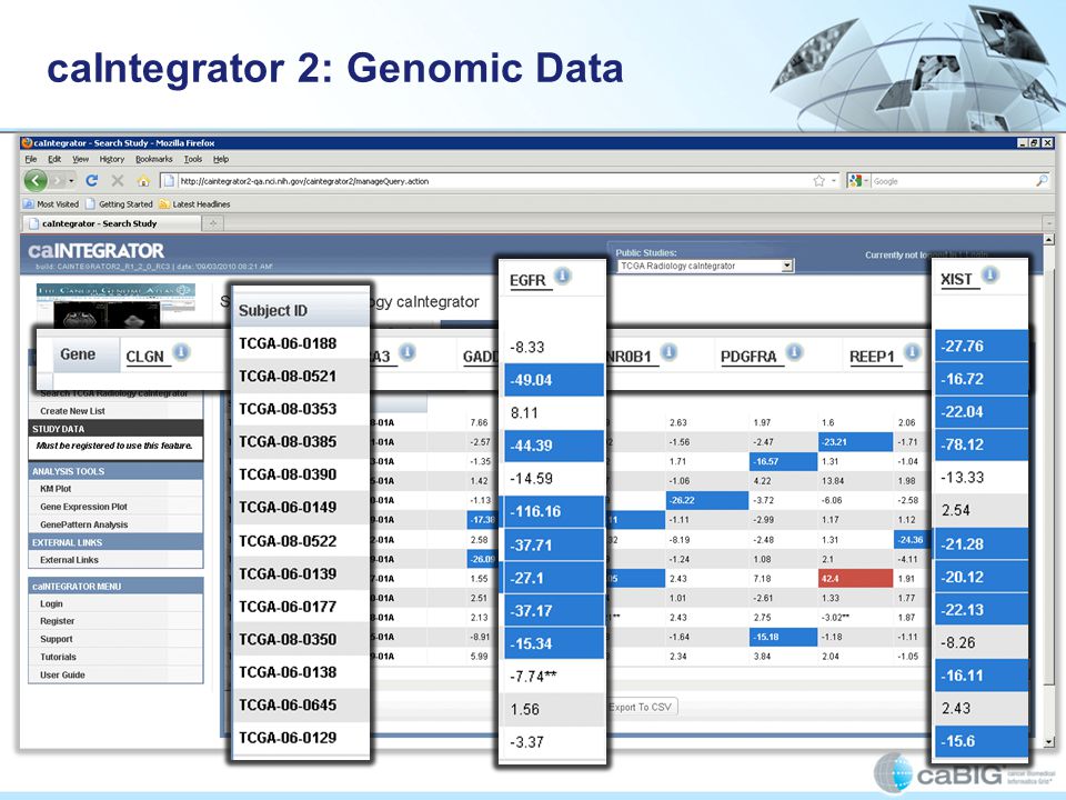
caIntegrator 2: Genomic Data

44
Patient Decision Support

46
Personalized Survival Curve Compared to All Patients

47
Thank you Adam Flanders CBITT Government Sponsors: Ed Helton Robert Shirley Mervi Heiskanen Juli Klemm In collaboration with: NCI Cancer Imaging Program Carl Jaffe John Freyman Justin Kirby Supported by: 5AM Booz Allen Hamilton Buckler Biomedical, LLC. Capability Plus Solutions ClearCanvas, Inc. Emory University Northwestern University SAIC Stanford University Thomas Jefferson University University of Maryland University of Virginia

Similar presentations


 v.4: Updating a Cancer Research Standard Ann Setser 1, Ranjana Srivastava 2, Lawrence Wright 1,>")


![Wrapup. NHGRI strategic plan What does the NIH think genomics should be for the next 10 years? [Nature, Feb. 2011]](/14/4306833/big_thumb.jpg "Wrapup. NHGRI strategic plan What does the NIH think genomics should be for the next 10 years? [Nature, Feb. 2011]>")
: Background & Introduction Timothy Hays, Ph.D. Project Manager, Knowledge Management for Disease Coding.>")


 Project Update Building queries one question mark at a time March, 2009.>")


 is to improve the role of quantitative imaging for clinical decision making in oncology.>")

