Download presentation
Presentation is loading. Please wait.

1
Determination of Sites of CYP1B1 Mutations in Aligned Sequences of Cytochrome P450 Family Members and 3D-Structural Model by: Betsabeh Khoramian Tusi

2
EYE

3
The human eye is filled with a liquid known as the aqueous humor. The aqueous humor is produced by the ciliary body, a stucture which lies behind the iris. This liquid circulates in the chambers of the eye, then gets drained away via a pathway which lies at the angle between the cornea and the iris (the drainage angle). Anything that block this exit path will result in liquid accumulation and produce high intraocular pressure (IOP), which, if left untreated, leads to optic-nerve damage and ultimately blindness. This condition is called Glaucoma.
. Anything that block this exit path will result in liquid accumulation and produce high intraocular pressure (IOP), which, if left untreated, leads to optic-nerve damage and ultimately blindness. This condition is called Glaucoma..")
4
Primary Congenital Glaucoma (PCG) PCG is a form of glaucoma, manifested during the neonatal or infantile period (prior to the age of 3). PCG is likely to be due to maldevelopment of the anterior chamber angle of the eye, thus interfering with the aqueous humor outflow. The incidence of this disease in the Middle East is approximately four times greater than in Western countries.

5
CYP1B1 Three loci for Primary Congenital Glaucoma have been identified through linkage analysis in affected families: GLC3A, GLC3B, GLC3C. (Locus is really,an approximate address of a gene but we don’t know the exact gene) Only the gene associated with GLC3A has been found: CYP1B1. It codes for the protein cytochrome P4501B1. Mutations in CYP1B1 account for approximately 1/3 of PCG cases in populations studied (very variable!!).
 Only the gene associated with GLC3A has been found: CYP1B1. It codes for the protein cytochrome P4501B1. Mutations in CYP1B1 account for approximately 1/3 of PCG cases in populations studied (very variable!!)..")
6
Why do mutations in this gene cause maldevelopment of the eye? Really, we don’t know !!!

7
But we do know: CYP1B1 is expressed in many tissues. CYP1B1 is a member of a superfamily of genes (many related genes) whose protein products are all oxygenases. Oxygenases have roles in detoxification reactions. Mutations in CYP1B1occur in many cancers. WE DO NOT KNOW WHY CYP1B1 DAMAGE SHOULD HAVE SPECIFIC EFFECTS IN THE EYE!!!
 whose protein products are all oxygenases. Oxygenases have roles in detoxification reactions. Mutations in CYP1B1occur in many cancers. WE DO NOT KNOW WHY CYP1B1 DAMAGE SHOULD HAVE SPECIFIC EFFECTS IN THE EYE!!!.")
8
CYP1B1 gene structure CYP1B1 has three exons,that only two of them are translated in to protein.(exon no.2& the first part of the exon no.3)
")
9
OUR RESEARCH 1.We have collected blood samples and isolated DNA from 100 Iranian PCG patients. 2.We sequenced the three exons and neighboring intronic sequences of the gene in 50 (exon1) to 70 (exon 3) of the patients. 3.We have identified 15 CYP1B1 mutations, five of which have not been previously reported: E173K, D291G, G329V, R368C, I399V. 4.We used bioinformatics tools and did protein homology modeling of the CYP1B1 protein using information at databases and located our mutations and other known CYP1B1 mutations in derived 3D-structure.
 to 70 (exon 3) of the patients. 3.We have identified 15 CYP1B1 mutations, five of which have not been previously reported: E173K, D291G, G329V, R368C, I399V. 4.We used bioinformatics tools and did protein homology modeling of the CYP1B1 protein using information at databases and located our mutations and other known CYP1B1 mutations in derived 3D-structure..")
10
The cytochrome P450 superfamily in the human genome has 22 members. The amino acid sequences of these 22 cytochromes were obtained from SwissProt and Genebank. We did multiple sequence alignment using the ClustalW program.

11
We determined the positions of putative disease mutations (including new mutations found in the Iranian population) in the multiple sequence alignment.
 in the multiple sequence alignment.")
12
Mutation spectrum: Only nucleotide substitution mutations were analyzed. i.e. deletions and insertions which cause frameshifts and are likely to be damaging wherever they occur, are not considered.

13
NEXT: Construction of a three dimensional model of CYP1B1 gene product by homology modeling. (homology modeling using the Swiss-Model Program)
.")
14
Protocol for Modeling 1. The complete protein sequence of CYP1B1 was screened against the PDB structure database in order to identify the template structures appropriate for modeling. 2. From a series of templates, we selected those of four proteins with the highest homology. The four proteins were: 1po5,1suo,1nr6 and 1n6b.(all of these proteins are kinds of cytochromeP450) 3. We entered our cytochrome P450 amino acid sequence and browsed the known 3D structures of the selected homologous proteins, and then asked SWISS-Model to construct a 3D- model for cytochrome P450.
 3. We entered our cytochrome P450 amino acid sequence and browsed the known 3D structures of the selected homologous proteins, and then asked SWISS-Model to construct a 3D- model for cytochrome P450..")
15
Results

16
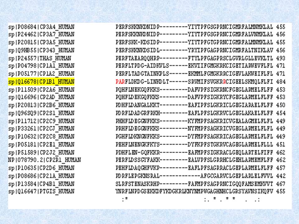
Mutation Sites Cytochrome P450 proteins all have approximately 500 aa’s; CYP1B1 has 543 aa’s. As the 22 cytochrome P450 proteins aligned are not very closely related, only 25 sites were well conserved and only 9 of these were strictly (100%) conserved. The 9 strictly conserved residues all lie within a 122 aa region. 12 of the 28 mutations analyzed lie within the highly conserved 122 aa region; five of these mutations lie at or adjacent to the strictly conserved positions (including one of the new Iranian mutations).
 conserved. The 9 strictly conserved residues all lie within a 122 aa region. 12 of the 28 mutations analyzed lie within the highly conserved 122 aa region; five of these mutations lie at or adjacent to the strictly conserved positions (including one of the new Iranian mutations)..")
17
Multiple sequence alignment for 22 different members of the cytochrome P450 superfamily

19
LOGO NB: Although the amino acids at many sites are not highly conserved, but the chemical properties of amino acids at most sites are well conserved e.g. hydrophobicity, hydrophilicity as shown here.

20
Three-dimensional model of the CYP1B1 protein

21
See rotating model of 3-D structure

22
Site of mutations in 3D-model of CYP1B1 protein

23
NOTE: 1.Sites of 12 mutations in conserved sequence region are within heme binding site. 2. Many of the remaining mutations are on part of surface region of the protein, suggesting this region has an important role----- perhaps interaction with other proteins. 3. Many of our novel mutations lie within the surface region.

24
Secondary structure derived from 3-D model of cytochrome p450 1B1 protein using DSSPSecondary structure (Definition of secondary structure of proteins) Structural alignment NB: Structural alignment shows that structural elements such as alpha helices and beta strands are more conserved than residue types and residue properties.
 Structural alignment NB: Structural alignment shows that structural elements such as alpha helices and beta strands are more conserved than residue types and residue properties.")
25
The End Thanks a lot for your attention

Similar presentations


.>")

 The Mechanics of Alignments.>")


 Sequence Information>")


