Download presentation
Presentation is loading. Please wait.

1
Muscle Physiology and Anatomy “The Last Chapter”!

2
ASSIGNMENT Read Pages 7:152-170 Answer: Content Review – page 191 Questions: 1-11

3
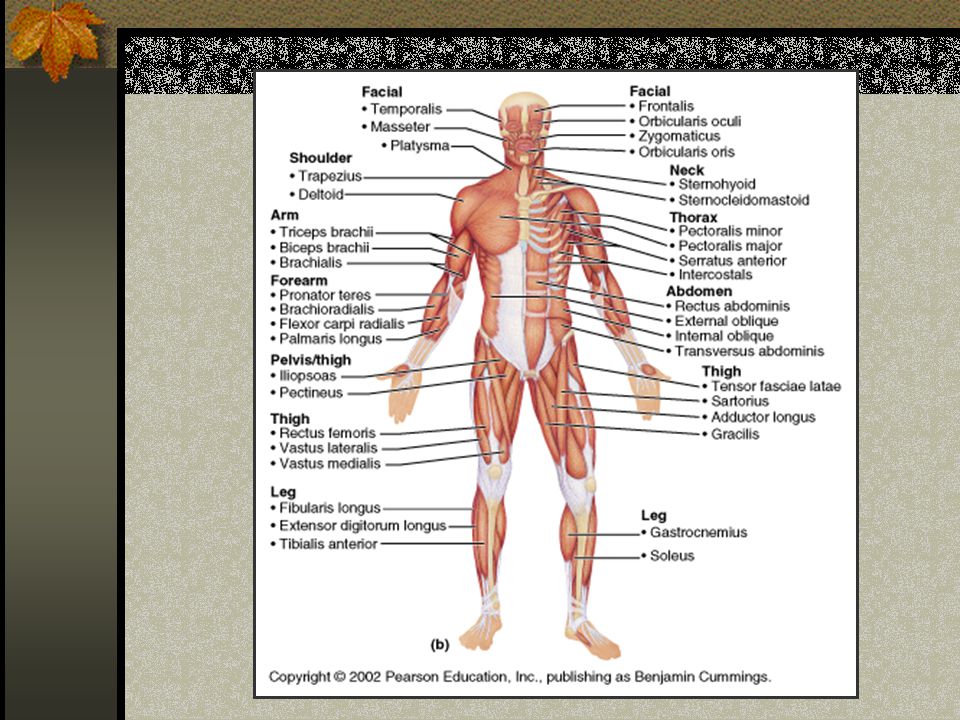
“Gross” Anatomy Requirements Identify 20 Major Muscles For Exam: Know 3 Muscles (of the 20 major muscles) Origin: Most stable attachment Insertion: Most mobile attachment Action: What movement(s) Exercise specific for that muscle
 Origin: Most stable attachment Insertion: Most mobile attachment Action: What movement(s) Exercise specific for that muscle")
5
Example: Biceps Brachii (p. 181): Origin: Scapula, Superior to glenoid fossa, and coracoid process Insertion: Radial Tuberosity Action: Flex and supinate arm and forearm Exercise: Curls
: Origin: Scapula, Superior to glenoid fossa, and coracoid process Insertion: Radial Tuberosity Action: Flex and supinate arm and forearm Exercise: Curls.")
6
Other “Gross” Terms Prime Mover: The major muscle in a movement Synergist: “Helpers” Antagonists: “Opposers” Fixators: Stabilizing the proximal joint

7
Functions: Muscular System Movement Posture Respiration Circulation Produce Heat Communication

8
Characteristics of Skeletal Muscle Tissue Contractility: Shorten with force Excitability: Respond to stimulus Extensibility: Limited stretch”ability” Elasticity: Recoil to resting length

9
Three Types of Muscle Tissue Smooth: Found in walls of hollow organs, blood vessels and glands Cardiac: Heart muscle Skeletal: Attached to bone * Compared by striations, shape, control, nuclei and function

10
Striations: “Stripes” Skeletal: YES Smooth: NO Cardiac: YES

11
Shape and Nucleus Skeletal: Long cylinder “fiber” Smooth: “spindle shape” Cardiac: Branched Multiple, peripheral Single, central

12
Control and Autorhythmicity Skeletal: Voluntary- NO Smooth: Involuntary- YES Cardiac: Involuntary-YES

13
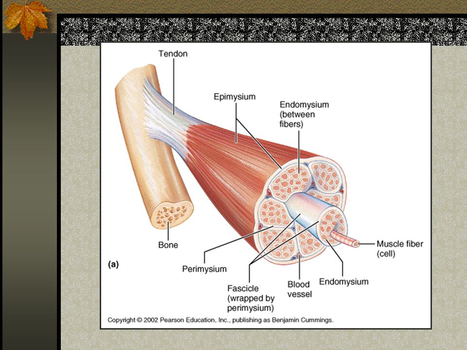
Muscle Structure Connective Tissue: Epimysium (Fascia) wraps muscle, Perimysium wraps fascicles, and endomysium wraps muscle fibers Bundles of Bundles: Muscle ->fascicles->fibers->myofibrils-> myofilaments
 wraps muscle, Perimysium wraps fascicles, and endomysium wraps muscle fibers Bundles of Bundles: Muscle ->fascicles->fibers->myofibrils-> myofilaments")
15
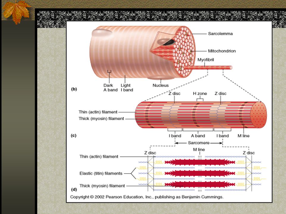
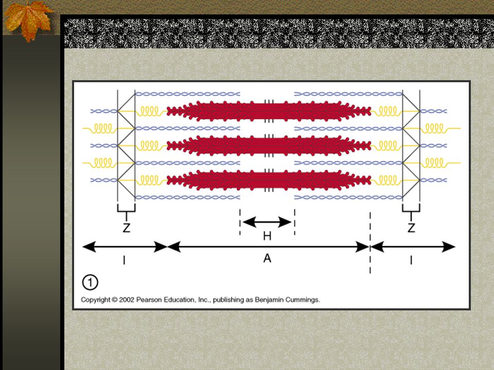
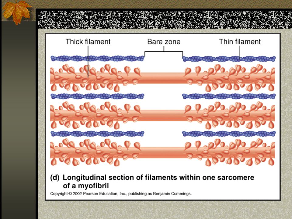
Muscle Cell (Fiber) Structure Sarcomeres: The real contractile elements of muscle cells Myofilaments: Thick (myosin) and Thin (actin, troponin, tropomyosin) overlap to create the “striations” visible in the microscope
 Structure Sarcomeres: The real contractile elements of muscle cells Myofilaments: Thick (myosin) and Thin (actin, troponin, tropomyosin) overlap to create the striations visible in the microscope")
19
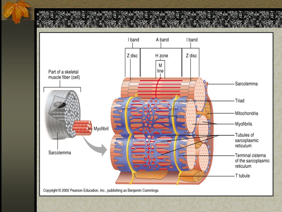
Muscle Cell Structure Nuclei: Multiple (many fused cells), peripheral Mitochondria: Many, near sarcomeres Transverse Tubules, Terminal Cisternae: Internal extension of cell membrane – Action Potential transmission
, peripheral Mitochondria: Many, near sarcomeres Transverse Tubules, Terminal Cisternae: Internal extension of cell membrane – Action Potential transmission")
21
Excitable Tissues Nervous and Muscular Respond to stimulus - transmitting electrical signal Special quality of membrane proteins: pumps and channels

22
Resting Potential Outside is More Positive than Inside K+: Inside > Outside Na+: Out > In

23
Excitable Cells: It’s All About Membranes! Membrane channels and Pumps keep Na + OUT: This makes the inside RELATIVELY Negative: Resting (waiting) Membrane Potential Resting Membrane Potential = -70 mV
 Membrane Potential Resting Membrane Potential = -70 mV.")
24
Depoloarization Rapid Charge reversal when stimulated Na+ channels open - flooding inside with Na+ K+ channels close

25
Time: msec Membrane Potential (inside) mV -65 TH 0 Depolarization RMP Threshold voltage Na + Channels Open
 mV -65 TH 0 Depolarization RMP Threshold voltage Na + Channels Open")
26
Repolarization Na+ Channels close K+ channels reopen Charge separation returns to resting values: Na+/K+ Pumps “kick out” leaking Na+ Inside becomes negative again

27
mV Time Repolarization Na channels CLOSE K channels OPEN K moves OUT

28
Action Potential FACTS: All or None Principle Refractory Period: During “recovery” from AP, cell cannot be re-stimulated Conduction along membrane is like “dominos” Entire Cell Depolarizes

29
Action Potential The “Domino Effect” of depolarization along an entire cell membrane Includes Depolarization and Repolarization to reestablish the Resting Potential

30
And Now: “Interactive Physiology Muscle Cell Anatomy

31
Have a Nice Week! Quiz on Tuesday

Similar presentations