Download presentation
Presentation is loading. Please wait.

1
Congenital Heart Defects

2
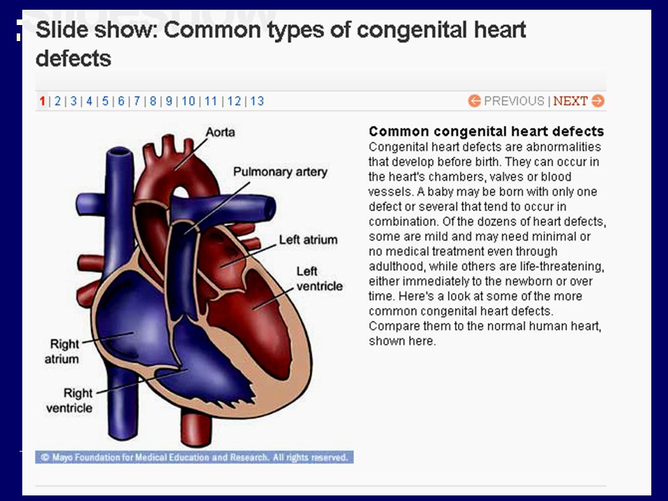
Eight out of every 1,000 infants have some type of structural heart abnormality at birth. Such abnormalities, known as congenital heart defects, may be so minor that they cause no symptoms or so severe that they're usually fatal. Congenital heart defects also vary widely in complexity, from a simple hole in the wall between two heart chambers to a complicated set of malformations, such as blood vessels in the wrong places and underdevelopment of one side of the heart.

3
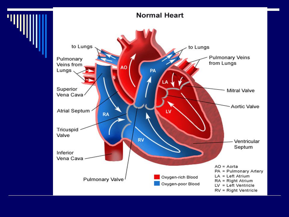
How the heart works: A normal heart is divided into four hollow chambers, two on the right and two on the left. In performing its basic job — pumping blood throughout the body — the heart uses its left and right sides for different tasks. The right side moves blood into vessels called pulmonary arteries, leading to the lungs. In the lungs, oxygen enriches the blood, which circulates to the heart's left side in blood vessels called pulmonary veins. The left side of the heart pumps blood into a large vessel called the aorta. Branching off from the aorta are numerous other vessels that circulate blood to the rest of the body.

4
How heart defects develop: A baby's heart starts beating just 22 days after conception. At that point, the heart has a simple tube shape. Between days 22 and 24, however, the heart begins to bend to the right and fold in on itself to form a loop. By 28 days after conception, looping is completed and the tube has a vaguely heart-like shape with structures corresponding to the heart's two sides and the large blood vessels that carry blood in and out of them. If the genetic messages that direct the growth and movement of early heart cells are scrambled, part of the heart muscle may fail to develop. Or if the process of bending and looping doesn't go exactly the way it's supposed to, the heart may form abnormal connections to the large vessels leading to and from the lungs.

5
Signs of trouble: Many heart defects can be detected before birth by a test called fetal echocardiography. It uses sound waves to create a picture of the baby's heart. If the heart is beating too slowly or quickly, medications can usually fix the problem before the heart starts to fail. If a heart defect can't be treated before birth, doctors can use the information from the ultrasound test to prepare for treatments that can be given immediately after birth if necessary. Serious heart defects usually become evident during the first few days, weeks and months of life. The infant's skin may lose its healthy color and look pale gray or blue. Swelling may develop in the legs, abdomen or areas around the eyes. Perhaps the baby has shortness of breath during feedings, which leads to poor weight gain. Although other conditions can cause such symptoms, they all may be signs of congenital heart defects.

6
In all, there are more than 35 common congenital heart defects, falling mainly into these categories.

7
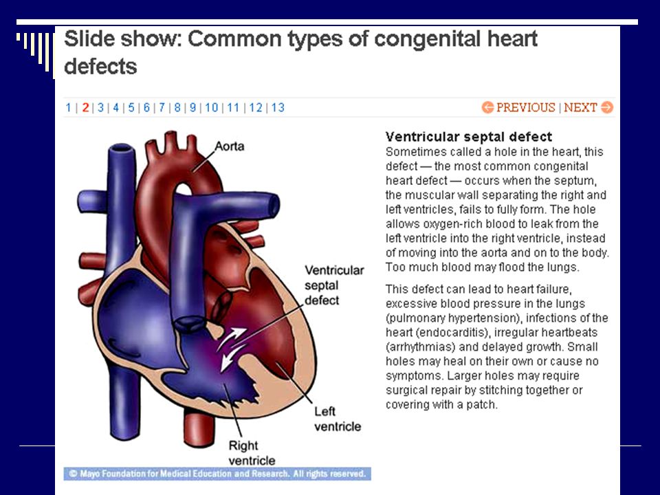
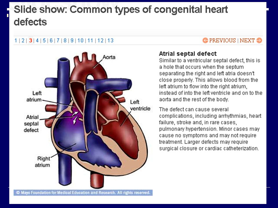
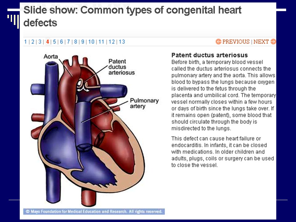
Holes in the heart Several defects can be thought of as holes in the walls between heart chambers or abnormal passageways between major blood vessels leaving the heart. These holes allow oxygen-rich and oxygen-poor blood to mix. If the holes are large and a lot of blood is mixed, the baby's skin or the area under the fingernails may be a slight bluish color. He or she may also develop signs and symptoms of congestive heart failure, such as shortness of breath, fatigue and leg swelling, because blood is flooding (overcirculating) the lungs. The most common examples of hole defects include: ventricular septal defect, which is a hole in the wall between the right and left ventricles, and patent ductus arteriosus (DUK-tus ahr-teer-e-O-sus), an opening between the pulmonary artery and the aorta. During fetal development, this opening allows blood in the fetus to bypass the lungs. Within a few hours after birth, however, it should close. If it doesn't, oxygen-rich blood intended for the body is directed back to the lungs.
 the lungs. The most common examples of hole defects include: ventricular septal defect, which is a hole in the wall between the right and left ventricles, and patent ductus arteriosus (DUK-tus ahr-teer-e-O-sus), an opening between the pulmonary artery and the aorta. During fetal development, this opening allows blood in the fetus to bypass the lungs. Within a few hours after birth, however, it should close. If it doesn t, oxygen-rich blood intended for the body is directed back to the lungs..")
8
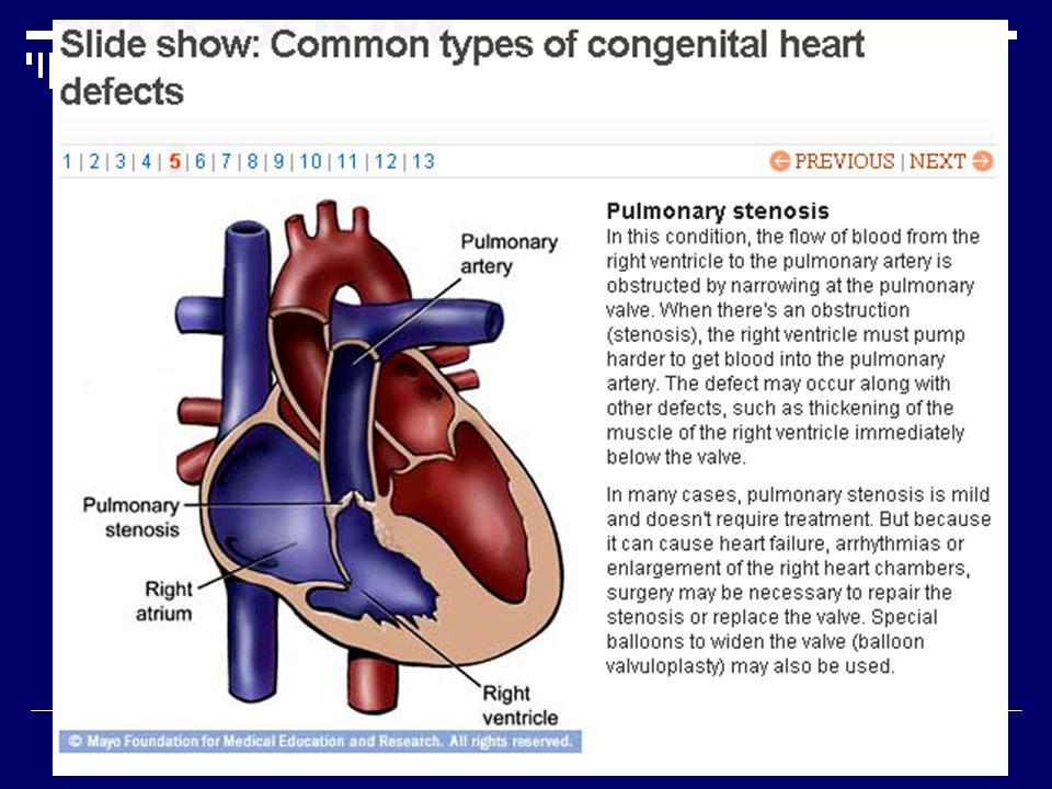
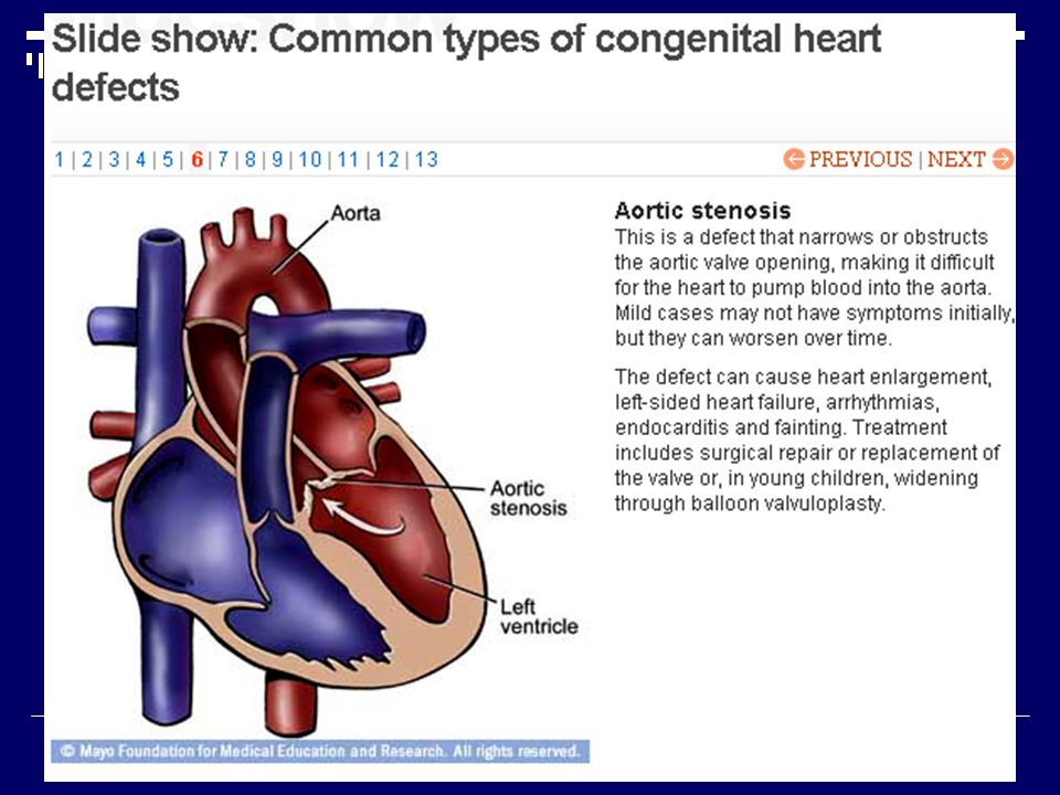
Obstructed blood flow When vessels or valves become narrowed, the heart must work harder to move blood through them. Imagine trying to squeeze water out of a small hole in a balloon, rather than a large hole, and you'll get the idea. Among the most common obstructive defects is pulmonary stenosis (stuh-NO-sis), a narrowing of the pulmonary valve, through which blood passes from the right ventricle to the pulmonary artery. Another obstructive defect, aortic stenosis, is a narrowing of the aortic valve, through which blood passes from the left ventricle into the aorta, eventually causing the heart muscle to thicken and the left ventricle to enlarge.
, a narrowing of the pulmonary valve, through which blood passes from the right ventricle to the pulmonary artery. Another obstructive defect, aortic stenosis, is a narrowing of the aortic valve, through which blood passes from the left ventricle into the aorta, eventually causing the heart muscle to thicken and the left ventricle to enlarge..")
9
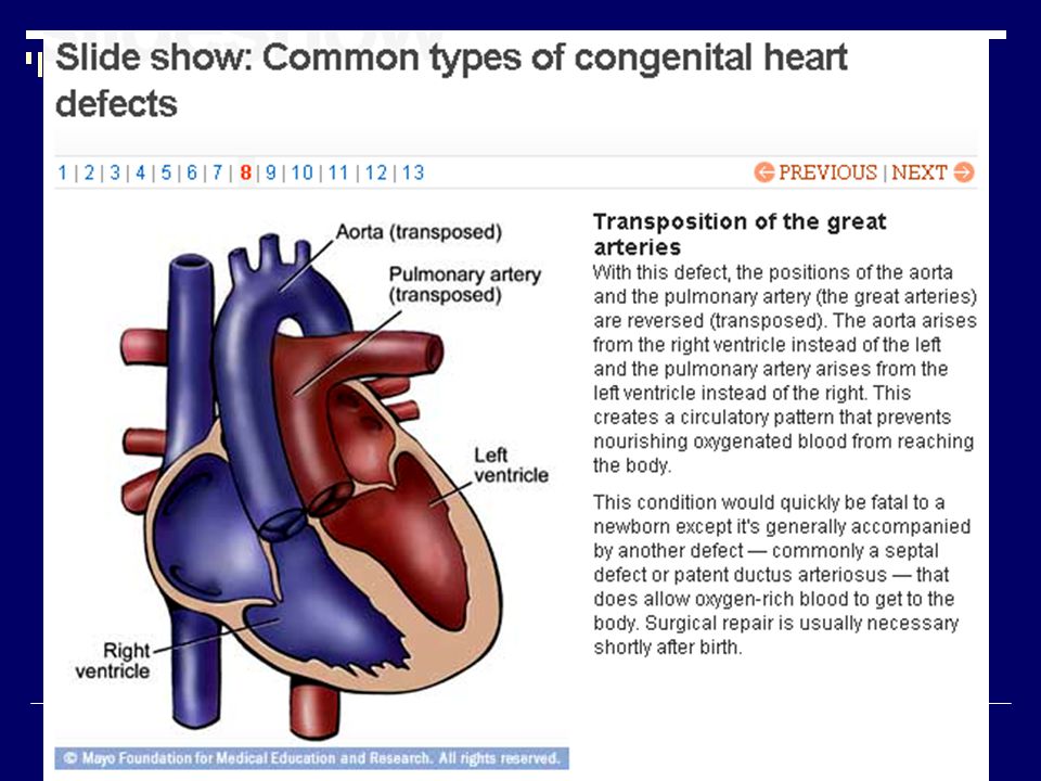
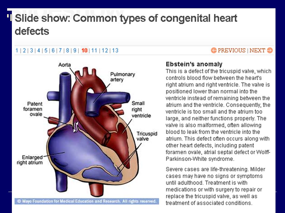
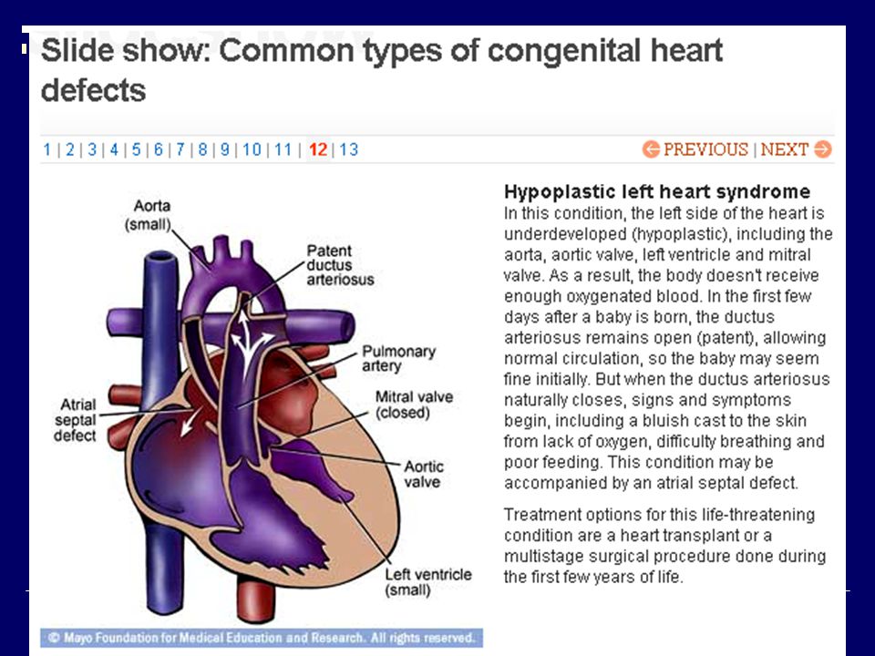
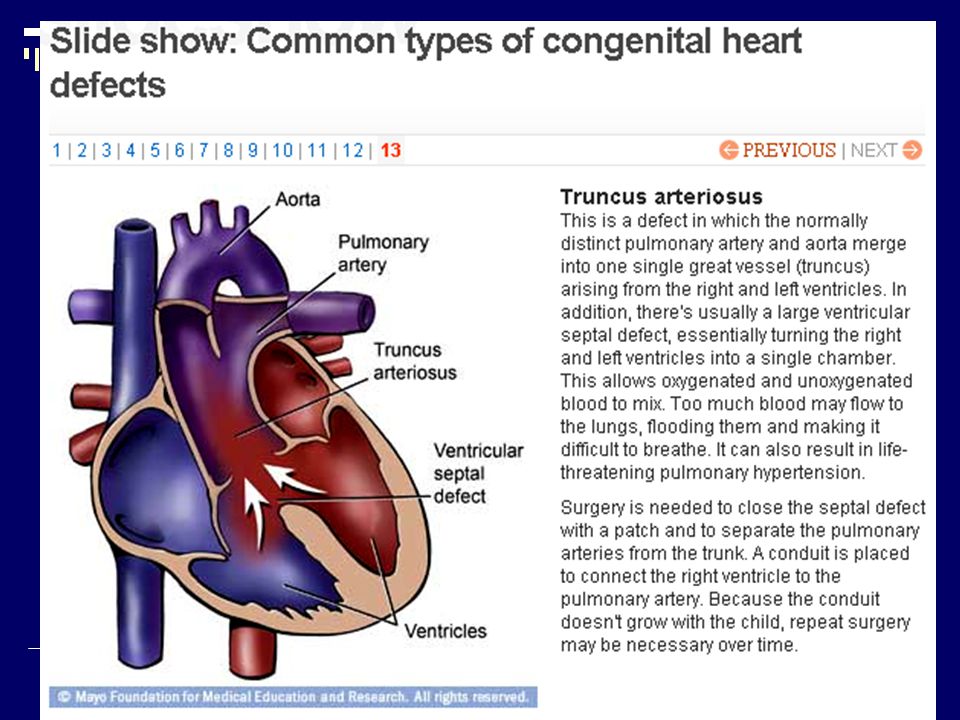
Abnormal blood vessels Several congenital heart defects involve incorrectly formed or positioned blood vessels going to and from the heart. For example, transposition of the great arteries occurs when the pulmonary artery and the aorta are on the wrong sides of the heart. This is a serious and immediately life-threatening defect. Heart valve abnormalities. If the heart valves can't open and close correctly, blood can't flow smoothly. Examples include Ebstein's anomaly, in which the tricuspid valve is malformed and often leaks, and pulmonary atresia, in which a solid sheet of tissue forms in place of the pulmonary valve and blocks normal blood flow to the lungs. Both defects prevent oxygen-poor blood from circulating to the lungs.

10
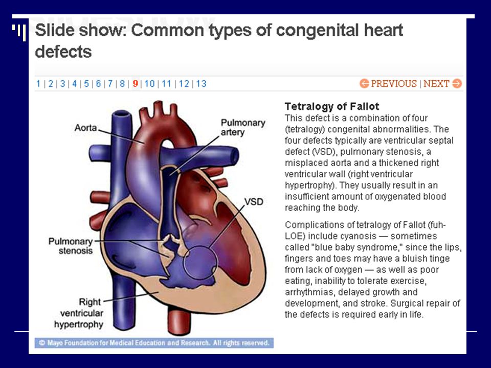
A combination of defects. Some congenital heart conditions are the result of not one but several defects. For example, tetralogy of Fallot is a combination of four defects: a hole in the ventricular septum, a narrowed passage between the right ventricle and pulmonary artery, a shift in the connection of the aorta to the heart, and thickened muscle in the right ventricle.

25
Reference 1998-2005 Mayo Foundation for Medical Education and Research (MFMER)
")
Similar presentations


 Bell Ringer: On a piece of paper, write your name and today’s date Do not use your notes!!! Write the process of how.>")


, nutrient molecules and waste materials (from the digestive system) 2.Regulates.>")


