Download presentation
Presentation is loading. Please wait.

1
Procaryotic Cell Structure and Function
Copyright © The McGraw-Hill Companies, Inc. Permission required for reproduction or display. Chapter 3 Procaryotic Cell Structure and Function

2
A Preview of Procaryotic Cell Structure and Function
Procaryotes differ from eucaryotes in many traits including size and lack of internal membrane systems Procaryotes are divided into Bacteria and Archaea Bacteria are divided into 2 groups based on their Gram stain reaction

4
Copyright © The McGraw-Hill Companies, Inc
Copyright © The McGraw-Hill Companies, Inc. Permission required for reproduction or display. Figure 3.2

5
Size, Shape, and Arrangement
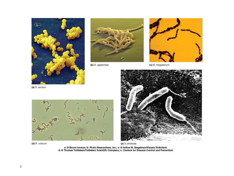
Cocci (coccus) – spheres Diplococci (diplococcus) – pairs Streptococci – chains Staphylococci – grape-like clusters Tetrads – 4 cocci in a square Sarcinae – cubic configuration of 8 cocci
 – spheres. Diplococci (diplococcus) – pairs. Streptococci – chains. Staphylococci – grape-like clusters. Tetrads – 4 cocci in a square. Sarcinae – cubic configuration of 8 cocci.")
6
Size, Shape, and Arrangement
Bacilli (bacillus) – rods -coccobacilli – very short rods -vibrios – resemble rods, comma shaped Spirilla (spirillum) – rigid helices Spirochetes – flexible helices Mycelium – network of long, multinucleate filaments
 – rods. -coccobacilli – very short rods. -vibrios – resemble rods, comma shaped. Spirilla (spirillum) – rigid helices. Spirochetes – flexible helices. Mycelium – network of long, multinucleate filaments.")
7
Size, Shape, and Arrangement
Pleomorphic – organisms that are variable in shape

8
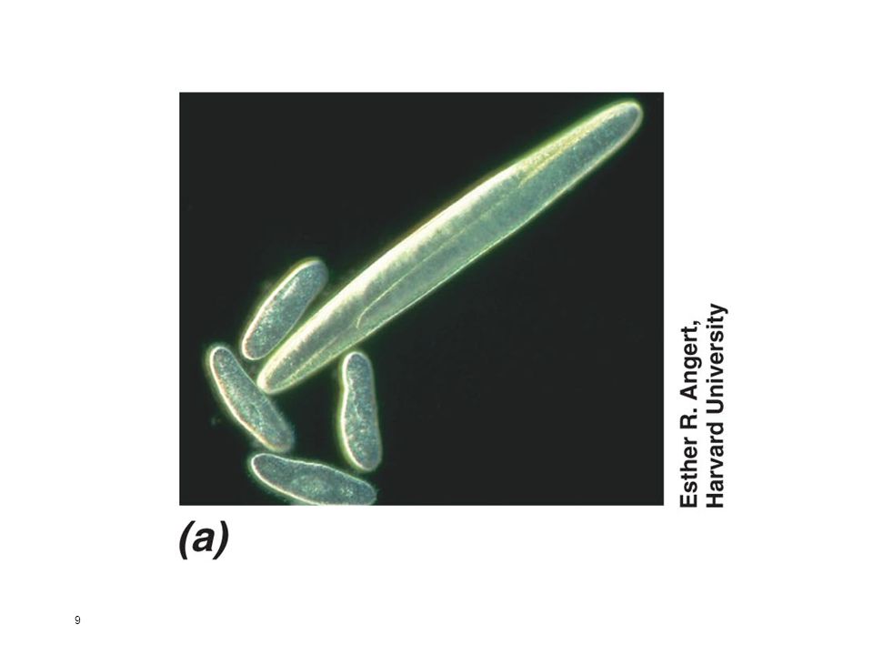
Size Size range for prokaryotes: 0.2 µm to > 700 µm in diameter
Most cultured rod-shaped bacteria are between 0.5 and 4.0 µm wide and < 15 µm long Few very large prokaryotes; examples include Epulopiscium fishelsoni Thiomargarita namibiensis Size range for eukaryotic cells: 10 to > 200 µm in diameter

10
largest – 50 μm in diameter smallest – 0.3 μm in

11
Figure 3.4

12
Procaryotic Cell Membranes
Membranes are an absolute requirement for all living organisms Plasma membrane encompasses the cytoplasm Some procaryotes also have internal membrane systems

13
Composition Phospholipids
Contain both hydrophobic and hydrophilic components Can exist in many different chemical forms due to variation in the groups attached to the glycerol backbone Fatty acids point inward to form hydrophobic environment

14
Bacterial Phopholipids
polar ends interact with water hydrophilic nonpolar ends insoluble in water hydrophobic Figure 3.6

15
General Structure of Phospholipids

16
Functions of the plasma membrane
Separation of cell from its environment Selectively permeable barrier some molecules are allowed to pass into or out of the cell transport systems aid in movement of molecules

17
More functions… Location of crucial metabolic processes Special receptor molecules in the membrane

18
Fluid Mosaic Model of Membrane Structure
Lipid bilayer in which proteins float Figure 3.5

19
Membrane proteins Peripheral proteins Integral proteins
Copyright © The McGraw-Hill Companies, Inc. Permission required for reproduction or display. Membrane proteins Peripheral proteins loosely associated with the membrane and easily removed Integral proteins embedded within the membrane and not easily removed

20
Bacterial Membranes differ from eucaryotes in lacking sterols
do contain hopanoids, sterol-like molecules a highly organized, asymmetric system which is also flexible and dynamic

21
Other membrane lipids Figure 3.7

22
Internal Membranous Structures
Plasma membrane infoldings Observed in many photosynthetic bacteria Nitrifying bacteria Methanotrophic bacteria

23
Plasma membrane infoldings: observed in many photosynthetic bacteria
Figure 3.8

24
Archaeal membranes Composed of unique lipids
Some have a monolayer structure instead of a bilayer structure

25
Figure 3.9

26
Figure 3.10

27
Figure 3.11

28
Cytoplasmic Matrix Substance in which nucleoid, ribosomes and inclusion bodies are suspended Lacks organelles bound by unit membranes Composed largely of water

29
Inclusion bodies Granules of organic or inorganic material that are stockpiled by the cell for future use Some are enclosed by a single-layered membrane

30
Organic inclusion bodies
Glycogen polymer of glucose units Poly-β-hydroxybutyrate (PHB) polymers of β-hydroxybutyrate
 polymers of β-hydroxybutyrate.")
31
Organic inclusion bodies
cyanophycin granules large polypeptides containing about equal quantities of arginine and aspartic acid carboxysomes contain the enzyme ribulose-1,5,-bisphosphate carboxylase (Rubisco), enzyme used for CO2 fixation
, enzyme used for CO2 fixation.")
32
Copyright © The McGraw-Hill Companies, Inc
Copyright © The McGraw-Hill Companies, Inc. Permission required for reproduction or display. Figure 3.13

33
Organic inclusion bodies
gas vacuoles found in cyanobacteria and some other aquatic procaryotes provide buoyancy aggregates of hollow cylindrical structures called gas vesicles

34
Copyright © The McGraw-Hill Companies, Inc
Copyright © The McGraw-Hill Companies, Inc. Permission required for reproduction or display. Freeze-fracture preparation of Anabaena flos-aquae; clusters of cigar-shaped gas vesicles form gas vacuoles Figure 3.14

35
Inorganic inclusion bodies
Polyphosphate granules linear polymers of phosphates Sulfur granules - source of energy (purple sulfur bacteria magnetosomes contain iron in the form of magnetite used to orient cells in magnetic fields

36
Copyright © The McGraw-Hill Companies, Inc
Copyright © The McGraw-Hill Companies, Inc. Permission required for reproduction or display. Aquaspirillum magnetotacticum; MP=magnetite particles; OM=outer membrane; P=periplasmic space; CM=cytoplasmic membrane TA 3.2 (a)
")
37
TA 3.2 (b) Isolated magnetosomes
Copyright © The McGraw-Hill Companies, Inc. Permission required for reproduction or display. Isolated magnetosomes TA 3.2 (b)
")
38
Copyright © The McGraw-Hill Companies, Inc
Copyright © The McGraw-Hill Companies, Inc. Permission required for reproduction or display. Bacteria migrating in waves when exposed to a magnetic field TA 3.2 (c)
")
39
Ribosomes Complex structures consisting of protein and RNA
Sites of protein synthesis Smaller than eucaryotic ribosomes procaryotic ribosomes 70S eucaryotic ribosomes 80S S = Svedburg unit

40
Copyright © The McGraw-Hill Companies, Inc
Copyright © The McGraw-Hill Companies, Inc. Permission required for reproduction or display. Figure 3.15

41
The Nucleoid Irregularly shaped region Location of chromosome
Copyright © The McGraw-Hill Companies, Inc. Permission required for reproduction or display. The Nucleoid Irregularly shaped region Location of chromosome usually 1/cell Not membrane-bound Figure 3.4

42
Copyright © The McGraw-Hill Companies, Inc
Copyright © The McGraw-Hill Companies, Inc. Permission required for reproduction or display. Procaryotic chromosomes are located in the nucleoid, an area in the cytoplasm Figure 3.14a: growing Bacillus cells stained with HCl-Giemsa and viewed with light microscope Figure 3.14b: section of growing E. coli immunostained specifically for DNA; viewed with TEM Figure 3.15c: model of 2 nucleoids in an actively growing E. coli cell Figure 3.16

43
The procaryotic chromosome
Closed circular, double-stranded DNA molecule Looped and coiled extensively Nucleoid proteins probably aid in folding nucleoid proteins differ from histones

44
Plasmids Closed circular or linear DNA molecules
Copyright © The McGraw-Hill Companies, Inc. Permission required for reproduction or display. Plasmids Closed circular or linear DNA molecules Replicate independently of chromosome Have relatively few genes present Genes on plasmids are not essential to host but may confer selective advantage (e.g., drug resistance)
")
45
Copyright © The McGraw-Hill Companies, Inc
Copyright © The McGraw-Hill Companies, Inc. Permission required for reproduction or display. Table 3.3

46
The Bacteria Cell Wall rigid structure that lies just outside the plasma membrane Figure 3.4

47
Functions of cell wall Provides characteristic shape to cell
Protects the cell from osmotic lysis Protects from toxic compounds and pathogens May also contribute to pathogenicity A site for antibiotic action Some organisms lack cell walls

48
Cell walls of Bacteria Bacteria are divided into two major groups based on the response to gram-stain procedure. gram-positive bacteria stain purple gram-negative bacteria stain pink Staining reaction due to cell wall structure

49
Copyright © The McGraw-Hill Companies, Inc
Copyright © The McGraw-Hill Companies, Inc. Permission required for reproduction or display. Figure 3.17

50
Peptidoglycan Figure 4.18

51
Peptidoglycan (Murein) Structure
Meshlike polymer composed of identical subunits Contains N-acetyl glucosamine and N-acetylmuramic acid and several different amino acids Chains of peptidoglycan subunits are cross linked by peptides

52
Copyright © The McGraw-Hill Companies, Inc
Copyright © The McGraw-Hill Companies, Inc. Permission required for reproduction or display. Top: E. coli peptidoglycan; direct cross-linking, typical of many gram-negative bacteria Bottom: Staphylococcus aureus peptidoglycan NAM= N-acetylglucosoamine; NAG= N-acetylmuramic acid Figure 3.20

53
Copyright © The McGraw-Hill Companies, Inc
Copyright © The McGraw-Hill Companies, Inc. Permission required for reproduction or display. Figure 3.21

54
Gram-Positive Cell Walls
composed primarily of peptidoglycan may also contain large amounts of teichoic acids some gram-positive bacteria have layer of proteins on surface of peptidoglycan Isolated cell wall from Bacillus megaterium; latex spheres are 0.25 micrometer in diameter Figure 3.22

55
Copyright © The McGraw-Hill Companies, Inc
Copyright © The McGraw-Hill Companies, Inc. Permission required for reproduction or display. Figure 3.23

56
teichoic acids polymers of glycerol or ribitol joined by
Copyright © The McGraw-Hill Companies, Inc. Permission required for reproduction or display. teichoic acids polymers of glycerol or ribitol joined by phosphate groups Figure 3.24

57
Gram-Negative Cell Walls
Consist of a thin layer of peptidoglycan surrounded by an outer membrane Outer membrane composed of lipids, lipoproteins, and lipopolysaccharide (LPS) No teichoic acids
 No teichoic acids.")
58
Gram-Negative Cell Walls
More complex than Gram + walls Peptidoglycan is ~2-5% of wall weight Periplasmic space differs from that in gram + cells may constitute 20-40% of cell volume many enzymes present in periplasm

59
Gram-Negative Cell Walls
Outer membrane lies outside the thin peptidoglycan layer Braun’s lipoproteins connect outer membrane to peptidoglycan

60
Copyright © The McGraw-Hill Companies, Inc
Copyright © The McGraw-Hill Companies, Inc. Permission required for reproduction or display. Figure 3.25

61
Lipopolysaccharides (LPSs)
Consists of three parts lipid A core polysaccharide O side chain (O antigen)
")
62
Copyright © The McGraw-Hill Companies, Inc
Copyright © The McGraw-Hill Companies, Inc. Permission required for reproduction or display. LPS from Salmonella: Abe=abequose; Gal=galactose; Glc=glucose; GlcN=glucosamine; Hep=heptulose; KDO=2-keto-3-deoxyoctonate; Man=mannose; NAG=N-acetyleglucosamine; P=phosphate; Rha=rhamnose Figure 3.27

63
Importance of LPS Protection from host defenses (O antigen)
Contributes to negative charge on cell surface (core polysaccharide) Helps stabilize outer membrane structure (lipid A) Can act as an exotoxin (lipid A)
 Helps stabilize outer membrane structure (lipid A) Can act as an exotoxin (lipid A)")
64
Other Characteristics of the Outer Membrane
More permeable than plasma membrane due to presence of porin proteins and transporter proteins porin proteins form channels through which small molecules ( daltons) can pass
 can pass.")
65
Copyright © The McGraw-Hill Companies, Inc
Copyright © The McGraw-Hill Companies, Inc. Permission required for reproduction or display. Figure 3.28

66
Molecular Model of Porin Proteins

67
Archaeal cell walls Lack peptidoglycan
Cell wall varies from species to species but usually consists of complex heteropolysaccharides Methanogens have walls containing pseudomurein

68
Copyright © The McGraw-Hill Companies, Inc
Copyright © The McGraw-Hill Companies, Inc. Permission required for reproduction or display. Figure 3.30

69
Copyright © The McGraw-Hill Companies, Inc
Copyright © The McGraw-Hill Companies, Inc. Permission required for reproduction or display. Figure 3.31

70
Protein Secretion in Procaryotes
Numerous protein secretion pathways have been identified four major pathways are: Sec-dependent pathway type II pathway type I (ABC) protein secretion pathway type III protein secretion pathway
 protein secretion pathway. type III protein secretion pathway.")
71
Overview of Bacterial Protein Secretion
Gram+ and Gram- bacteria have different problems secreting proteins based on the differences between the structure of their walls Both use Sec-dependent pathway All protein secretion systems require energy

72
Sec-Dependent Pathway
Also called general secretion pathway Translocates proteins from cytoplasm across or into plasma membrane Secreted proteins synthesized as preproteins having amino-terminal signal peptide signal peptide delays protein folding chaperone proteins keep preproteins unfolded

73
Sec-Dependent Pathway
Copyright © The McGraw-Hill Companies, Inc. Permission required for reproduction or display. Sec-Dependent Pathway secA translocates preprotein through the plasma membane When preprotein emerges from plasma membrane a signal peptidase removes the signal peptide

74
Sec-Dependent Pathway
Copyright © The McGraw-Hill Companies, Inc. Permission required for reproduction or display. Sec-Dependent Pathway Sec-dependent pathway of E. coli Figure 3.32

75
Protein Secretion in Gram- bacteria
Copyright © The McGraw-Hill Companies, Inc. Permission required for reproduction or display. Protein Secretion in Gram- bacteria 5 secretion pathways have been identified in gram- bacteria type II and V pathways transport proteins across outer membrane that were translocated across plasma membrane by Sec-dependent pathway types I and III pathways are Sec-independent type IV pathway usually functions independently of the Sec pathway

76
Type II Protein Secretion Pathway
Copyright © The McGraw-Hill Companies, Inc. Permission required for reproduction or display. Type II Protein Secretion Pathway Present in a number of plant and animal pathogens transports proteins from periplasmic across outer membrane observed in some gram-negative bacteria, including some pathogens complex systems consisting of up to proteins most are integral membrane proteins

77
Type V Protein Secretion Pathway
Copyright © The McGraw-Hill Companies, Inc. Permission required for reproduction or display. Type V Protein Secretion Pathway Most recently discovered protein secretion system Rely on Sec-dependent pathway to move proteins across plasma membrane When proteins are in periplasmic space many can form a channel in outer membrane through which they transport themselves; hence they are called autotransporters

78
ABC Protein Secretion Pathway
Copyright © The McGraw-Hill Companies, Inc. Permission required for reproduction or display. ABC Protein Secretion Pathway also called Type I Protein Secretion Pathway ubiquitous in procaryotes transports proteins from cytoplasm across both plasma membrane and outer membrane secreted proteins have C-terminal secretion signals Gram+ bacteria use a modified version of this pathway to translocate proteins across the plasma membrane

79
Type III Protein Secretion Pathway
Copyright © The McGraw-Hill Companies, Inc. Permission required for reproduction or display. Type III Protein Secretion Pathway secretes virulence factors of gram-negative bacteria from cytoplasm, across both plasma membrane and outer membrane, and into host cell some type III secretion machinery is syringe-shaped secreted proteins thought to move through a translocation channel

80
Type IV Protein Secretion Pathway
Copyright © The McGraw-Hill Companies, Inc. Permission required for reproduction or display. Type IV Protein Secretion Pathway Type IV pathways are unique because they secrete proteins and transfer DNA during conjugation Type IV systems are made of many different proteins, some of which form a syringe-like structure

81
Protein Secretion Systems of Gram-Negative Bacteria
Copyright © The McGraw-Hill Companies, Inc. Permission required for reproduction or display. Protein Secretion Systems of Gram-Negative Bacteria Figure 3.33

82
Components External to Cell Wall
Figure 3.4

83
Capsules, Slime Layers, and S-Layers
usually composed of polysaccharides well organized and not easily removed from cell Slime layers similar to capsules except diffuse, unorganized and easily removed

84
Bacterial Capsules Figure 3.34

85
Bacterial capsules

86
Glycocalyx network of polysaccharides extending from the surface of the cell a capsule or slime layer composed of polysaccharides can also be referred to as a glycocalyx

87
Bacterial Glycocalyx Figure 3.35

88
Capsules, Slime Layers, and S-Layers
regularly structured layers of protein or glycoprotein in bacteria the S layer is external to the cell wall common among Archaea, where they may be the only structure outside the plasma membrane

89
Copyright © The McGraw-Hill Companies, Inc
Copyright © The McGraw-Hill Companies, Inc. Permission required for reproduction or display. S-layer of Deinococcus radiodurans; electron micrograph after shadowing Figure 3.36

90
Functions Protection from host defenses (e.g., phagocytosis)
Protection from harsh environmental conditions (e.g., desiccation) Attachment to surfaces
 Attachment to surfaces.")
91
More functions… Protection from viral infection or predation by bacteria Protection from chemicals in environment (e.g., detergents) Facilitate motility of gliding bacteria Protection against osmotic stress

92
Pili and Fimbriae Fimbriae (fimbria) Sex pili (pilus)
short, thin, hairlike, proteinaceous appendages up to 1,000/cell mediate attachment to surfaces some (type IV fimbriae) required for twitching motility or gliding motility that occurs in some bacteria Sex pili (pilus) similar to fimbriae except longer, thicker, and less numerous (1-10/cell) required for mating
 required for twitching motility or gliding motility that occurs in some bacteria. Sex pili (pilus) similar to fimbriae except longer, thicker, and less numerous (1-10/cell) required for mating.")
93
Figure 3.37

94
Flagella and Motility Figure 3.4

95
Patterns of Flagella Distribution
Monotrichous – one flagellum Polar flagellum – flagellum at end of cell Amphitrichous – one flagellum at each end of cell Lophotrichous – cluster of flagella at one or both ends Peritrichous – spread over entire surface of cell

96
Flagella Distribution
Copyright © The McGraw-Hill Companies, Inc. Permission required for reproduction or display. Figure 3.38

97
Flagellar Ultrastructure
negatively stained flagella from E. coli; arrows indicate curved hooks and basal bodies Figure 3.39 (a) and (b)
 and (b)")
98
Flagellar Ultrastructure
negatively stained flagella from E. coli; arrows indicate curved hooks and basal bodies Figure 3.39 (c)
")
99
The filament Extends from cell surface to the tip Hollow, rigid cylinder Composed of the protein flagellin Some procaryotes have a sheath around filament

100
The Hook and Basal Body hook basal body links filament to basal body
series of rings that drive flagellar motor basal body of E. coli flagellum; upper most arrow indicates junction of hook and filament Figure 3.39d

101
Flagellar Synthesis an example of self-assembly
Copyright © The McGraw-Hill Companies, Inc. Permission required for reproduction or display. Flagellar Synthesis an example of self-assembly complex process involving many genes and gene products new molecules of flagellin are transported through the hollow filament growth is from tip, not base

102
Copyright © The McGraw-Hill Companies, Inc
Copyright © The McGraw-Hill Companies, Inc. Permission required for reproduction or display. Export of flagellar components is carried out by an apparatus in the basal body that appears to be closely related to the type III protein secretion system. Figure 3.40

103
The Mechanism of Flagellar Movement
Copyright © The McGraw-Hill Companies, Inc. Permission required for reproduction or display. The Mechanism of Flagellar Movement flagellum rotates like a propeller in general, counterclockwise rotation causes forward motion (run) in general, clockwise rotation disrupts run causing a tumble (twiddle)
 in general, clockwise rotation disrupts run causing a tumble (twiddle)")
104
Copyright © The McGraw-Hill Companies, Inc
Copyright © The McGraw-Hill Companies, Inc. Permission required for reproduction or display. Figure 3.41

105
Copyright © The McGraw-Hill Companies, Inc
Copyright © The McGraw-Hill Companies, Inc. Permission required for reproduction or display. Rotor portion of motor is primarily rod, M ring, and C ring. Stator portion of motor is Mot A and Mot B. Figure 3.42

106
Other Types of Motility
Copyright © The McGraw-Hill Companies, Inc. Permission required for reproduction or display. Other Types of Motility spirochetes exhibit flexing and spinning movements of axial filaments which are composed of periplasmic flagella gliding motility cells coast along solid surfaces no visible motility structure has been identified

107
Copyright © The McGraw-Hill Companies, Inc
Copyright © The McGraw-Hill Companies, Inc. Permission required for reproduction or display. Chemotaxis movement towards a chemical attractant or away from a chemical repellent concentrations of chemical attractants and chemical repellents detected by chemoreceptors on surfaces of cells

108
Copyright © The McGraw-Hill Companies, Inc
Copyright © The McGraw-Hill Companies, Inc. Permission required for reproduction or display. Figure 3.38: On left is positive chemotaxis by E. coli; outer ring is composed of bacteria consuming serine and the second ring was formed by cells consuming aspaartate, a less powerful attractant; top right colony is composed of motile but nonchemotactic mutant; bottom right colony is composed of nonmotile bacteria. Figure 3.39: negative chemotaxis by E. coli in response to acetate; bright disks are plugs of agar containing acetate; acetate concentration increases from 0 at the top right to 3 M at top left; as acetate concentration increases, size of bacteria-free zone increases Figure 3.43 Figure 3.44

109
Chemotaxis Towards Attractant
Copyright © The McGraw-Hill Companies, Inc. Permission required for reproduction or display. Chemotaxis Towards Attractant in presence of attractant (b) tumbling frequency is reduced and runs in direction of attractant are longer Figure 3.45
 tumbling frequency is reduced and runs in direction of attractant are longer. Figure")
110
Chemotaxis away from repellent
Copyright © The McGraw-Hill Companies, Inc. Permission required for reproduction or display. Chemotaxis away from repellent involves similar but opposite responses

111
The Bacterial Endospore
Copyright © The McGraw-Hill Companies, Inc. Permission required for reproduction or display. The Bacterial Endospore formed by some bacteria dormant resistant to numerous environmental conditions heat radiation chemicals desiccation

112
Copyright © The McGraw-Hill Companies, Inc
Copyright © The McGraw-Hill Companies, Inc. Permission required for reproduction or display. Figure 3.46

113
Copyright © The McGraw-Hill Companies, Inc
Copyright © The McGraw-Hill Companies, Inc. Permission required for reproduction or display. EX=exosprium SC=spore coat: composed of several protein layers; impermeable to many toxic molecules; thought to contain enzymes involved in germination CX=cortex: made of peptidoglycan that is less cross-linked than that in vegetative cells CW=core wall (spore cell wall) CR=core: has normal cell structures but is metabolically inactive Figure 3.47
 CR=core: has normal cell structures but is metabolically inactive. Figure")
114
What makes an endospore so resistant?
Copyright © The McGraw-Hill Companies, Inc. Permission required for reproduction or display. What makes an endospore so resistant? calcium (complexed with dipicolinic acid) acid-soluble, DNA-binding proteins dehydrated core spore coat DNA repair enzymes
 acid-soluble, DNA-binding proteins. dehydrated core. spore coat. DNA repair enzymes.")
115
Sporogenesis Also called endospore formation or sporulation
Copyright © The McGraw-Hill Companies, Inc. Permission required for reproduction or display. Sporogenesis Also called endospore formation or sporulation normally commences when growth ceases because of lack of nutrients complex multistage process

116
Copyright © The McGraw-Hill Companies, Inc
Copyright © The McGraw-Hill Companies, Inc. Permission required for reproduction or display. Figure 3.49

117
Transformation of endospore into vegetative cell
Copyright © The McGraw-Hill Companies, Inc. Permission required for reproduction or display. Transformation of endospore into vegetative cell germination complex, multistage process Clostridium pectinovorum emerging from the spore Figure 3.50

118
Transformation of dormant spores into active vegetative cells
Copyright © The McGraw-Hill Companies, Inc. Permission required for reproduction or display. Transformation of dormant spores into active vegetative cells activation prepares spores for germination often results from treatments like heating germination spore swelling rupture of absorption of spore coat loss of resistance increased metabolic activity outgrowth emergence of vegetative cell

Similar presentations


 Flagella 1) Functions in movement of the cell 2) 3 components.>")