Download presentation
Presentation is loading. Please wait.

1
Chapter 5 Urinary system
By Dr. Amr A. Abd-Elghany

2
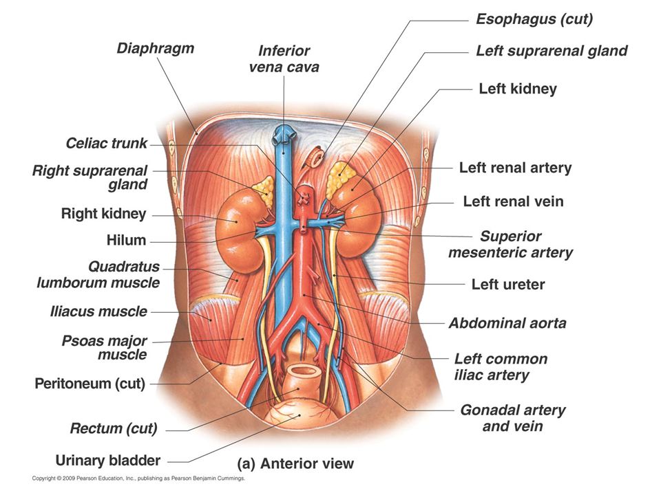
Anatomy and physiology
Paired kidneys A ureter for each kidney Urinary bladder Urethra

3
Main Functions of Urinary System
Kidneys filter blood to keep it pure Toxins Metabolic wastes Excess water Excess ions Dispose of nitrogenous wastes from blood Urea Uric acid Creatinine Regulate the balance of water and electrolytes, acids and bases

5
Transverse sections show retroperitoneal position of kidneys

7
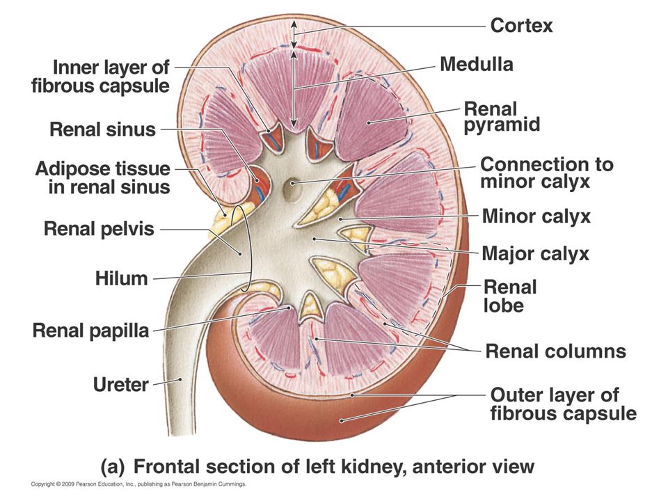
For studying Parts of the kidney: 1. Renal pyramid 2. Efferent vessel 3. Renal artery 4. Renal vein 5. Renal hilum 6. Renal pelvis 7. Ureter 8. Minor calyx 9. Renal capsule 10. Inferior renal capsule 11. Superior renal capsule 12. Afferent vessel 13. Nephron 14. Minor calyx 15. Major calyx 16. Renal papilla 17. Renal column

8
Ureters

9
IMAGING MODALITIES Plain kidney, ureter, and bladder (KUB)
Intravenous Urogram (IVU) US CT MRI Nuclear medicine Interventional (invasive) techniques.
 US. CT. MRI. Nuclear medicine. Interventional (invasive) techniques.")
10
Plain UT (KUB) Simple. Require adequate preparation. Useful for:
Radio-opaque stones, gas pattern, Calcifications, Organomegaly, Bone abnormalities.

11
IVU (x-ray + contrast media)
Requirements: Fasting 4-6 hours, good hydration is essential. Renal function test before. Non ionic contrast media is used in case of hypersensitivity of ionic contrast. Diagnostic value: Show renal function. UT obstraction. Renal or bladder masses Congenital anomalies. Contraindicated in patients with: Renal impairment. Hypersensitivity to contrast media.

12
IVU Plain x-ray is important before IVU to see the stones because the contrast media will hide the stone.

13
Cystoureathrography Contrast is inserted into the bladder and images are obtained. The patient is asked to void the bladder and images are also taken. Diagnostic value: Urethral lesion. Vesicourethral reflux

14
Ultrasound Image features: Fasting 4-6 hrs. Operator dependant.
Projectional image. Good resolution. Used for stone, hydronephrosis, focal lesion. No contrast media. No bowel preparation. No radiation hazards.

15
CT Fasting 4-6 hrs. May need bowel preparation. More precise. Costly.
+/- contrast. Useful for trauma, stone, tumor, infection. Consider radiation hazards.

16
CT Diagnostic value Detection of UT calculi. Sections each 3mm through kidney till the end of urinary bladder (without no preparation). UT obstruction. Renal or bladder masses. Congenital anomalies. Differentiates cystic from solid masses. CT angiography.

17
MRI Used in case of no possibility to give the patient contrast.
Functional imaging. No bowel preparation. Better evaluation of soft tissue. Expensive. Useful for soft tissue pathology: tumor, infection.

18
Diagnosis of UT Pathology
Stone disease. (Radiolucent, radiopaque) UT neoplasm. UT infections. UT trauma. Miscellaneous lesions (congenital anomalies, vesico- ureteric reflux, urethral lesions) .
 UT neoplasm. UT infections. UT trauma. Miscellaneous lesions (congenital anomalies, vesico- ureteric reflux, urethral lesions) .")
19
Renal masses Benign, malignant

20
Simple Renal cyst Image features: Water filled vesicle.
Causing stretching of the calyces.

21
Renal cell carcinoma Filled with soft tissue. Lesion is solid.
If you have cystic lesion + solid= consider all is solid and the cystic part is necrotic tissue.

22
Wilm tumor In children. e.g. nephroblastoma
One of the famous causes of abdominal masses.

23
Transitional cell carcinoma TCC
The most common renal pelvic tumor. Multiple lesions in about 30% of cases. Filling defects in the pelvis. Detected by IVU, CT, MRU.

24
Bladder carcinoma IVU of patient with hematuria clearly shows a large, irregular filling defect within the bladder caused by cancer.

25
Bladder carcinoma

26
UT trauma Perinephric hematoma
Accidental crash followed by hematuria. The best technique to detect trauma is CT using contrast media. Perinephric hematoma Perinephrium: is the connective and fatty tissue surrounding a kidney. Kidney is intact. There is a hemorrhage around the kidney.

27
Renal laceration The renal parenchyma is divided into two or more parts.

28
Avulsion of the vascular pedicle
One kidney secrete and the other not due to rupture of the renal artery.

29
UT infections Pyelonephritis
Common manifestations in acute renal conditions: 1. Swollen kidney 2. Poor renal function Dirty perinephric fat Pyelonephritis CT scan demonstrates swollen kidney with patchy striated nephrogram. The most common UT infections. Bacterial infection of the kidney generally produces fever, flank pain, proteinuria.

30
Renal abscess With thick wall uptake the contrast media but cyst thin wall..

31
Renal Tuberculosis The presence of calcification inside the renal parenchyma. Usually above the age of 50y.

32
Congenital lesions Ectopic pelvic kidney
The kidney is located out of the normal site.

33
Duplex kidney and ureter
2 kidneys and ureters

34
Diverticula of the bladder
May be congenital, but more often result from a portion of the mucosa pooching out with chronic obstruction.

35
Bilateral ureteroceles
Congenital dilatation of the distal part of the ureter. This produces a typical cobra head deformity. Ureterocele can be unilateral or bilateral and is prone to calculi formation.

36
Bilateral ureteroceles

37
Vesicoureteric reflux
The urine is returned back to the ureter from the bladder. This pathology is detected by Ascending Cystourethrography.

38
Vesicoureteric reflux
Grade III and Grade V

39
Urethral stricture Traumatic rupture of the Urethra
Can be detected by Ascending Cystourethrography. Narrowing of the Urethra. Traumatic rupture of the Urethra Can be detected by Ascending Cystourethrography. Leaking of the contrast media from the urethra.

40
Kidney failure (renal failure)
End stage kidney disease is terminal stage of destruction of kidney to totally affect kidney function. Patient presents with: • Uremia (excess urea in blood), • High blood pressure, • Anuria (no urine) • Oliguria (little urine). Types of renal failure Acute: Usually presents as oliguria (less than 500 mL /day). Chronic renal failure: Is the end result of irreversible kidney damage from any cause. • Fatal disease, and requires continuous dialysis.
, • High blood pressure, • Anuria (no urine) • Oliguria (little urine). Types of renal failure. Acute: Usually presents as oliguria (less than 500 mL /day). Chronic renal failure: Is the end result of irreversible kidney damage from any cause. • Fatal disease, and requires continuous dialysis.")
41
Urinary System Terminology:
Dysuria: Difficult or painful urination. • Polyuria: Excessive urination. • Nocturia: Excessive urination during the night. • Uremia: (uremic poisoning) Excessive urea and other waste products in the bloodstream. • Anuria (anuresis): Complete suppression of urine excretion.
 Excessive urea and other waste products in the bloodstream. • Anuria (anuresis): Complete suppression of urine excretion.")
Similar presentations