Download presentation
Presentation is loading. Please wait.

1
URORADIOLOGY Ayşegül SARSILMAZ M.D. Radiology

2
Radiological Modalities
Ultrasound Intravenous Pyelography Computed Tomography Magnetic Resonance Imaging Radionuclide Scanning

3
Ultrasound CT (-) Anatomical info. MRI Radionuclide Functional info. IVP Anatomical + Fnx.

4
ULTRASOUND First line investigation !!!
Provides anatomical information without ionizing radiation No need for intravenous contrast !!!

5
INDICATIONS (USG) İnvestigate patients with symptoms thought to arise from UT Size of the kidneys Presence of hydronephrosis Renal tumors, cysts, abcesses Assess bladder and prostate

6
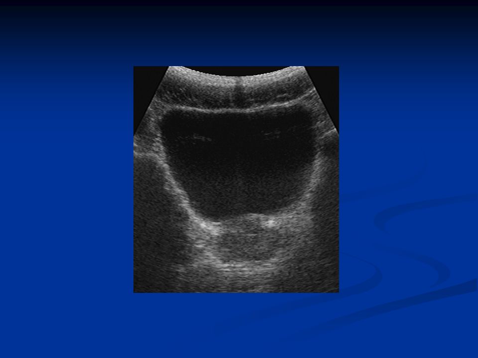
Normal Findings Kidneys; smooth in outline
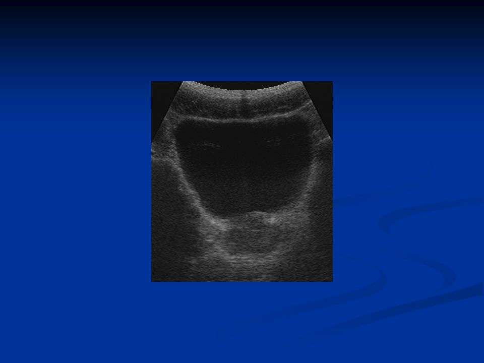
Central echogenic region (renal sinus) Renal cortex (hypoechoic) Pyramids (triangular hypoechoic areas) Size : mm Urinary Bladder : examined in distended state, imperceptible walls, anechoic lumen
 Renal cortex (hypoechoic) Pyramids (triangular hypoechoic areas) Size : mm. Urinary Bladder : examined in distended state, imperceptible walls, anechoic lumen.")
10
Intravenous Pyelography
Largely replaced by US ml of contrast is injected intravenously Carried via blood to the kidneys, passes through the glomerular filtrate collecting systems Main indications: Detailed demonstration of PCS and ureters Acute ureteric colic Investigation of renal calculi Investigation of hematuria

11
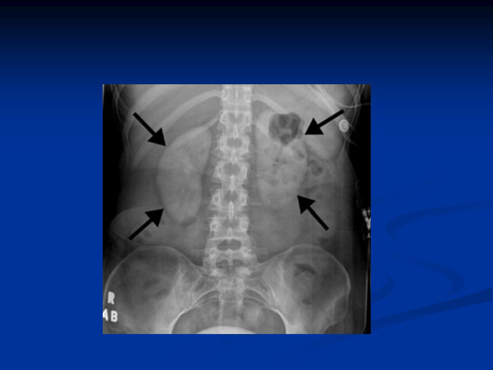
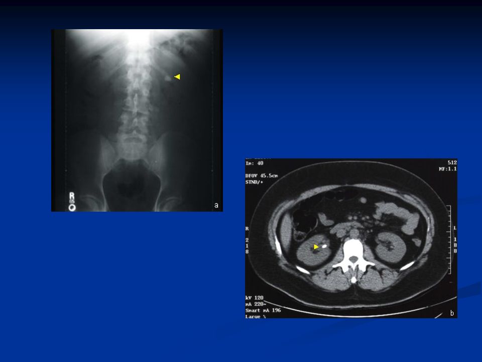
Identify all calcifications !!!
First step: Plain film Look at renal contour Identify all calcifications !!! Urinary calculi (kidney,ureter,bladder) Nephrocalcinosis Prostatic calcification
 Nephrocalcinosis. Prostatic calcification.")
16
Kidneys: position , length , contours
Calices: should be symmetrical, cup-shaped. If dilated club-shaped (due to obstruction or destruction of papilla) Renal pelvis and ureters: normal pelvis is funnel-shaped, ureters are seen in only part of their length due to peristaltism. Bladder: centrally located, smooth outline, should be empty after micturition
 Renal pelvis and ureters: normal pelvis is funnel-shaped, ureters are seen in only part of their length due to peristaltism. Bladder: centrally located, smooth outline, should be empty after micturition.")
18
Computed Tomography Indications:
To characterize renal masses and stage tumors To diagnose or exclude renal trauma To demonstrate stones To assess acute ureteric colic To delineate renal vascular anatomy

19
Normal Findings Renal sinus ; low attenuation in the center
There should not be any calcification Ureters are seen as dots in cross section lying on the psoas muscles Bladder has a smooth outline, thin wall, anechoic urine. Axial images may be reformatted in the coronal and sagittal planes

21
MRI Used in selected circumstances
(renal artery stenosis, IVCal extension of renal tumors)
")
23
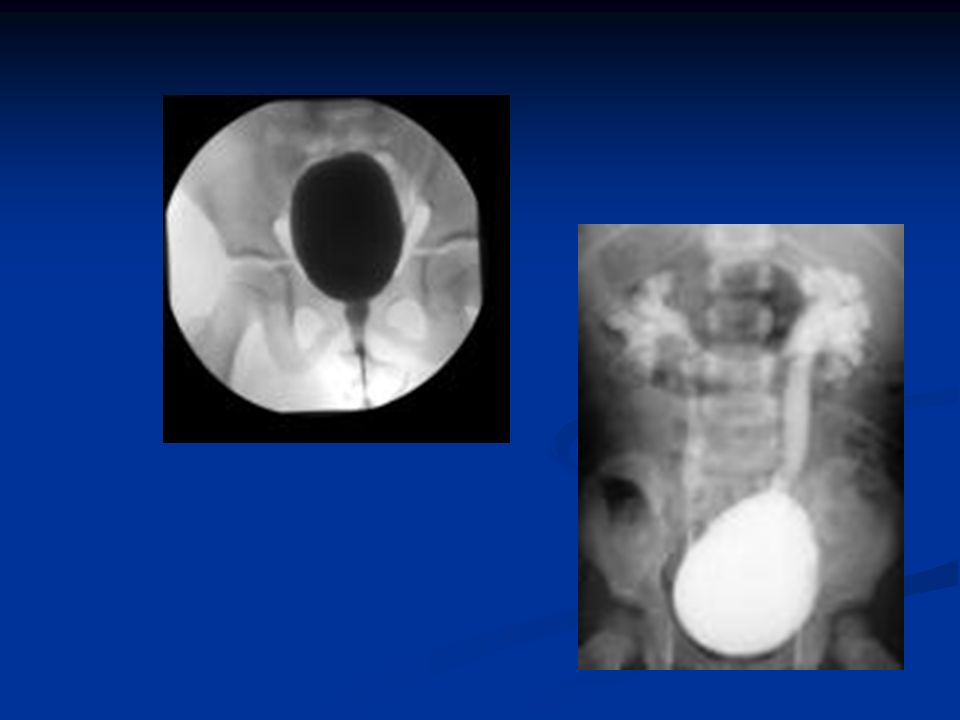
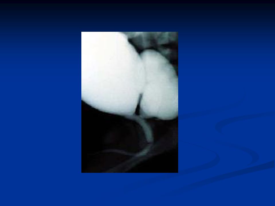
Voiding Cystourethrogram
Bladder is filled with contrast medium through a catheter and films are taken during voiding Observed fluoroscopically to identify reflux of contrast medium from bladder to upper UT. Risk of urinary tract infection, chronic pyelonephritis and renal scarring is increased in VUR.

24
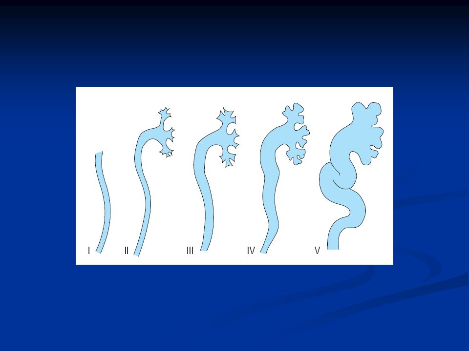
Grading of VUR Grade 1 reflux of urine only into the ureter
Grade 2 reflux into the pelvis and calices, no dilatation Grade 3 mild to moderate dilatation of the ureters and renal pelvis Grade 4 moderate dilatation and tortuosity of the ureters,pelvis,calices Grade 5 gross dilatation and tortuosity of the ureters,pelvis and calices

27
UPPER URINARY TRACT DISORDERS
Urinary Calculi Urinary Tract Obstruction Renal Parenchymal Masses Urothelial Tumors Acute Pyelonephritis, Perinephric abscess Chronic Pyelonephritis Congenital Anomalies

28
LOWER URINARY TRACT DISORDERS
Bladder Tumors Bladder Diverticula Prostatic Enlargement

29
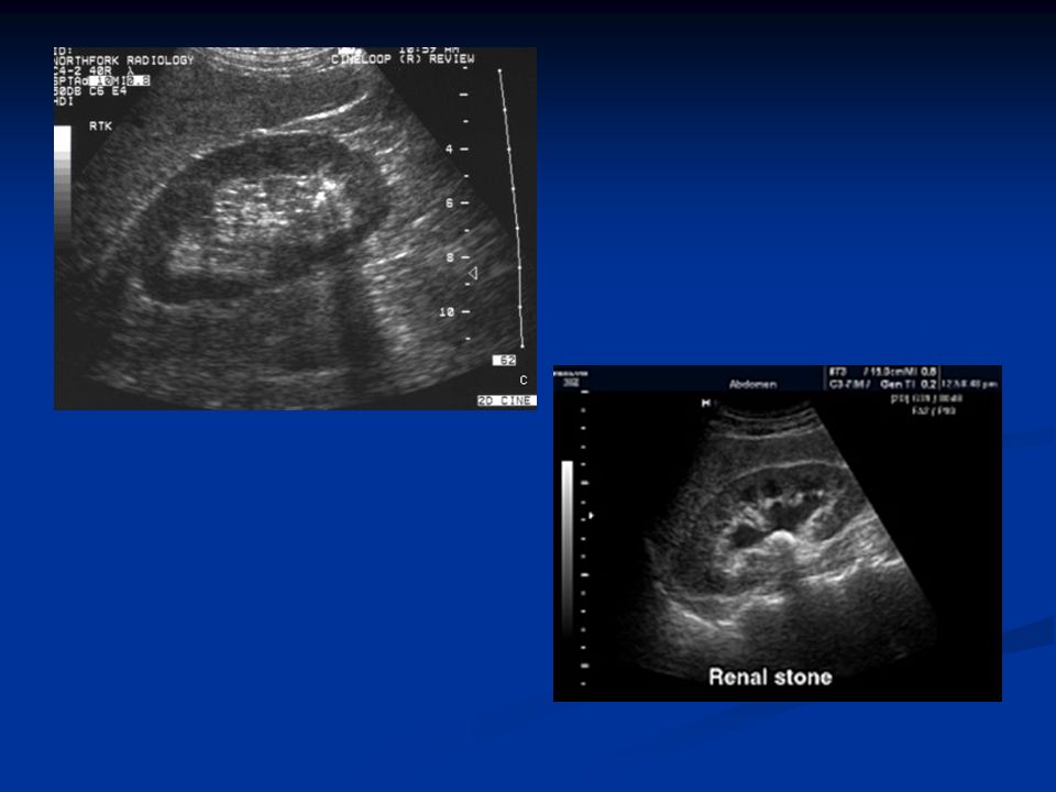
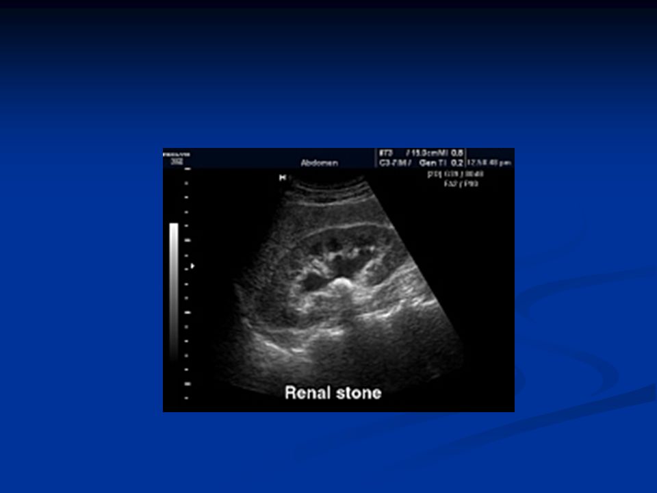
Urinary Calculi Most calculi are calcified and can be seen as radiodense on plain x-ray. Only pure uric acid and xanthine stones are radiolucent on plain radiography, and they can be identified at CT or US. US ; hyperechoic with posterior acoustic shadowing CT ; hyperdense

32
UPPER URINARY TRACT DISORDERS
Urinary Calculi Urinary Tract Obstruction Renal Parenchymal Masses Urothelial Tumors Acute Pyelonephritis, Perinephric abscess Chronic Pyelonephritis Congenital Anomalies

33
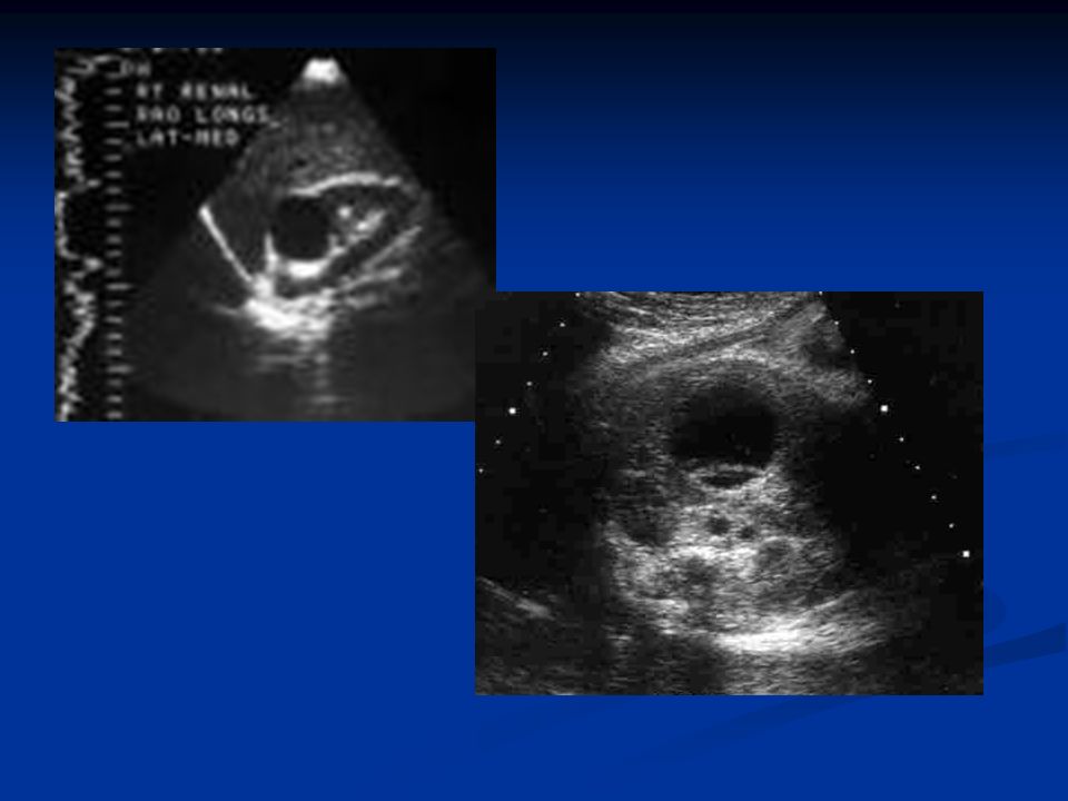
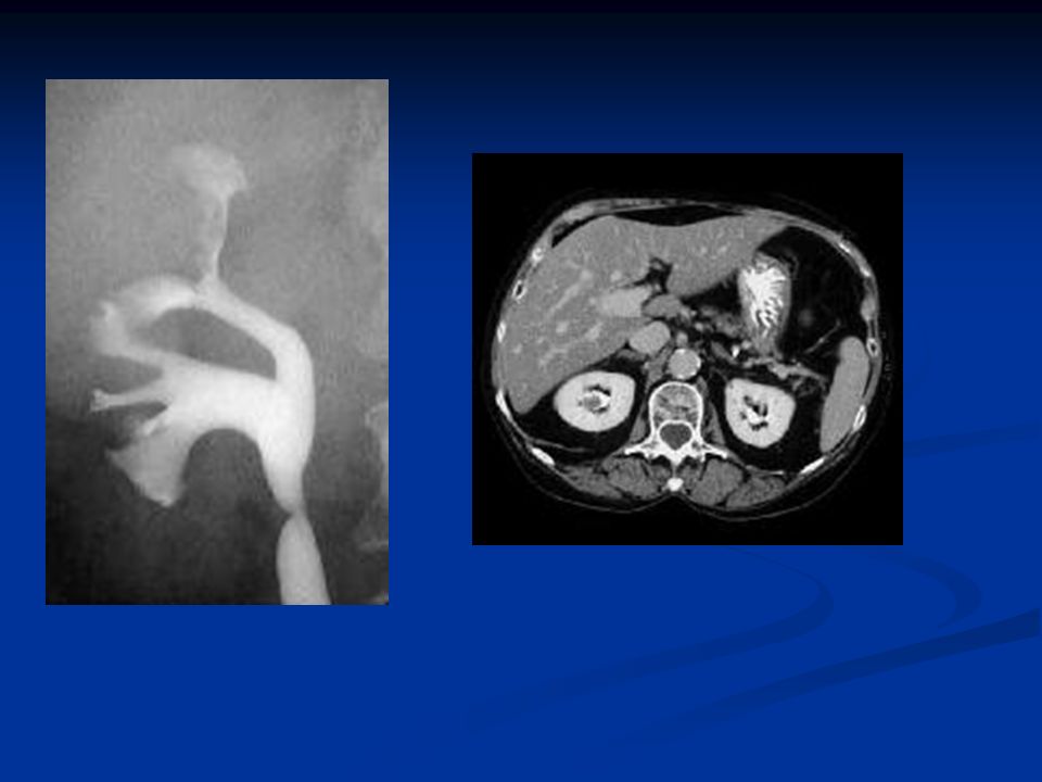
Urinary Tract Obstruction
The main feature is dilatation of the pelvicaliceal system and ureters Main causes : Calculi Blood clot Sloughed papilla Tumors US, IVP and CT

34
US : dilatation of PCS is seen as multiloculate fluid collection in the central echo complex (caused by pooling of urine within the distended pelvis and calices). Proximal ureteric dilatation can also be demonstrated but overlying bowel gas obscures dilatation of the mid and distal ureter.

36
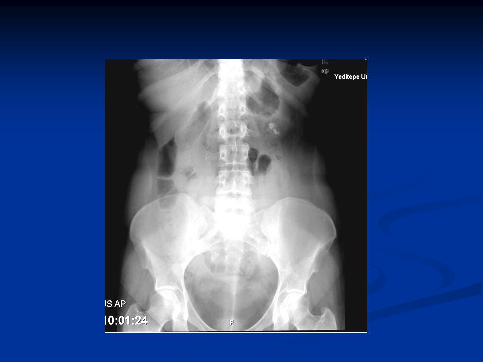
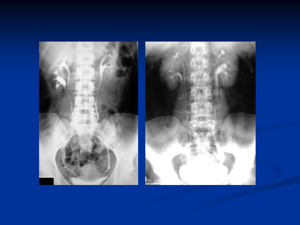
IVP Plain films may be helpful to demonstrate the calculus
Delayed films are essential Filling of the pelvicaliceal system with contrast medium is greatly delayed.

37
CT In acute obstruction, non-contrast CT demonstrates the calculi.

38
UPPER URINARY TRACT DISORDERS
Urinary Calculi Urinary Tract Obstruction Renal Parenchymal Masses Urothelial Tumors Acute Pyelonephritis, Perinephric abscess Chronic Pyelonephritis Congenital Anomalies

39
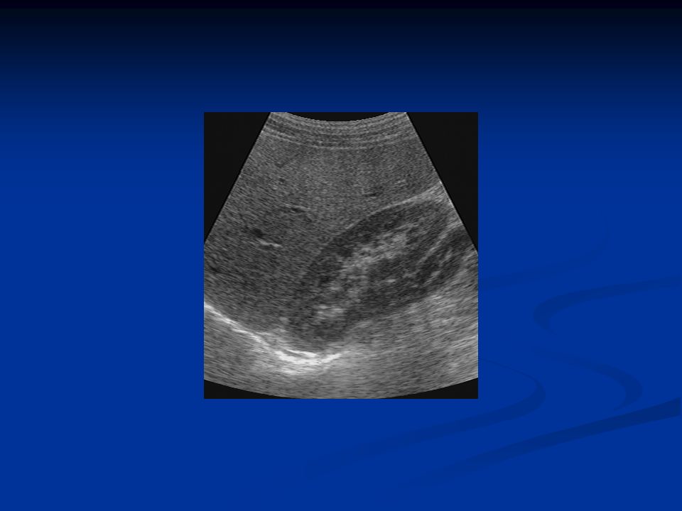
Renal Parencymal Masses
Causes: cyst, benign tumor (angiomyolipoma), renal cell carcinoma, metastases, abscess US usually renal masses are first detected by US. Cystic versus Solid Simple cyst: common in elderly, solitary or multiple, unilocular or septated. Acoustic enhancement.
, renal cell carcinoma, metastases, abscess. US. usually renal masses are first detected by US. Cystic versus Solid. Simple cyst: common in elderly, solitary or multiple, unilocular or septated. Acoustic enhancement.")
41
Angiomyolipoma: small echogenic masses.

42
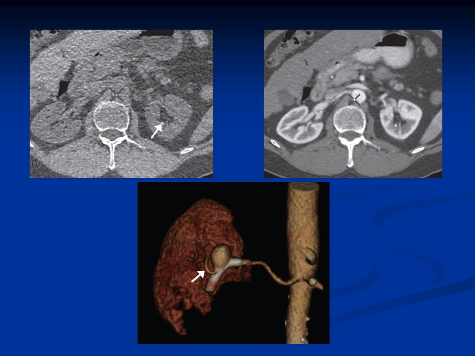
Renal Cell Carcinoma RCCs account for 86% of all primary renal parenchymal tumors. On US; solid tumors May be iso, hypo or hyperechoic. When a tumor is demonstrated, extension into the renal vein and inferior vena cava should be assessed.

43
CT Useful for diagnosis and staging of renal tumors
Shows local direct spread, enlargement of lymph nodes, liver or other organ metastases, renal vein and IVC involvement.

45
UPPER URINARY TRACT DISORDERS
Urinary Calculi Urinary Tract Obstruction Renal Parenchymal Masses Urothelial Tumors Acute Pyelonephritis, Perinephric abscess Chronic Pyelonephritis Congenital Anomalies

46
Urothelial tumors are seen as filling defects in the renal pelvis and ureters
Filling defects in the collecting system: calculi, blood clot, tumor They may obstruct the ureter and cause hyrdonephrosis.

48
Congenital Anomalies Bifid Collecting System Ectopic Kidney
Horseshoe kidney Renal Agenesis

49
Bifid Collecting System
Most frequent congenital variation Unilateral or bilateral Bifid pelvis ureteric duplication

51
Ectopic Kidney During fetal development kidneys ascend in the abdomen
Ectopic kidney results when this ascent is halted In some cases kidneys lie on the same side and are fused (crossed fused ectopia)
")
52
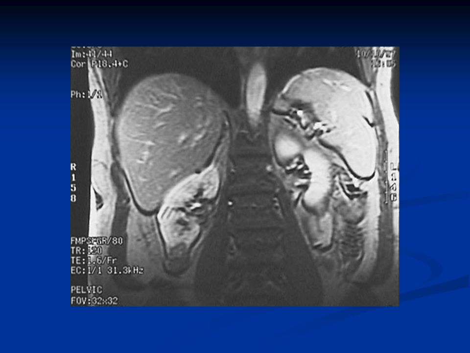
Horseshoe Kidney The kidneys may fail to seperate, giving rise to a horseshoe kidney Lower poles are fused by parenchyma or fibrous tissue

54
LOWER URINARY TRACT DISORDERS
Bladder Tumors Bladder Diverticula Prostatic Enlargement

55
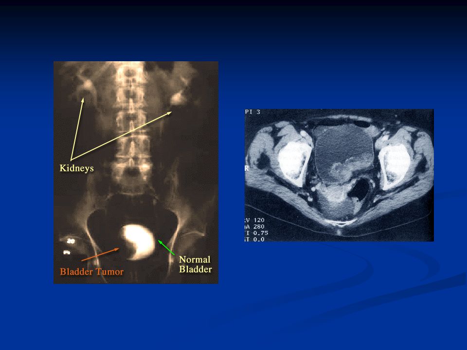
Bladder Tumor Most frequent site for neoplasms of the urinary tract
On US; bladder tumors are seen as soft tissue masses protruding into the bladder. IVP; filling defect in the bladder On CT and MRI; soft tissue mass projecting from the bladder wall

58
LOWER URINARY TRACT DISORDERS
Bladder Tumors Bladder Diverticula Prostatic Enlargement

59
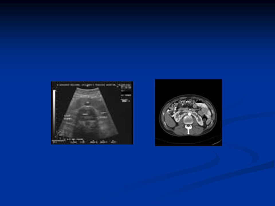
Bladder Diverticula May be congenital or secondary to chronic obstruction Demonstrated by US, CT or MRI.

62
LOWER URINARY TRACT DISORDERS
Bladder Tumors Bladder Diverticula Prostatic Enlargement

63
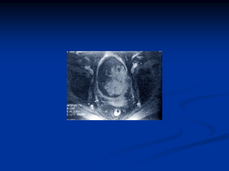
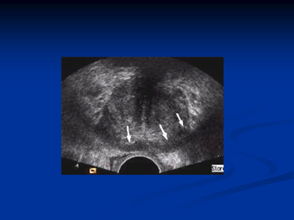
Prostate Imaging Prostatic enlargement : benign prostatic hypertrophy, prostatic carcinoma Prostatic ultrasound; transducer introduced into the rectum TRUS guided biopsy MRI ; tumor is seen mostly in the peripheral zone as hypointense on T2

67
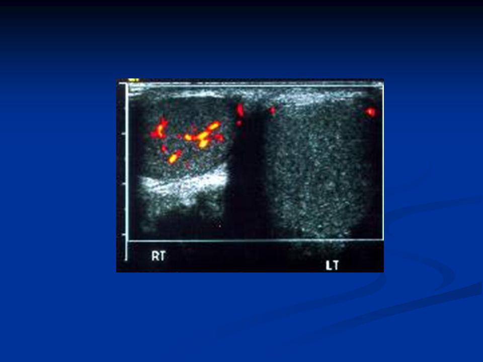
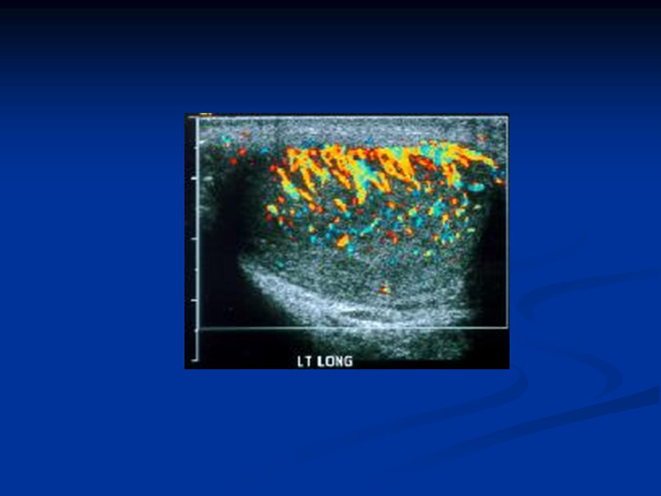
Scrotal Swelling US, Color Doppler US, MRI
Testicular tumor, orchitis-epididymo-orchitis, testicular torsion, hydrocele Doppler ; used in acute scrotum; to differentiate between epididiymo-orchitis and testicular torsion Epididiymo-orchitis ; medical treatment Torsion ; surgery

Similar presentations


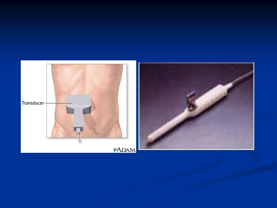


 Transducer placed on patient’s body Sound waves echo.>")


 are variable, occur in 1 of 500 newborns; predisposing to development of hypertension,>")


