Download presentation
Presentation is loading. Please wait.

1
Approach to Abdominal Plain Film Radiology Nalin Amin, MD, CCFP, FRCSC Assistant Professor Dept. Of Surgery, McMaster University

2
Objectives What is the use of plain xrays anymore? What is the use of plain xrays anymore? Three views? Three views? Radiation concern? Radiation concern? Flat Plate? KUB? Decub? Flat Plate? KUB? Decub? Develop approach Develop approach CASES CASES

3
What is the use? What is the use? Still good screening for: Still good screening for: Bowel obstruction Bowel obstruction Ileus Ileus Free air (on upright views) Free air (on upright views) NGT placement NGT placement Stones follow-up Stones follow-up etc etc
 Free air (on upright views) NGT placement NGT placement Stones follow-up Stones follow-up etc etc.")
4
3 views 3 views Upright/Standing Upright/Standing Supine (upper) Supine (upper) Supine (lower) Supine (lower)
 Supine (upper) Supine (lower) Supine (lower)")
5
Upright Supine (upper) Supine (lower)
 Supine (lower)")
6
Flat plate Flat plate b/c used ‘glass plate’ in old days b/c used ‘glass plate’ in old days Now digital technology Now digital technology KUB KUB ‘Kidneys/Ureters/Bladder’ ‘Kidneys/Ureters/Bladder’ Supine view with field-of-view from Supine view with field-of-view from kidney to bladder +/- IV contrast +/- IV contrast Assess for radiopaque calculi Assess for radiopaque calculi Decub Decub Lying on side Lying on side Good for free air if cannot do upright Good for free air if cannot do upright

7
Radiation 1 CXR ~ 0.1 mSv ≈ 1 cigarette ( risk of ca) 1 CXR ~ 0.1 mSv ≈ 1 cigarette ( risk of ca) 1 abdo XR ~ 0.7 mSv ≈ 7 cigs 1 abdo XR ~ 0.7 mSv ≈ 7 cigs 1 pelvis XR ~ 0.6 mSv 1 pelvis XR ~ 0.6 mSv CT abdo/pelvis ~14 mSv ≈ 140 cigs CT abdo/pelvis ~14 mSv ≈ 140 cigs CT chest ~7 mSv CT chest ~7 mSv
 1 CXR ~ 0.1 mSv ≈ 1 cigarette ( risk of ca) 1 abdo XR ~ 0.7 mSv ≈ 7 cigs 1 abdo XR ~ 0.7 mSv ≈ 7 cigs 1 pelvis XR ~ 0.6 mSv 1 pelvis XR ~ 0.6 mSv CT abdo/pelvis ~14 mSv ≈ 140 cigs CT abdo/pelvis ~14 mSv ≈ 140 cigs CT chest ~7 mSv CT chest ~7 mSv")
8
APPROACH ABCS! ABCS! A = air A = air B = bowel B = bowel C = calcifications C = calcifications S = soft tissues S = soft tissues

9
A A ir

10
B B owel LB SB Stomach

11
renal stone chole clips anastomotic staple pelvic clips C C alcifications 12 th rib phlebolith L-Spine w/DDD

12
liver edge S S oft Tissues spleen margin properitoneal fat left kidney shadow

13
S S oft Tissues bladder male pt XY

14
Diff SB vs LB Small BowelLarge Bowel Valvulae conniventesHaustra CompleteIncomplete Central Peripheral (Picture frame) Smaller calibre (max 3 cm) Larger calibre ( LB 6 cm, cecum 9 cm)
 Smaller calibre (max 3 cm) Larger calibre ( LB 6 cm, cecum 9 cm)")
15
CASES Case 1 Case 1

17
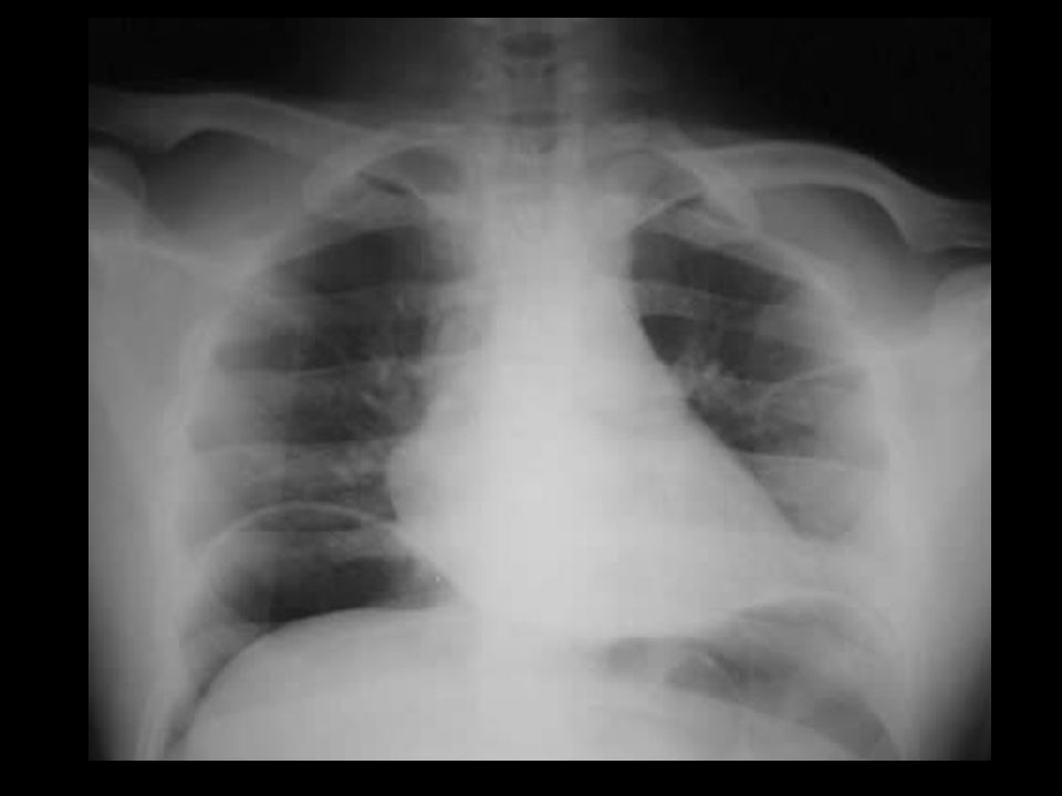
Free air (on upright) Free air (on upright)
 Free air (on upright)")
19
CASES Case 2 Case 2

21
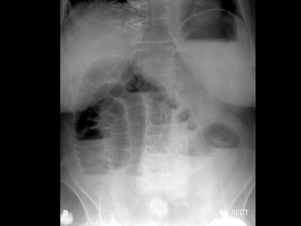
Free air (on supine) Free air (on supine) harder to see harder to see need significant amount more to see need significant amount more to see Football sign Football sign falciform is the laces falciform is the laces Rigler’s sign Rigler’s sign bowel wall outlined by free air bowel wall outlined by free air normally not mesenteric side aspect seen normally not mesenteric side aspect seen
 Free air (on supine) harder to see harder to see need significant amount more to see need significant amount more to see Football sign Football sign falciform is the laces falciform is the laces Rigler’s sign Rigler’s sign bowel wall outlined by free air bowel wall outlined by free air normally not mesenteric side aspect seen normally not mesenteric side aspect seen")
23
CASES Case 3 Case 3

24
CASES

25
left renal calculi left renal calculi 80% radioopaque 80% radioopaque ca2+ oxalate, phosphate ca2+ oxalate, phosphate struvite struvite 20% radiolucent 20% radiolucent uric acid (+ve on CT) uric acid (+ve on CT) cystine (+ve on CT) cystine (+ve on CT) HIV indinavir (-ve on CT) HIV indinavir (-ve on CT)
 uric acid (+ve on CT) cystine (+ve on CT) cystine (+ve on CT) HIV indinavir (-ve on CT) HIV indinavir (-ve on CT)")
26
CASES

27
CASES Case 4 Case 4

29
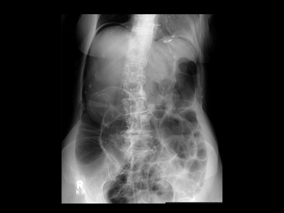
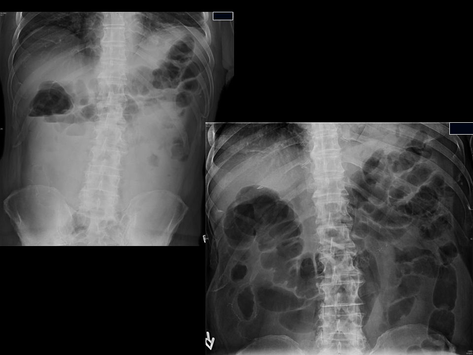
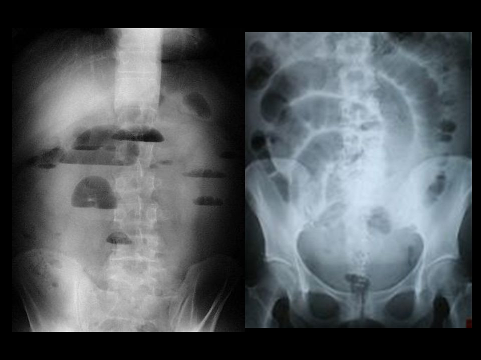
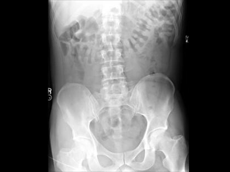
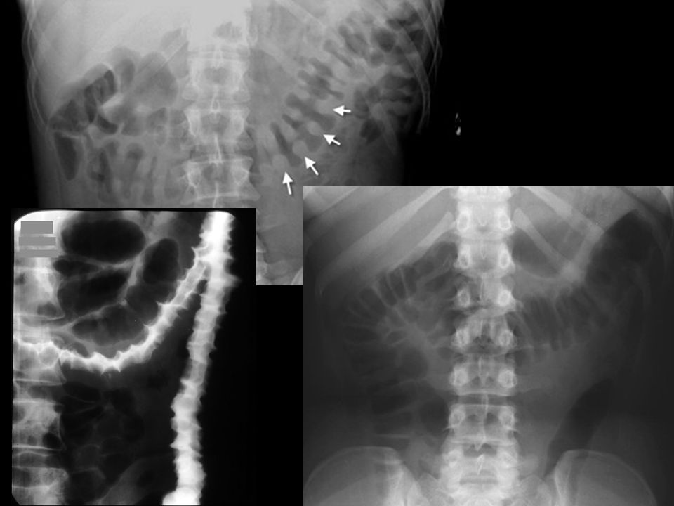
SBO SBO multiple AF levels multiple AF levels varying heights varying heights “string-of-pearls” “string-of-pearls” low grade vs high grade low grade vs high grade early/partial vs complete early/partial vs complete if gas is seen distally (early/partial) if gas is seen distally (early/partial) decompressed distally (complete/high grade) decompressed distally (complete/high grade)
 if gas is seen distally (early/partial) decompressed distally (complete/high grade) decompressed distally (complete/high grade)")
31
SBO causes SBO causes adhesions (50%) adhesions (50%) hernias (15%) hernias (15%) ca (1º and mets) (15%) ca (1º and mets) (15%)
 adhesions (50%) hernias (15%) hernias (15%) ca (1º and mets) (15%) ca (1º and mets) (15%)")
32
CASES Case 5 Case 5

34
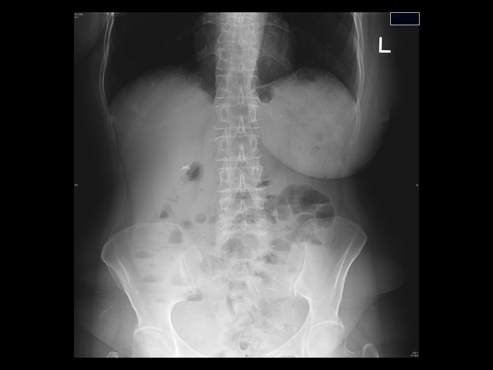
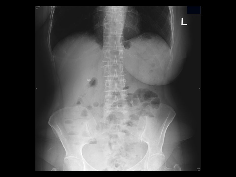
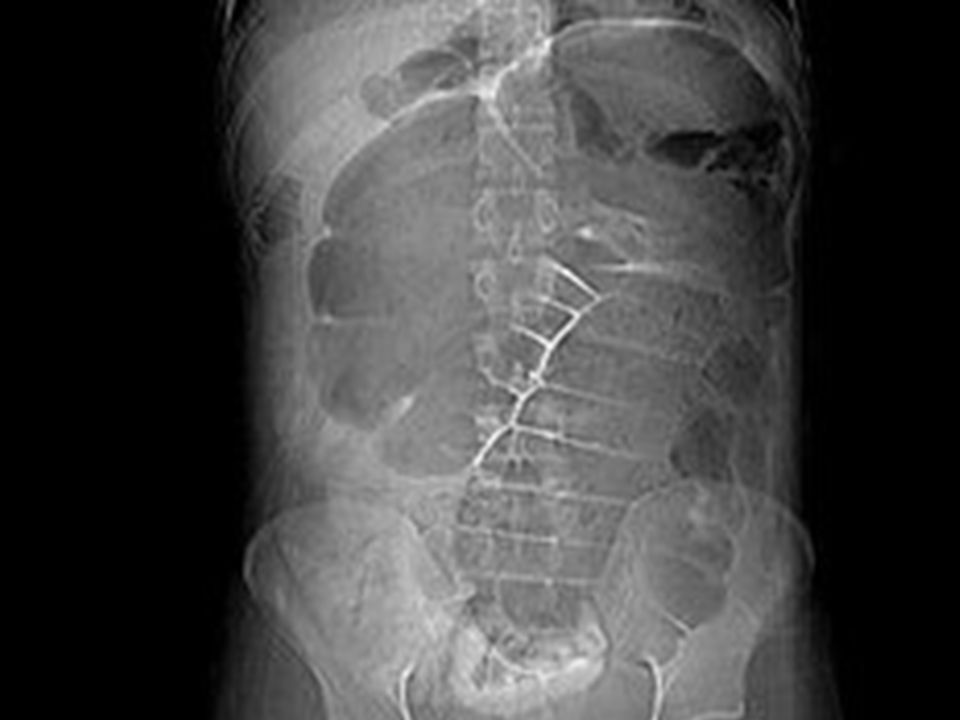
LBO LBO

35
‘applecore’ carcinoma proximal LBO

36
LBO causes LBO causes Cancer Cancer Diverticulitis Diverticulitis Volvulus Volvulus Hernia Hernia note if IC valve competent or incompetent note if IC valve competent or incompetent this case, competent this case, competent ie. no SBD ie. no SBD

37
non-dilated SB loops

38
CASES Case 6 Case 6

40
paralytic ileus paralytic ileus hard to discriminate from BO hard to discriminate from BO ileusbowel obstruction tends to involve SB & LB usually only SB or part LB (unless distal LBO) uniform dilated calibreprogressive dilatation AFL at same heightAFL at varying heights clinical history (meds, post-op, tinkle, etc.) clinical history (bowel sounds, etc.)
 uniform dilated calibreprogressive dilatation AFL at same heightAFL at varying heights clinical history (meds, post-op, tinkle, etc.) clinical history (bowel sounds, etc.)")
42
CASES Case 7 Case 7

44
gallstone ileus gallstone ileus

45
gallstone dilated SB loops pneumobilia

46
triad triad gallstone gallstone SBO or ileus SBO or ileus pneumobilia pneumobilia central central as opposed to PV gas (peripheral) as opposed to PV gas (peripheral)
 as opposed to PV gas (peripheral)")
47
CASES Case 8 Case 8

49
thumbprinting bowel wall edema DDx (4 I’s) ischemia infectious colitis PMC inflammatory (UC/IBD) infiltrative other: edema, Rn, tumor, hemorrhage
 ischemia infectious colitis PMC inflammatory (UC/IBD) infiltrative other: edema, Rn, tumor, hemorrhage")
51
CASES Case 9 Case 9

53
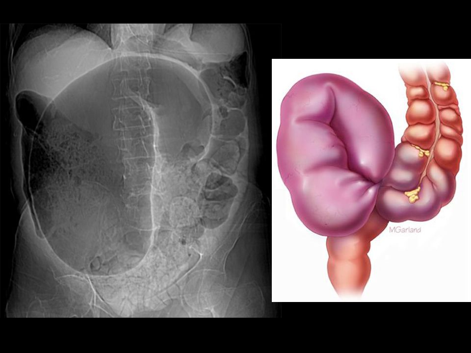
sigmoid volvulus sigmoid volvulus flips into RUQ flips into RUQ flips into LUQ cecal volvulus flips into LUQ kidney bean shaped kidney bean shaped midline crease – mesenteric vessels midline crease – mesenteric vessels coffee bean cecal volvulus coffee bean

55
CASES Case 9a Case 9a

57
cecal volvulus cecal volvulus

58
CASES Case 10 Case 10

60
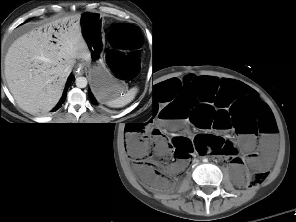
Bad News Bad News Pneumatosis Pneumatosis Portal venous gas Portal venous gas pre-morbid pre-morbid

61
portal venous gas pneumatosis

62
CASES Case 10 Case 10

64
THE END

Similar presentations


![James Zeng. Bowel Obstruction A blockage of bowel lumen prohibiting the passage of materials[1] 8% of abdo pain in ED (3 rd leading cause)[2] 24% require.](/20/6022766/big_thumb.jpg "James Zeng. Bowel Obstruction A blockage of bowel lumen prohibiting the passage of materials[1] 8% of abdo pain in ED (3 rd leading cause)[2] 24% require.>")