Download presentation
Presentation is loading. Please wait.

1
Abdominal Imaging

2
The abdominal radiograph

3
Why do we see any structure on a normal abdominal radiograph? It has to be surrounded by tissue of different attenuation (x-ray stopping power). we therefore see margins delineated by air (the mucosal surface of air filled bowel) Things outlined by fat (usually the kidneys. the psoas margins,the bladder and the abdominal wall).
. we therefore see margins delineated by air (the mucosal surface of air filled bowel) Things outlined by fat (usually the kidneys. the psoas margins,the bladder and the abdominal wall)..")
4
We can only see gas soft tissue (effectively water density)- viscera, bowel and bowel wall, bladder. bone and calcium-stones, calcified lymph nodes,stones(90% renal, 10% gallstones) Fat
 Fat.")
5
Properitoneal fat Transversus abdominis

10
A FEW INTERESTING RADIOGRAPHS

11
Gas outside the bowel. Outling both the inside and outside of the bowel. Free gas

12
DILATED LARGE BOWEL WITH NO BOWEL WALL THICKENING Turned out to be a sigmoid tumour.

14
Gross large bowel wall thickening. Secondary to ulcerative colitis

15
Causes of colitis Inflammatory bowel disease Ischaemic colitis Pseudomembranous colitis Amoebic,schistosoma etc

16
Gas outside the bowel. Outling both the inside and outside of the bowel. Free gas

17
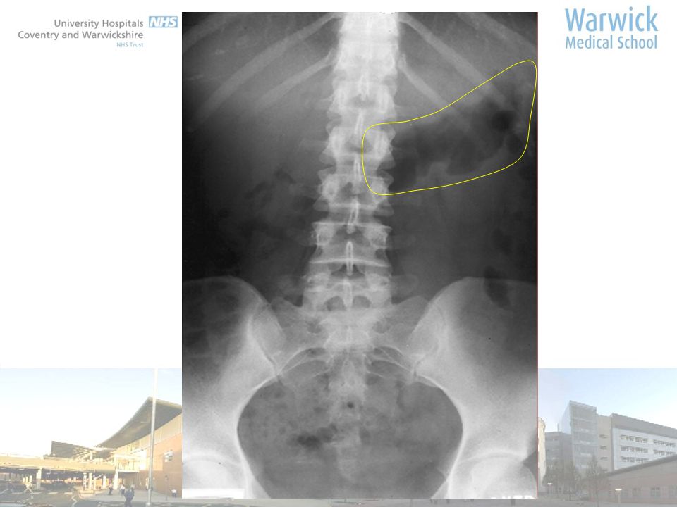
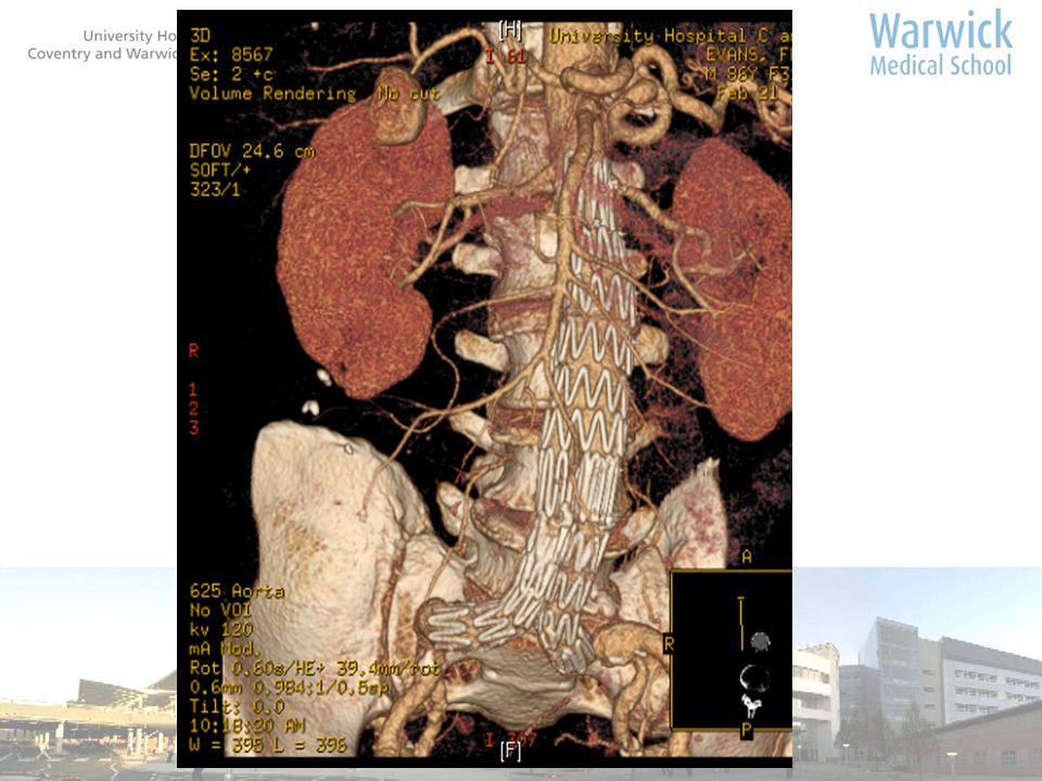
Aortic aneurysm

20
Basic CT Anatomy

21
mhv rhv IVC anterior posterior right left

23
gb rk ra cl (1) stomach anterior posterior right left spleen pv ha
 stomach anterior posterior right left spleen pv ha")
32
anterior posterior right left rk lk pancreas sv d cd

33
Fluid in lesser sac gallbladder

35
Renal pelvis Renal artery Renal vein

36
uu anterior posterior right left

37
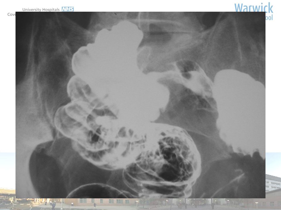
u u anterior posterior Right left

38
anterior posterior right left u u bladder nav

41
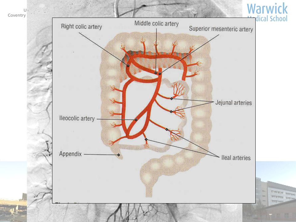
Left gastric A Gastroduodenal A Common Hepatic A Splenic A Pancreaticoduodenal A Right gastric –small, from proper hepatic or Lt gastric

44
Image of an aneurysm to discuss suitability

Similar presentations


>")


