Download presentation
Presentation is loading. Please wait.

1
Anatomy and Physiology I
Tissue Types

2
Key Terms Histology: the study of tissues. Tissues:
groups of cells which are similar in structure and which perform common or related functions.

3
Four Basic Kinds of Tissues
Epithelial Tissue Connective Tissue Muscle Tissue Nervous Tissue

4
Epithelial Tissue Epithelial Tissue Locations:
Covers the body Lines the cavities, tubes, ducts and blood vessels inside the body Covers the organs inside body cavities Epithelial Tissue Functions: Protection from physical & chemical injury, Protection against microbial invasion, Contains receptors which respond to stimuli, Filters, secretes & reabsorbs materials and Secretes serous fluids to lubricate structures.

5
Examples of Epithelium Tissue
Lines the respiratory tract Lines the digestive system Skin Surrounding internal organs Characteristics of Epithelium Tissue Cells are tightly packed Regenerate easily Avascular – lack blood vessels apical surface – exposed surface Basement membrane – anchors down cells

6
Connective Tissue Connective Tissue: Connective Tissue Functions:
Most abundant & widely distributed tissue Connective Tissue Functions: Connects, binds and supports structures, Tendons, ligaments, etc. Protects & cushions organs and tissues, Insulates (fat) and Transports substances (blood).
 and. Transports substances (blood).")
7
Examples of Connective Tissue
Bones Cartilage Tendons Blood Fat Characteristics of Connective Tissue Most are well vascularized – have a good blood supply Some have very poor blood supply being avascular. Extracellular matrix – nonliving substance found outside the cells

8
Extracellular Matrix explained
Nonliving material between cells Produced by the cells and then extruded Responsible for the strength Two components Ground substance Of fluid, adhesion proteins, proteoglycans Liquid, semisolid, gel-like or very hard Fibers: collagen, elastic or reticular

9
Epithelial Tissue in Detail

10
Classification of epithelia
According to thickness “simple” - one cell layer “stratified” – more than one layer of cells (which are named according to the shape of the cells in the apical(base) layer) According to shape “squamous” – wider than tall “cuboidal” – as tall as wide “columnar” - taller than wide
 layer) According to shape. squamous – wider than tall. cuboidal – as tall as wide. columnar - taller than wide.")
11
Classes of Epithelia Simple: just one layer or cell shape
Stratified: multiple layers and cell shapes Human Anatomy, Larry M. Frolich, Ph.D.

12
Human Anatomy, Larry M. Frolich, Ph.D.
Simple Epithelia Human Anatomy, Larry M. Frolich, Ph.D.

13
to protect

14
where diffusion is important
where tissues are involved in secretion and absorption: larger cells because of the machinery of production, packaging, and energy requirements

16
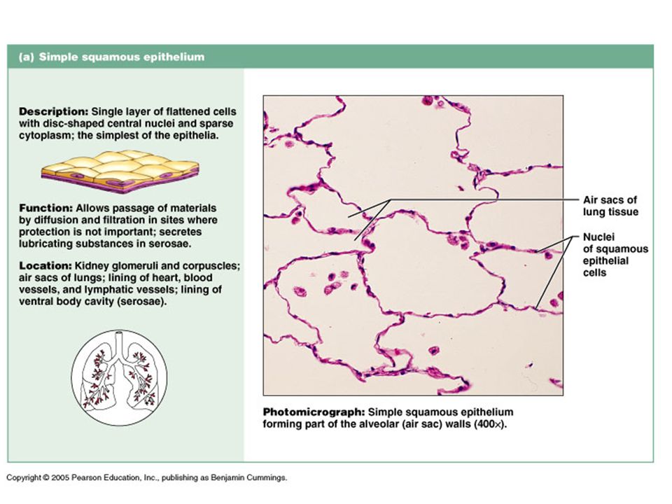
Simple Squamous Epithelium
Descriptions: Single layer, flattened cells, simplest of epithelia Function: Allows passage of materials by diffusion and filtration Secretes lubricating substances Location: Air sacs of lungs, lining of heart, blood vessels, lining of ventral body cavity

18
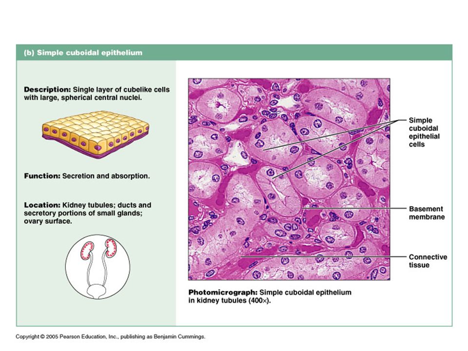
Simple Cuboidal epithelium
Descriptions: Single layered, cube like, large spherical central nuclei Function: Secretion and absorption Location: Kidney tubules, ovary surfaces

19
“ciliated” literally = eyelashes
(see next page)
")
20
Simple Columnar Epithelium
Descriptions: Single layer, tall, round or oval nuclei, some bear cilia, may contain mucus-secreting glands(goblet cells) Function: Absorption, secretion of mucus and enzymes, cilia propels mucus Location: Nonciliated lines the digestive tract Ciliated lines small bronchi, uterine tubes, parts of uterus.
 Function: Absorption, secretion of mucus and enzymes, cilia propels mucus. Location: Nonciliated lines the digestive tract. Ciliated lines small bronchi, uterine tubes, parts of uterus.")
22
Pseudostratified Columnar Epithelium
Descriptions: Single layer w/ differing heights, nuclei at different levels, may have goblet cells or cilia Function: Secretion(especially mucus), propulsion of mucus by ciliary action Location: Nonciliated in sperm carrying ducts Ciliated lines the trachea and upper respiratory tract.
, propulsion of mucus by ciliary action. Location: Nonciliated in sperm carrying ducts. Ciliated lines the trachea and upper respiratory tract.")
23
Stratified: regenerate from below

24
Stratified Squamous Epithelium
Descriptions: (most common stratified) Several layers, surface cells are squamous, basal cells are cuboidal or columnar Function: Protects underlying tissues in areas of abrasion Location: Esophagus, the mouth, outer portion of skin
 Several layers, surface cells are squamous, basal cells are cuboidal or columnar. Function: Protects underlying tissues in areas of abrasion. Location: Esophagus, the mouth, outer portion of skin.")
25
Rare…

26
Stratified Cuboidal Epithelium
Descriptions: (rare) Usually two layers of cube-like cells Function: Protection Location: Rare Largest ducts of sweat glands, mammary glands, and salivary glands.
 Usually two layers of cube-like cells. Function: Protection. Location: Rare. Largest ducts of sweat glands, mammary glands, and salivary glands.")
27
Rare…

28
Stratified Columnar Epithelium
Descriptions: (rare) Several layers thick, columnar with varying in size and shape Function: Protection, secretion Location: Rare Large ducts of glands
 Several layers thick, columnar with varying in size and shape. Function: Protection, secretion. Location: Rare. Large ducts of glands.")
30
Transitional Epithelium
Descriptions: Highly modified, several layers of cuboidal or columnar cells Function: Stretches readily, allowing distention of urinary organs Location: Lines the ureters, bladder, parts of urethra

31
Human Anatomy, Larry M. Frolich, Ph.D.

32
Can You Identify the Classes of Epithelium?
Quiz!! E Can You Identify the Classes of Epithelium? D A B C Human Anatomy, Larry M. Frolich, Ph.D.

33
Specific Connective Tissue In Detail

35
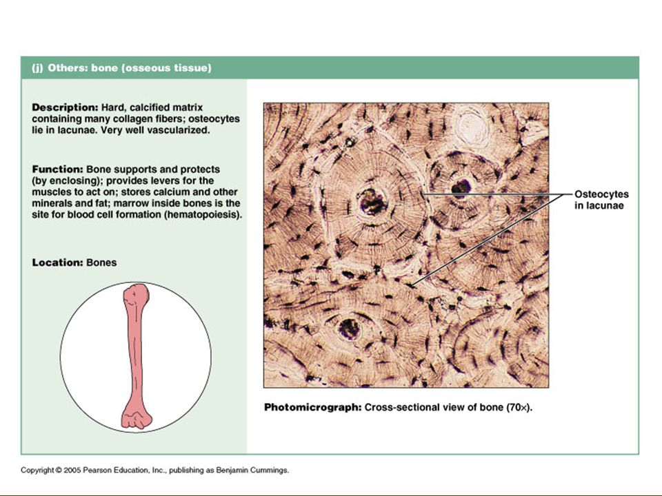
Bone Descriptions: Aka: osseous tissue Function: Location:
Hard calcified matrix, surrounded by layers of calcium salts in additions to lots of collagen fibers Cells sit in cavities called “lacunae” Function: Protection and support Location: Skeletal system

37
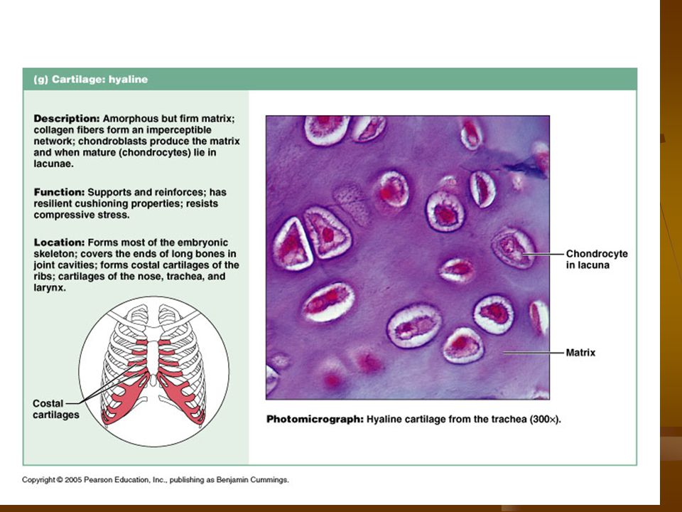
Hyaline Cartilage Descriptions: (hyalin=glass) Function: Location:
Most common and widespread type Collagen fibers with a rubbery matrix Function: Supports/reinforces, resilient cushioning Location: Covers ends of bones in joints, nose, trachea, larynx, embryonic skeleton

39
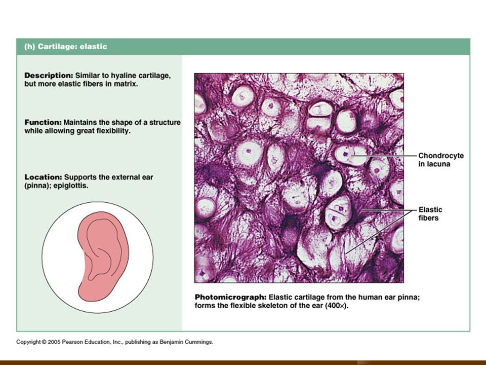
Elastic Cartilage Descriptions: Function: Location:
Collagen fibers with a rubbery matrix Very elastic Function: Maintains shape of structure gives flexibility Location: External ear epiglottis

41
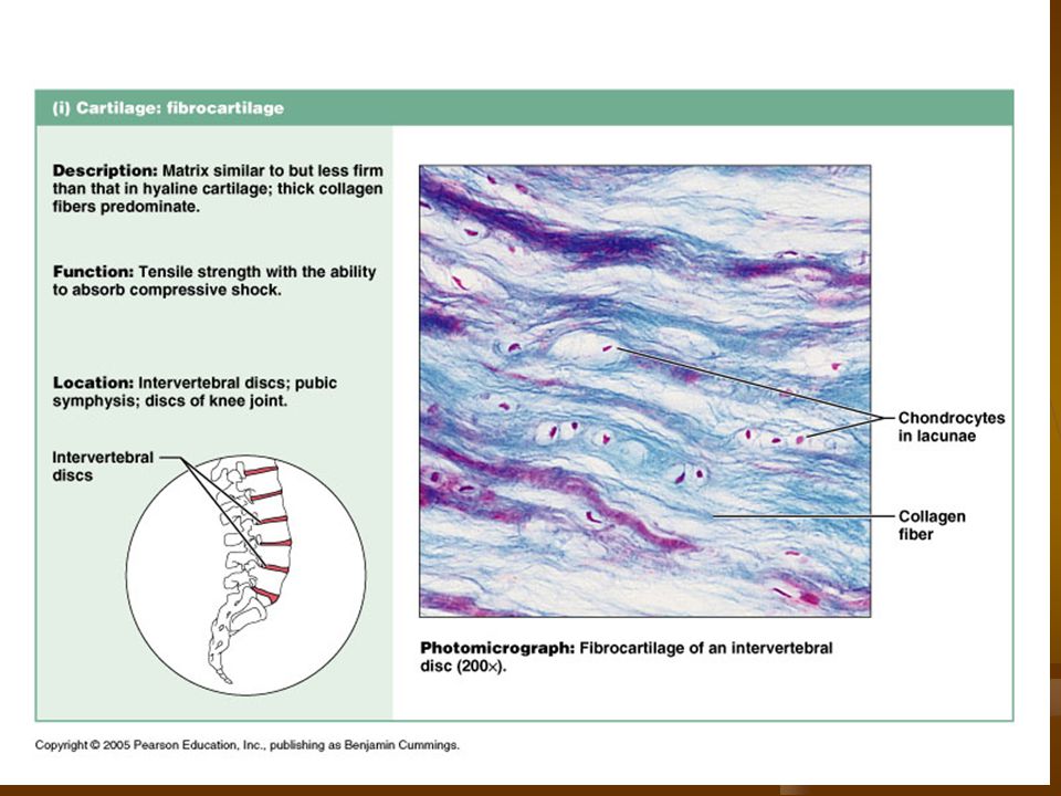
Fibrocartilage Descriptions: Function: Location:
Similar to hyaline but less firm Much like a cushion Function: Absorbs compressive shock Location: Intervertebral discs, discs of knee joint

44
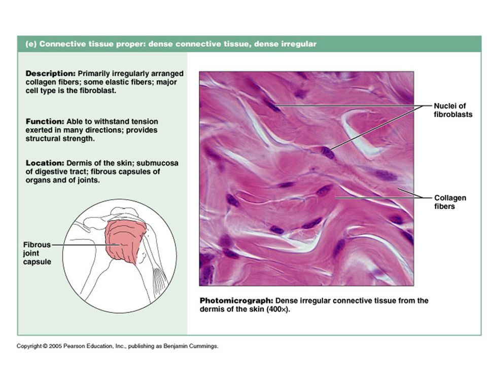
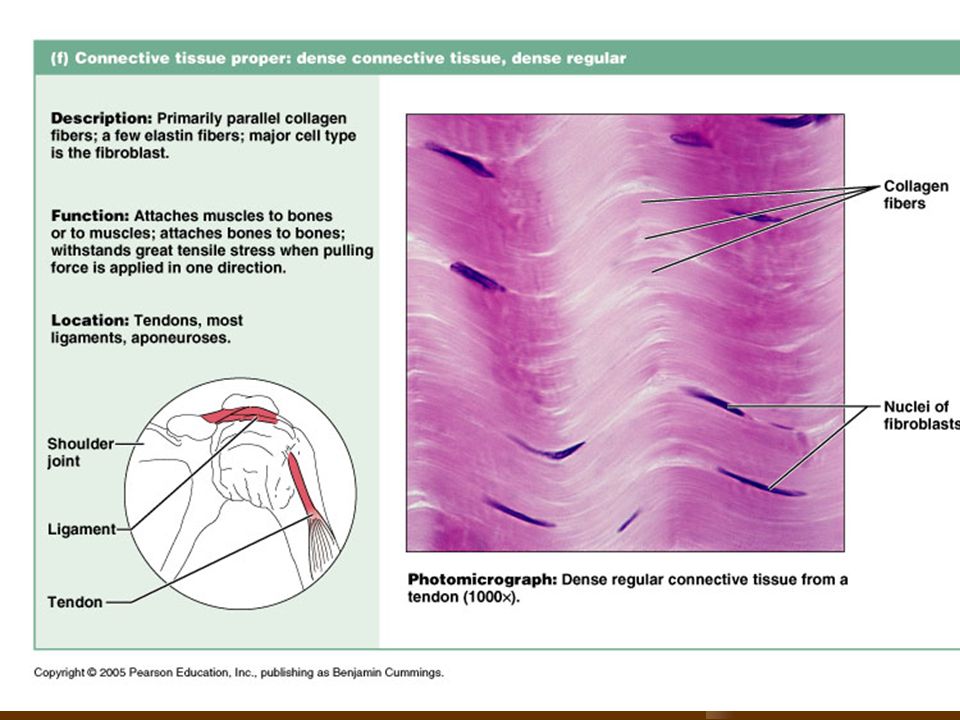
Dense Connective Tissue
Descriptions: Collagen fibers, fibroblasts fill gaps between collagen Strong ropelike structures like tendons and ligaments Function: Connect muscles to bones, connect bones at joints, lots of tensile strength Location: joints

45
Vocab Fibroblasts – cells that make collagen fibers
Tendons – attach skeletal muscles to bones Ligaments - connect bones to bones at joints. More stretchy and contain more elastic fibers than tendon

47
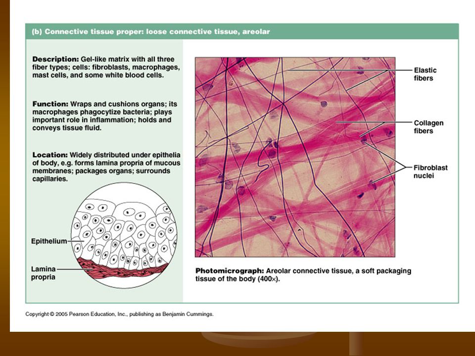
Loose CT: Areolar Tissue
Descriptions: Gel-like matrix w/ all three fiber types Very loose network w/ lots of open space Function: Wraps and cushions organs, a reservoir of water and salts for surrounding tissues, important for swelling Location: Under epithelia, around organs, surrounds capillaries

49
Loose CT: Adipose Tissue
Descriptions: (fat) Very similar to areolar tissue Fat cells predominate Function: Insulates against heat loss, supports and protects organs, reserve food fuel Location: Under skin, around kidneys and eyeballs, abdomen and breast
 Very similar to areolar tissue. Fat cells predominate. Function: Insulates against heat loss, supports and protects organs, reserve food fuel. Location: Under skin, around kidneys and eyeballs, abdomen and breast.")
51
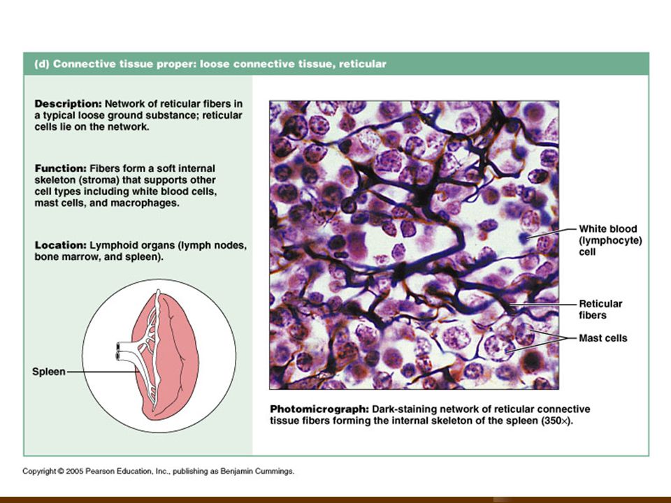
Loose CT: Reticular CT Descriptions: Function: Location:
Network of reticular fibers (similar to fibroblast) Delicate network of interwoven fibers Function: Form soft internal skeleton that supports other cells such as white blood cells, mast cells Location: Lymphoid organs (lymph nodes, bone marrow, spleen)
 Delicate network of interwoven fibers. Function: Form soft internal skeleton that supports other cells such as white blood cells, mast cells. Location: Lymphoid organs (lymph nodes, bone marrow, spleen)")
53
Blood Descriptions: (vascular tissue) Function: Location:
Red and white blood cells in a fluid matrix (plasma) Fibers are soluble only seen during clotting Function: Transportation of gases, nutrients, wastes ect. Location: Everywhere
 Fibers are soluble only seen during clotting. Function: Transportation of gases, nutrients, wastes ect. Location: Everywhere.")
54
Muscle Tissue Muscle Tissue: Muscle Tissue Functions:
Associated with the bones of the skeleton, the heart and in the walls of the hollow organs of the body. Made to contract Called muscle fibers because they are elongated to provide a long axis for contraction Muscle Tissue Functions: Movement Maintains posture Produces heat Facial expressions Pumps blood Peristalsis

55
Specific Muscle Tissue Types:
skeletal muscle Cardiac muscle Smooth muscle

57
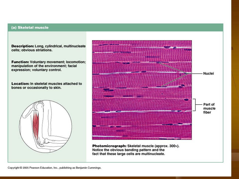
Skeletal Muscle Descriptions: Function: Location:
Long, cylindrical, obvious striations, multinucleated Function: Voluntary movement, manipulation of the environment, facial expression Gross body movement Location: Attached to bones

59
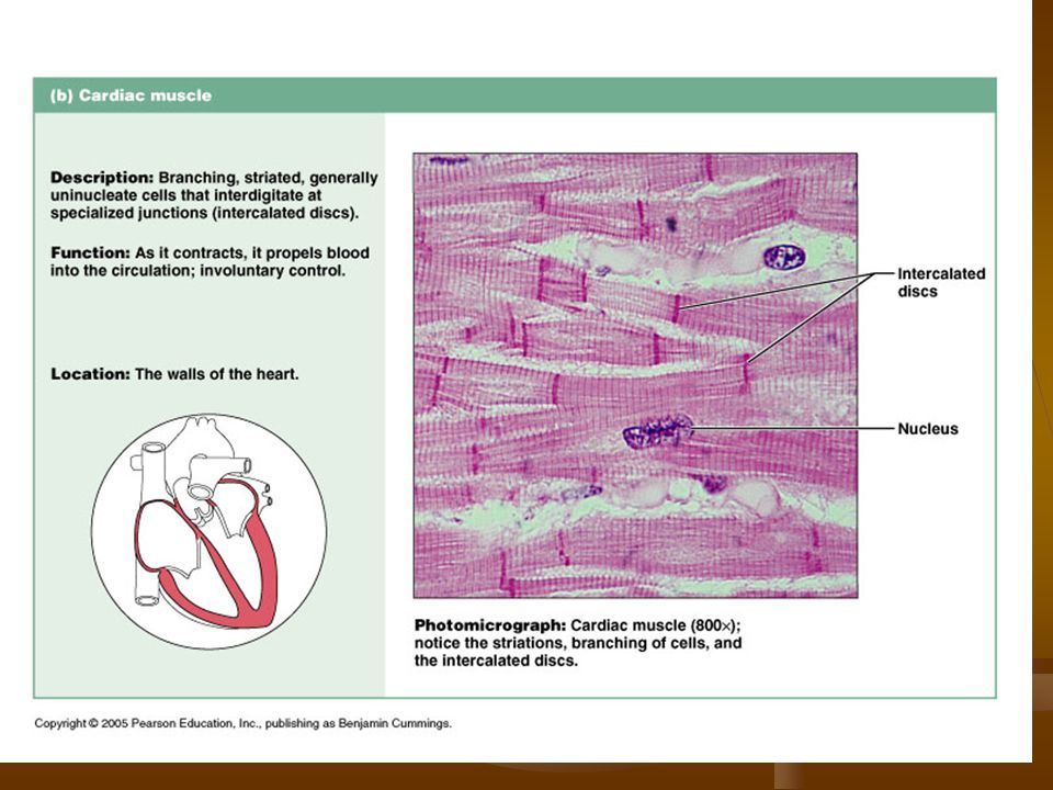
Cardiac Muscle Descriptions: Function: Location:
Branching, striated, uninucleated cells that fit tightly together at junctions called “intercalated disks” Function: Involuntary control, propels blood into circulation. Location: Walls of the heart

61
Smooth Muscle Descriptions: (visceral) Function: Location:
No striations, spindle-shaped w/ central nuclei Arranged closely to form sheets Function: Propelles substances along internal passageways, involuntary control Peristalsis – wavelike motion Location: Walls of the stomach, bladder, uterus and blood vessels

62
Nervous Tissue Nervous Tissue: Nervous Tissue Functions:
Main component of the nervous system, ie., brain, spinal cord & nerves. Nervous Tissue Functions: Regulates & controls body functions Generates & transmits nerve impulses Supports, insulates and protects impulse generating neurons.

64
Nervous Tissue Descriptions: (Neurons) Function: Location:
Branching cells, has long extended parts Irritability and conductivity Have “supporting cells” that insulate, support and protect neurons. Function: Transmits electrical signals from sensory receptors to effectors that control their activity Location: Brain, Spinal cord and nerves

65
Muscle - Skeletal Muscle fibers (cells) long, parallel & cylindrical
With many nuclei (multinucleate) Striations (cross stripes run perpendicular to the cells Produce voluntary movement Locomotion Heat
 Striations (cross stripes run perpendicular to the cells. Produce voluntary movement. Locomotion. Heat.")
66
Specific Nervous Tissue Types Nervous – Neuron
Branching cells with many long processes Large central nucleus Transmit impulses from one area of the body to other areas Regulate activities through neuron impulses

Similar presentations


 Connective tissue Muscle tissue.>")


