Download presentation
Presentation is loading. Please wait.

1
Ureteropelvic junction obstruction 報告者 : Intern 黃暉程 Supervisor: 主治醫師 : 邱元佑

2
Identification Name: 黃小弟 Birth date: 05/31/03 → 19 d/o G2P2, NSD, Apgar score: 9’ → 10’ GA 41weeks, BW: 3000g(10~25%) BL: 52.5cm(10~25%), HC: 35cm(10~25%) DOIC(-), PROM(-)
 BL: 52.5cm(10~25%), HC: 35cm(10~25%) DOIC(-), PROM(-)")
3
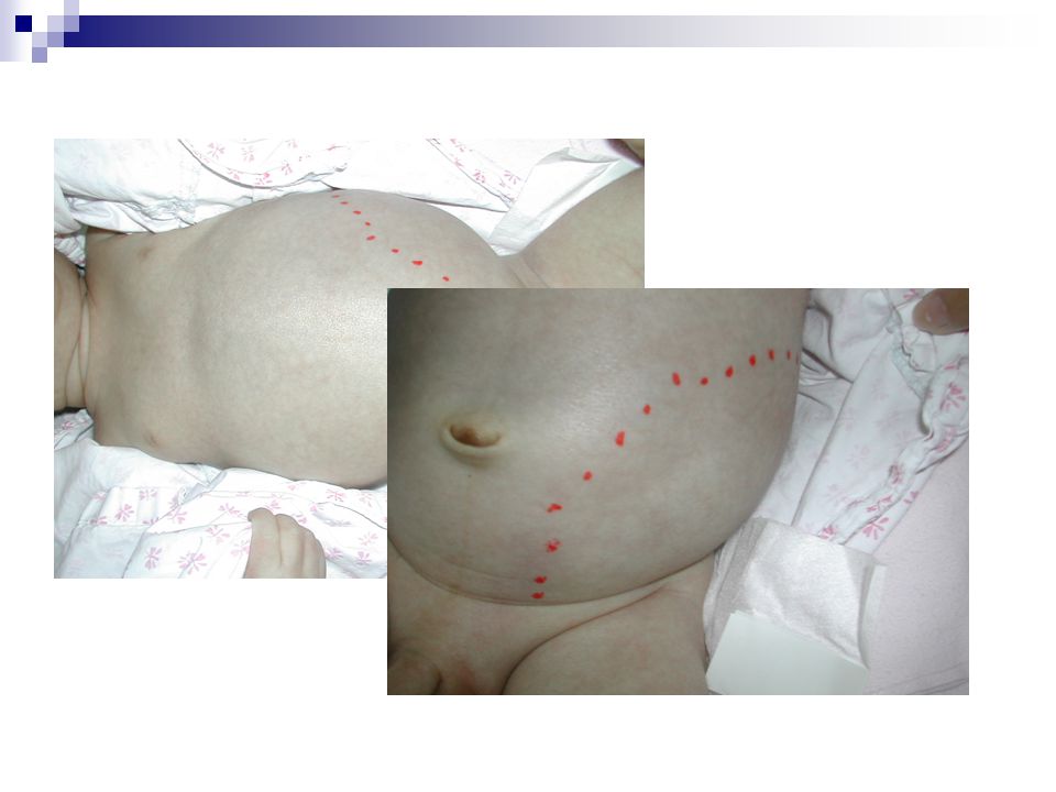
Chief complaint: left abdominal mass for 2 days

4
Present Illness GA 28-30wks Prenatal exam at 姚博琳 ’s clinic: Left hydronephrosis; Oligohydromino(-); Other abnormality(-) GA 28-30wks Prenatal exam at 姚博琳 ’s clinic: Left hydronephrosis; Oligohydromino(-); Other abnormality(-) 92/05/31 GA 41wks, NSD: Renal echo at 姚博琳 ’s clinic: Hydronephrosis is not identified GA 41wks, NSD: Renal echo at 姚博琳 ’s clinic: Hydronephrosis is not identified 92/06/16 One mass over left abdomen noted by his mother 92/06/18 Brought to Dr. 邱 : A mass over LUQ palpable Brought to Dr. 邱 : A mass over LUQ palpable

6
One 11x7cm soft mass over left abdomen; percussion: spongy-filling

7
Abdominal mass ~ approach Inspection, Percussion, Palpation

8
Abdominal mass by age Age-group Newborns 1 m~1 yr After 1 yr

9
Differential diagnosis Non-urologic Abdominal distention, pyloric stenosis, hepatosplenomegaly, intestinal obstruction, malignany, feces Urologic Hydronephrosis, cystic disease, Wilms’ tumor, neuroblastoma, distended bladder

10
Renal echo (Jun 18) Left severe hydronephrosis Cortex thickness: about 0.2cm AP diameter: 4.48cm (>1.5cm) Right moderate hydronephrosis No parenchyma involvement AP diameter: 1.2cm (>1cm) Imp: suspect left ureteropelvic junction obstruction
 Left severe hydronephrosis Cortex thickness: about 0.2cm AP diameter: 4.48cm (>1.5cm) Right moderate hydronephrosis No parenchyma involvement AP diameter: 1.2cm (>1cm) Imp: suspect left ureteropelvic junction obstruction")
11
Present Illness 92/06/18 Admission PE LAB:CBC/DC, Biochemistry, U/A Admission PE LAB:CBC/DC, Biochemistry, U/A 92/06/19 Left PCN 92/06/27 92/06/23 (1) VCUG (2) Antegrade pyelography 92/7/2: discharge (1) Left dismembered pyeloplasty (2) Pathologic Dx: Muscular hyperplasia and fibrosis, compatible with stenosis
 VCUG (2) Antegrade pyelography 92/7/2: discharge (1) Left dismembered pyeloplasty (2) Pathologic Dx: Muscular hyperplasia and fibrosis, compatible with stenosis")
12
Indication of PCN Obstruction with infection Obstruction without infection Stone disease Prelude to endoscopic/ interventional procedures Delivery of medications/ chemotherapy Urinary leaks Urinary diversion for hemorrhagic cystitis

13
VCUG (Jun 23) Imp: No evidence of vesico- ureteral reflux
 Imp: No evidence of vesico- ureteral reflux")
14
Antegrade pyelography: Left UPJ stenosis is considered

15
Present Illness 92/06/19 Left PCN 92/06/27 92/06/23 (1) VCUG (2) Antegrade pyelography 92/7/2 (1) Left dismembered pyeloplasty : UPJ obstruction, high insertion (2) Pathologic Dx: Muscular hyperplasia and fibrosis, compatible with stenosis Discharge!
 VCUG (2) Antegrade pyelography 92/7/2 (1) Left dismembered pyeloplasty : UPJ obstruction, high insertion (2) Pathologic Dx: Muscular hyperplasia and fibrosis, compatible with stenosis Discharge!")
16
Whitaker test during operation Measure the pressure gradient between the pelvis & the bladder under fixed infusion rate Less than 12 mmHg: no obstruction Above 20 mmHg: obstruction Pressure gradient was 14~15 mmHg → 1. intermediate 2. good compliance of pelvis and ureter

17
Diagnosis Left UPJ stenosis

18
Discussion UPJ obstruction

19
generally a congenital condition male, left-sided lesions predominating most frequently diagnosed cause of urinary obstruction in children causes hydronephrosis which may damage the kidney

20
Pathology Various interpretations- Preponderance of longitudinal muscle fibers Excessive collagen fibers in & around muscle bundles Compromised or attenuated muscle bundles Our case: moderately lymphocytic infiltration & focal suppurative inflammation

21
Symptoms & signs Back or flank pain UTI with fever Hematuria Abdominal mass → infants } old children

22
Diagnosis & tests Prenatal Maternal pregnancy ultrasound: hydronephrosis Postnatal Ccr, BUN, electrolytes, AP, DTPA, MAG3, VCUG

23
Etiology Intrinsic: Narrowed, dysfunctional or adynamic segments Extrinsic: Upper ureter is angulated, kinked or compressed by bands or adhesions

24
Intrinsic obstruction mechanical: narrowed → incomplete embryological ureteric bud recanalization; muscular invaginations overdevelop as flaps or valves functional: adynamic or dysfunctional segment → inability to initiate, form or conduct peristaltic waves across the UPJ

25
Extrinsic obstruction vessel or fibrous band may pass anterior to the pelvis & ureter: most common may secondary to intrinsic disturbance which produces pelvic overdistension & rotation high insertion of the ureter into the pelvis

26
Extrinsic ~ High insertion

27
Whitaker test: flow across UPJ obstructions Pressure dependent Volume dependent Intrinsic obstruction Extrinsic obstruction

28
Treatment influenced by renal function, infection surgical correction of the obstruction infants: dismembered pyeloplasty adults: percutaneous or endoscopic technique a nephrostomy stent is placed to drain urine until the patients heals

29
Surgical indication Bilateral UPJO Palpable mass Unilateral UPJO with hydronephrosis Grade 4 (Massive pelvic & calyceal dilatation with thinned parenchyma); DTPA 10% in f/u
; DTPA 10% in f/u")
30
Prognosis ~ pyeloplasty Author and YearPatients/KidneysSuccess (%) Poulsen et al, 198735 100 O ’ Reilly, 198930 83 – 93 MacNeily et al, 199375 85 Shaul et al, 199432/33 (<2 mo old) 97 30/33 (>2 mo old) 93 Salem et al, 1995100 98 McAleer and Kaplan, 1999 79 90 Austin et al, 2000135/137 91 Houben et al, 2000186/203 93
 Poulsen et al, O ’ Reilly, – 93 MacNeily et al, Shaul et al, /33 (<2 mo old) 97 30/33 (>2 mo old) 93 Salem et al, McAleer and Kaplan, Austin et al, / Houben et al, /203 93")
31
Prognosis ~ pyeloplasty

32
Expectantions Rapid decompression of the kidney immediately following birth can substantially improve kidney function in an infant with UPJ obstruction diagnosed before the child is born. Most patients do well with no long-term consequences

33
Complications Permanent loss of kidney function-renal failure require dialysis at some point in their lives as a result of this problem

34
Thanks for your attention!

Similar presentations


 Incidence: 1:188 Approximately 50% of antenatal scans are normal postnatally Posterior urethral.>")


