Download presentation
Presentation is loading. Please wait.

1
Anatomy of the Cranial Nerves (I, II, III, IV, VI)
NEUROANATOMY Lecture : 8 Peripheral Nervous System Anatomy of the Cranial Nerves (I, II, III, IV, VI) Prepared and presented by: Dr. Iyad Mousa Hussein, Ph.D in Neurology Head of Neurology Department Nasser Hospital
 Prepared and presented by: Dr. Iyad Mousa Hussein, Ph.D in Neurology. Head of Neurology Department. Nasser Hospital.")
2
LECTURE OBJECTIVES: Classification of the Peripheral Nervous System.
Structures of the Peripheral Nerve, and Nerve Fibers. Classification of the Cranial Nerve. Function of the Olfactory Nerve, and Pathway of the Smell. Lesion of the Olfactory Nerve. Function of the Optic Nerve, and Pathway of the Vision. Lesion of the Optic Nerve. Function, Branches, and Lesion of the Oculomotor Nerve. The actions of the ocular muscles. Function, and Lesion of the Trochlear Nerve. Function, and Lesion of the Abducent Nerve.

3
Classification of the Peripheral Nervous System
The peripheral nervous system (PNS) formed of the: 1. Somatic (voluntary or craniospinal) NS: which controls the skeletal muscles: a. Cranial nerves. b. Spinal nerves. 2. Autonomic NS: which controls the smooth and cardiac muscles: a. Sympathetic nervous system. b. Parasympathetic nervous system.
 formed of the: 1. Somatic (voluntary or craniospinal) NS: which. controls the skeletal muscles: a. Cranial nerves. b. Spinal nerves. 2. Autonomic NS: which controls the smooth and. cardiac muscles: a. Sympathetic nervous system. b. Parasympathetic nervous system.")
4
The Peripheral Nervous System
The peripheral nervous system consists of the cranial and spinal nerves and their associated ganglia. There are 12 pairs of cranial nerves, which leave the brain and pass through foramina in the skull. There are 31 pairs of spinal nerves, which leave the spinal cord and pass through intervertebral foramina in the vertebral column.

5
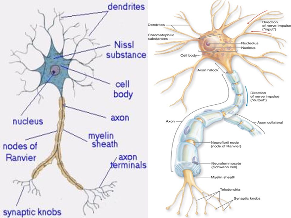
The Nerve Fibers Definition: it is an axon or a dendrite of a nerve cell. Structure of the Nerve Fiber: 1. Schwann cell. 2. Node of Ranvier. 3. Myelin sheath. 4. Mesaxon.

7
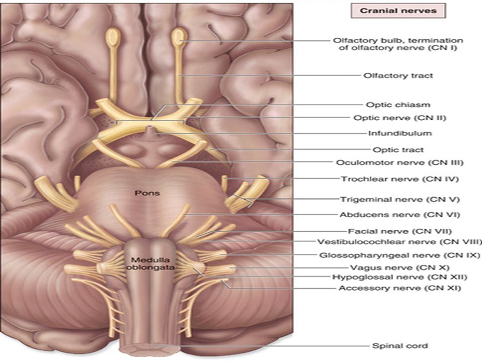
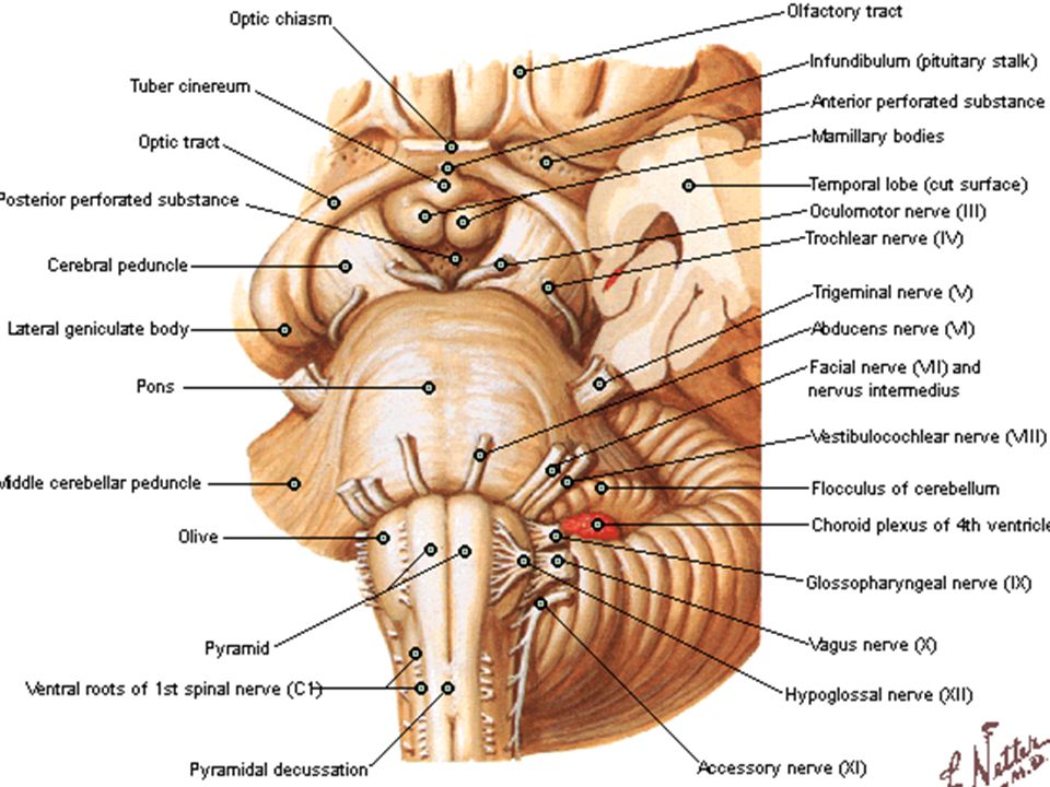
The Cranial Nerves Definition: The cranial nerves are consist of 12 pairs of nerves that arise from the brain. They exit/enter the cranium through openings in the skull.

8
The olfactory, optic and vestibulocochlear nerves are purely v
sensory nerves (128). The oculomotor, trochlear, abducent, accessory and hypoglossal nerves are purely motor nerves. The trigeminal, facial, glossopharyngeal and vagus nerves are mixed nerves (1975). There are three cranial motor nerves (III, IV, & VI nerves), which supply the muscles of the eye. There are three cranial mixed nerves (VII, IX, & X nerves), which have the same plan: each contains three types of fibers: motor, sensory and parasympathetic.
. The oculomotor, trochlear, abducent, accessory and. hypoglossal nerves are purely motor nerves. The trigeminal, facial, glossopharyngeal and vagus nerves are mixed nerves (1975). There are three cranial motor nerves (III, IV, & VI nerves), which supply the muscles of the eye. There are three cranial mixed nerves (VII, IX, & X nerves), which have the same plan: each contains three types of fibers: motor, sensory and parasympathetic.")
9
Classification of the Cranial Nerves
Opening in skull Site of nucleus Function Name № Cribriform plate Temporal Lobe Smell Olfactory 1 Optic canal Occipital lobe Vision Optic 2 Superior orbital fissure Midbrain Motor Oculomotor 3 Trochlear 4 Foramen rotundum Foramen ovale Mixed Trigeminal: Ophthalmic Maxillary Mandibular 5 Pons Abducent 6 Internal acoustic meatus, Facial canal, Stylomastoid foramen Facial 7 Internal acoustic meatus Hearing and balance Cochleo-Vestibular 8 Jugular nerve Medulla oblongata Glosso-pharyngeal 9 Jugular foramen Vagus 10 C1-C4 AHC of spinal cord Spinal Accessory 11 Hypoglossal canal Hypoglossal 12

12
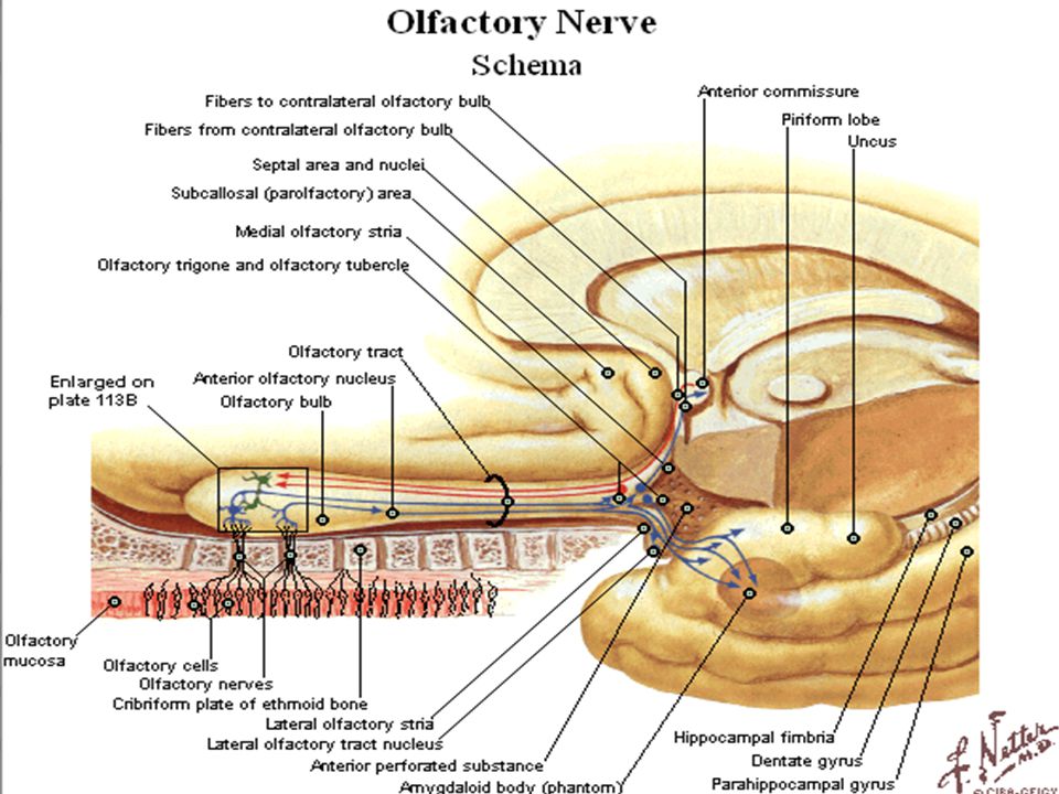
The Olfactory Nerve (I)
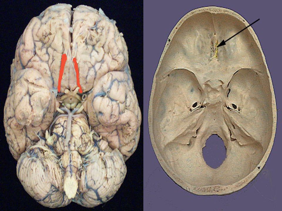
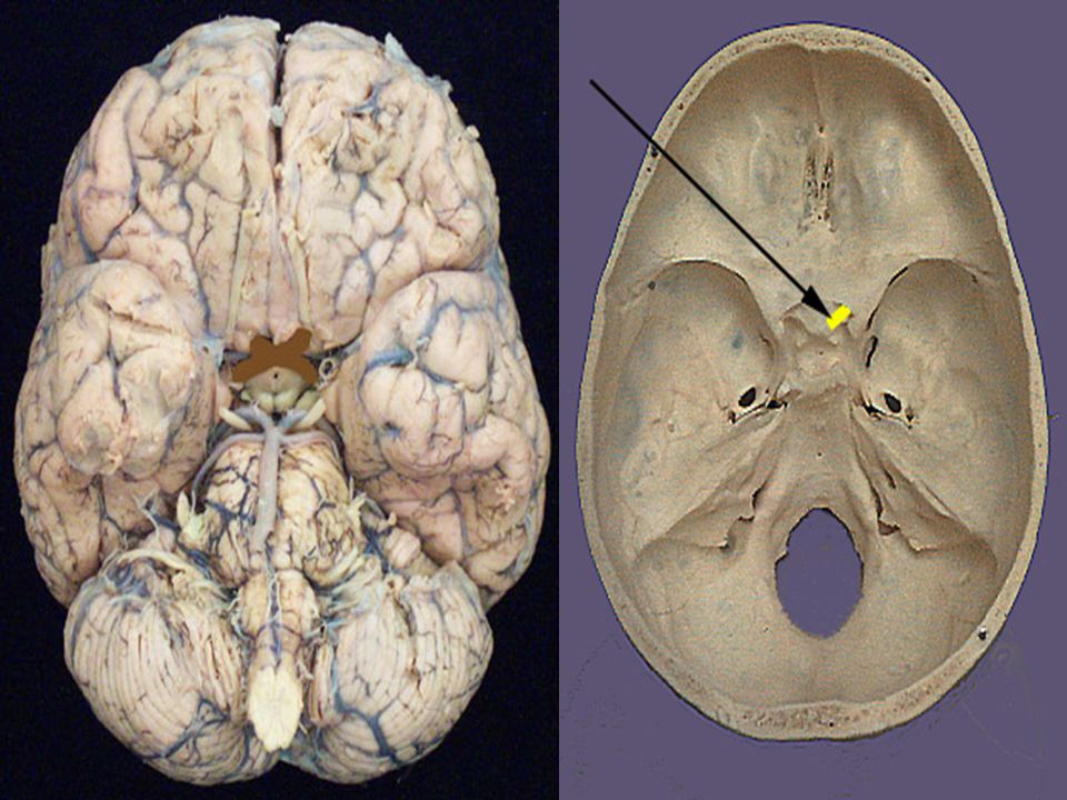
Olfactory Nerve: (Latin for "to smell"). Function: Special sensory nerve (smell). Olfactory nerve located in the anterior cranial fossa and attached to the under surface of the frontal lobe.
. Function: Special sensory nerve (smell). Olfactory nerve located in the anterior cranial fossa and attached to the under surface of the frontal lobe.")
13
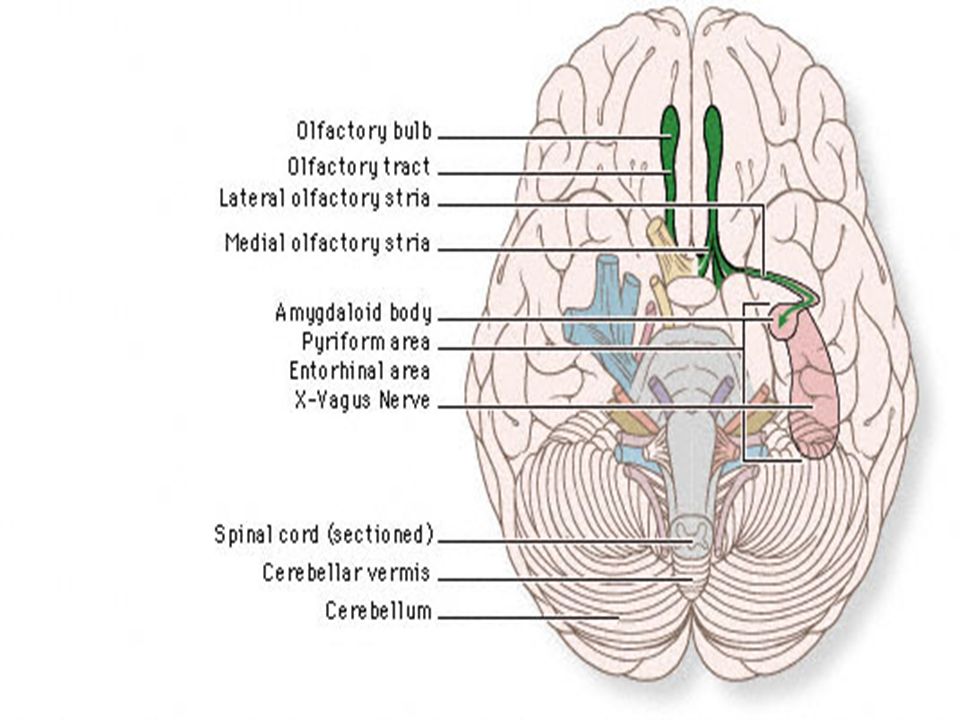
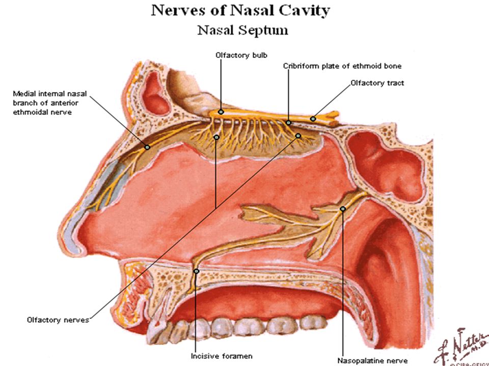
Pathway of the Smell From receptors in olfactory mucous membranes of the nasal cavity → Fibers of olfactory nerve (consist of about 20 small filaments on each side → The cribriform plate of the ethmoid bone → Olfactory groove → olfactory bulb → Olfactory tract → direct and indirect (around corpus callosum) to terminate in olfactory sensory area ( in the uncus of temporal lobe). Note: Smell as well bilateral represented.
 to terminate in olfactory sensory area ( in the uncus of temporal lobe). Note: Smell as well bilateral represented.")
18
Lesion of the Olfactory Nerve
Unilateral anosmia. Bilateral anosmia. Parosmia (Smell hallucination) Cacosmia (bad smell in chronic sinusitis). Olfactory agnosia (higher-order loss of olfactory discrimination).
 Cacosmia (bad smell in chronic sinusitis). Olfactory agnosia (higher-order loss of olfactory discrimination).")
20
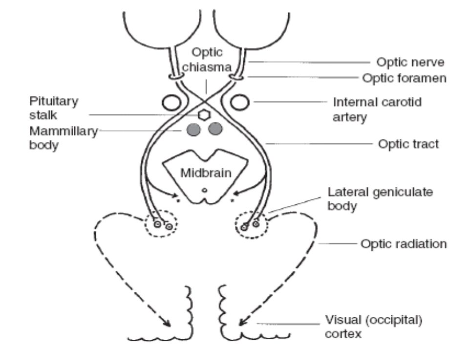
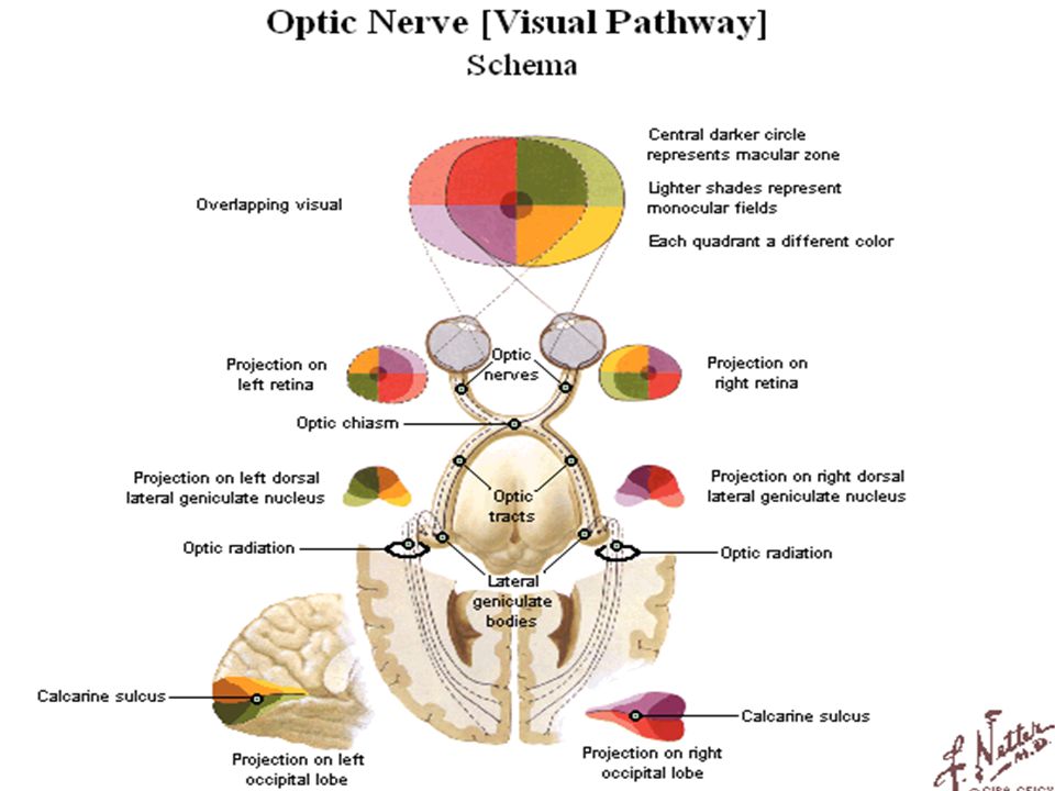
The Optic Nerve (II) Optic Nerve: (Latin for "to see").
Function: Special sensory nerve (Sense of vision).
.")
21
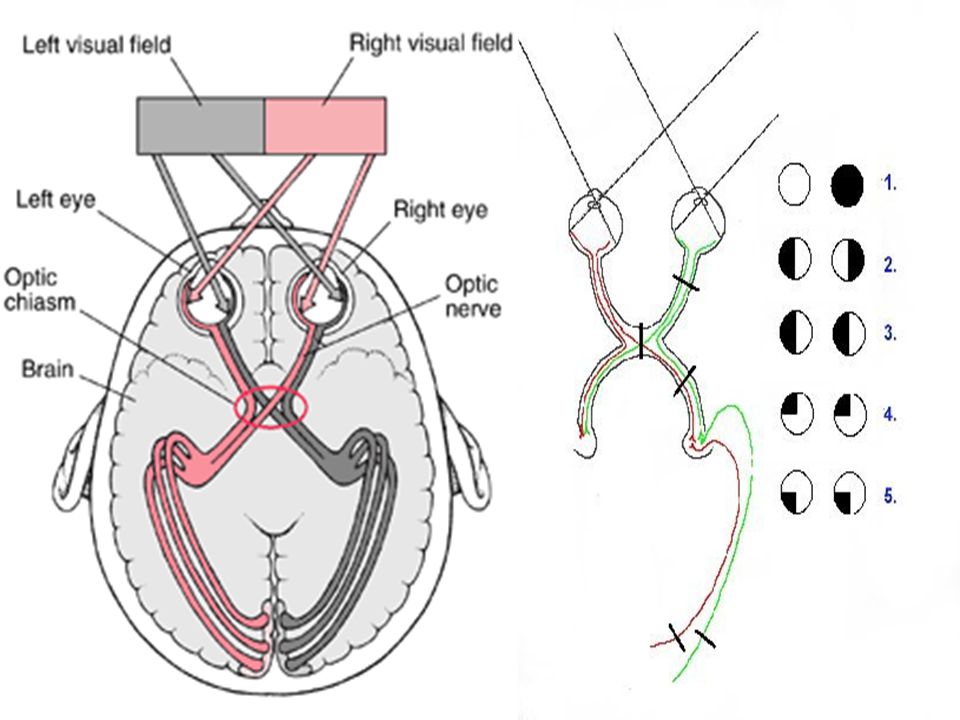
Pathway of the Vision Receptors of light are rods and cones of retina → Optic nerve → Optic canal of the sphenoid bone → Optic chiasma (the nasal or medial fibers decussate to the opposite optic tract, while the temporal or lateral fibers continue in the same optic tract → Optic tract → Relay in the lateral geniculate body → Posterior limb of internal capsule → Optic radiation → End in area 17, 18, 19 of occipital lobes.

26
Lesion in the Vision Pathway
Lesion in the optic nerve or retina: ipsilateral loss or decrease of vision (blindness). Lesion in the optic chiasma: bitemporal hemianopsia. Lesion in the optic tract: contralateral homonymous hemianopsia with preserved direct and indirect light reflexes. Lesion in the lateral geniculate body, internal capsule and optic radiation: contralateral homonymous hemianopsia with preserved direct and indirect light reflexes. Lesion in the occipital lobe: a. Irritative: Visual hallucination. b. Destructive: contralateral homonymous hemianopsia with macular sparing and visual agnosia (the patient can see but does not recognize objects).
. Lesion in the optic chiasma: bitemporal hemianopsia. Lesion in the optic tract: contralateral homonymous hemianopsia with preserved direct and indirect light reflexes. Lesion in the lateral geniculate body, internal capsule and optic radiation: contralateral homonymous hemianopsia with preserved direct and indirect light reflexes. Lesion in the occipital lobe: a. Irritative: Visual hallucination. b. Destructive: contralateral homonymous hemianopsia with macular sparing and visual agnosia (the patient can see but does not recognize objects).")
29
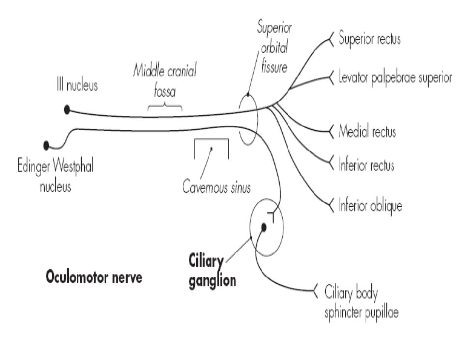
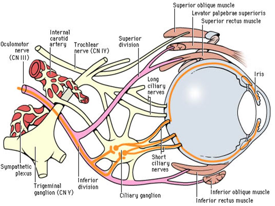
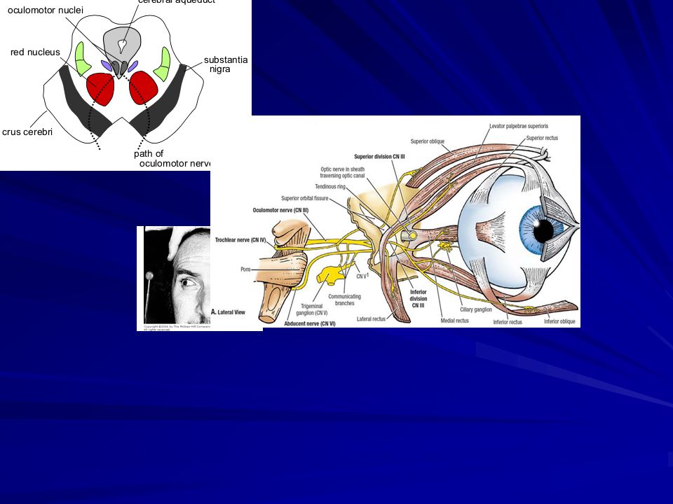
The oculomotor Nerve (III)
Oculomotor nerve: (Latin for "eye" and "moving"). Function: 1. Motor function: movement of the eye ball. 2. Autonomic (parasympathetic): pupillary reaction. Site of nucleus: it lies in the tegmentum of midbrain.
. Function: 1. Motor function: movement of the eye ball. 2. Autonomic (parasympathetic): pupillary reaction. Site of nucleus: it lies in the tegmentum of midbrain.")
30
Structure of oculomotor Nuclei
The nucleus of oculomotor nerve formed of 3 main parts: 1. The Edinger-Westphal nucleus: parasympathetic function which supplies two intra-ocular muscles: a. Constrictor pupillae muscles → Miosis. b. Ciliary muscles → Responsible for light and accommodation reflexes. 2. The lateral cell mass (motor function): which is divided into five parts which supplies five extra ocular muscles which are from above downwards: a. Levator palpebrea superior. b. Superior recti muscles. c. Medial recti muscles. d. Inferior oblique muscles. e. Inferior recti muscles. 3. The central nucleus of Perlia: motor function which supplies the medial recti muscles of the two eyes allowing to contract together when both eyes converge to look to a near object.
: which is divided into. five parts which supplies five extra ocular muscles which are. from above downwards: a. Levator palpebrea superior. b. Superior recti muscles. c. Medial recti muscles. d. Inferior oblique muscles. e. Inferior recti muscles. 3. The central nucleus of Perlia: motor function which supplies. the medial recti muscles of the two eyes allowing to contract. together when both eyes converge to look to a near object.")
31
The oculomotor nerve supplies all the intra- and extraocular muscles except:
Superior oblique muscles → from the trochlear nerve – IV. Lateral recti muscles → from the abducent nerve –VI. Dilator pupillae muscles → from the sympathetic fibers.

35
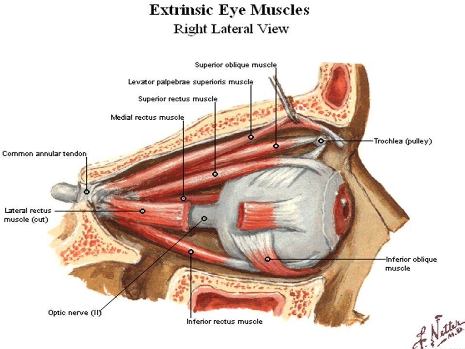
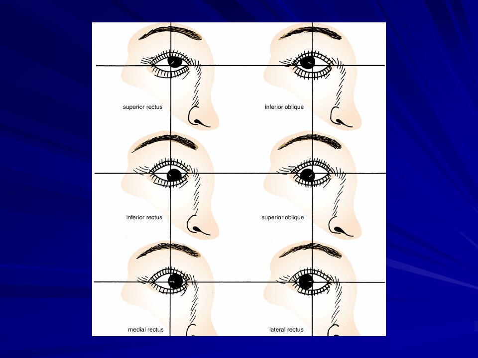
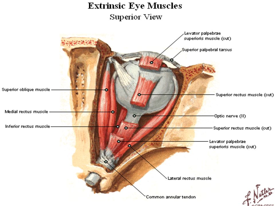
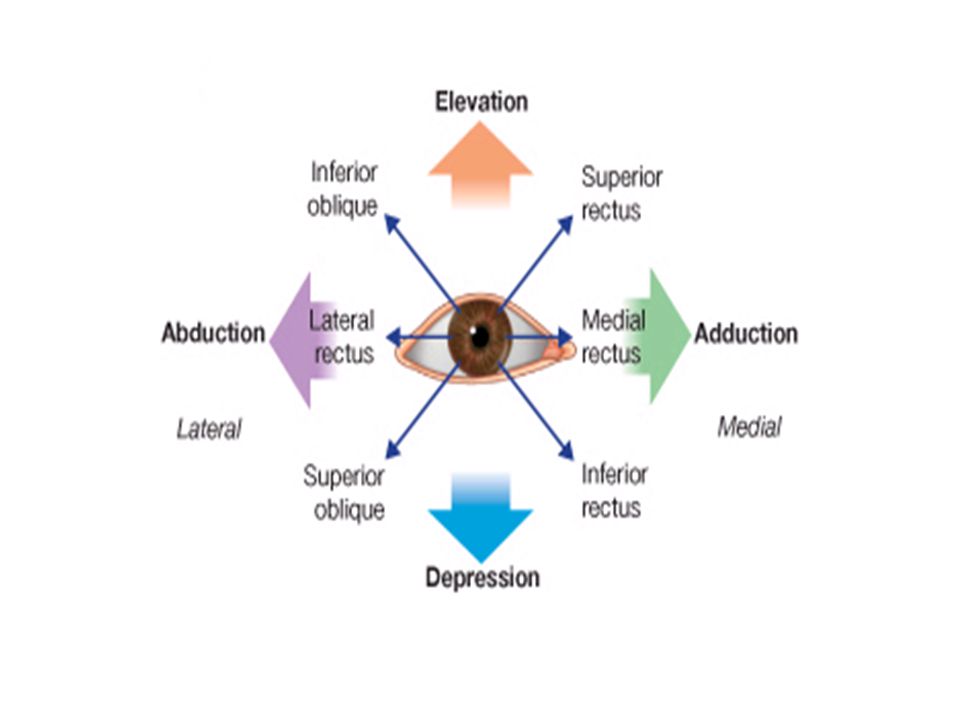
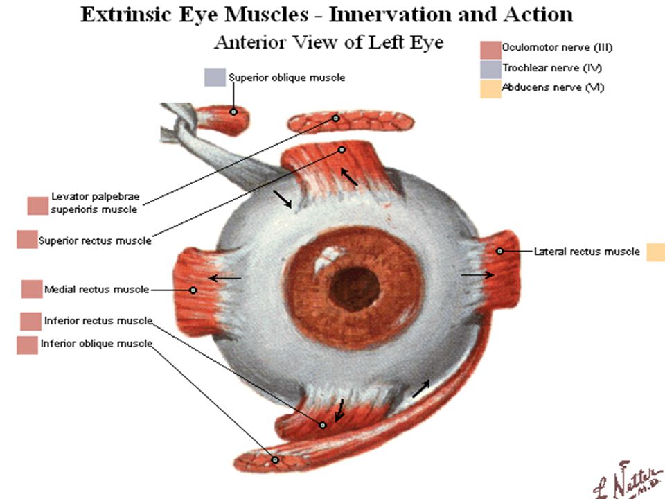
The Actions of the Ocular Muscles
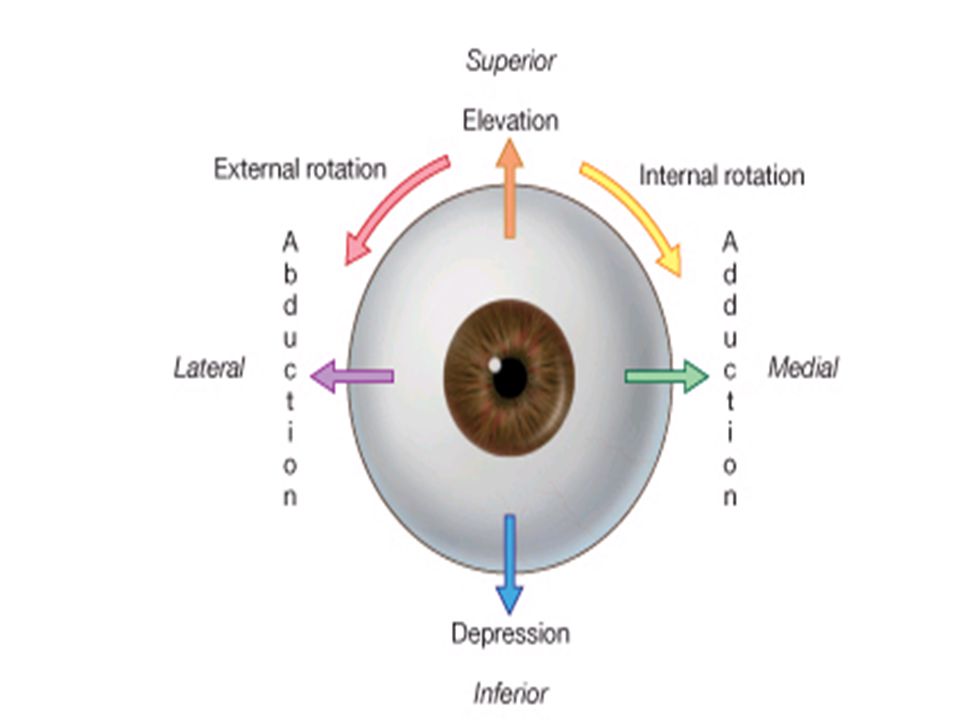
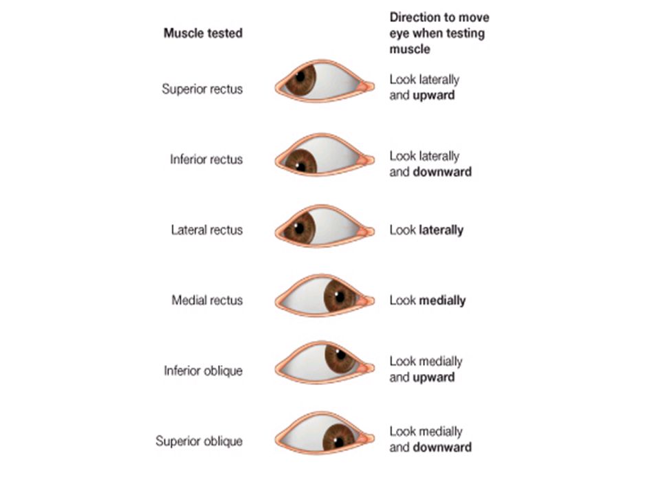
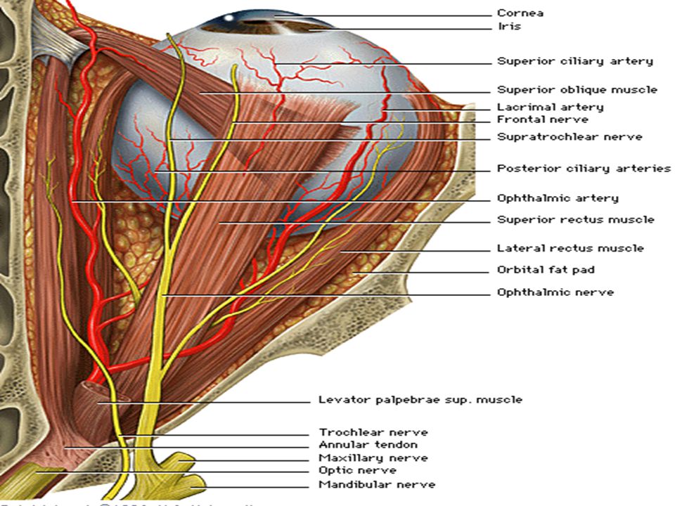
The Lateral Rectus Muscle: abducts (laterally) the eye ball. The Medial Rectus Muscle: adducts (medially) the eye ball. The Superior Rectus Muscle: elevates, adducts and rotates medially. The Inferior Rectus Muscle: depresses, adducts and rotates medially. The Superior Oblique Muscle: depresses, adducts and rotates laterally. The Inferior Oblique Muscle: elevates, adducts and rotates laterally.
 the eye ball. The Medial Rectus Muscle: adducts (medially) the eye ball. The Superior Rectus Muscle: elevates, adducts and rotates medially. The Inferior Rectus Muscle: depresses, adducts and rotates medially. The Superior Oblique Muscle: depresses, adducts and rotates laterally. The Inferior Oblique Muscle: elevates, adducts and rotates laterally.")
40
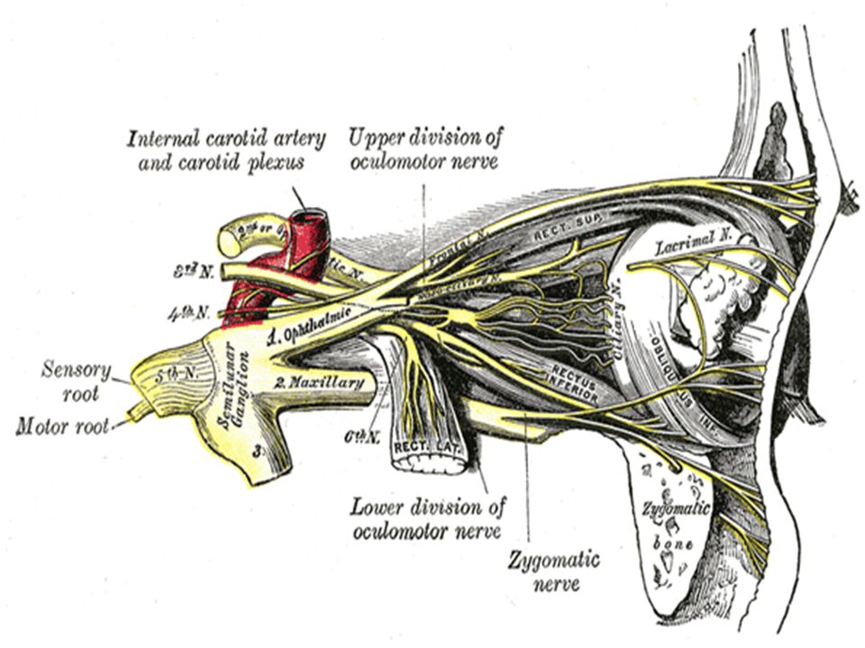
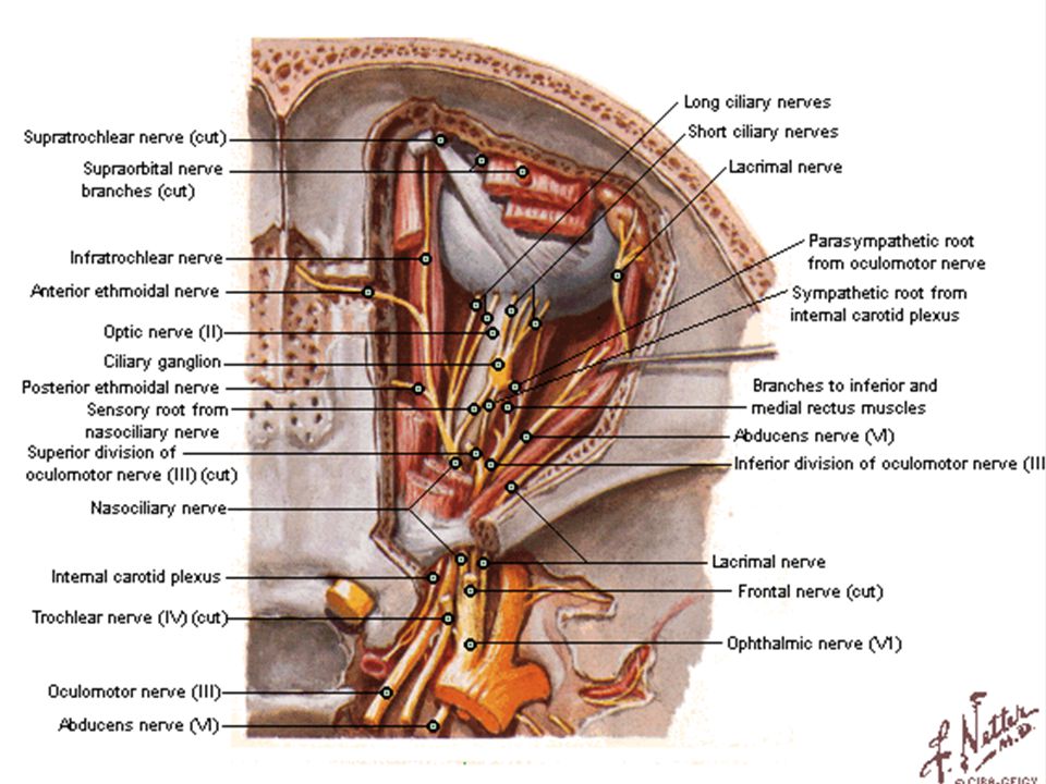
The oculomotor nerve emerges from a groove on the
medial aspect of midbrain (ventral surface). The nerve passes through the two layers of the dura mater including the lateral wall of the cavernous sinus and then enters the superior orbital fissure to access the orbit.
. The nerve passes through the two layers of the dura. mater including the lateral wall of the cavernous sinus and. then enters the superior orbital fissure to access the orbit.")
44
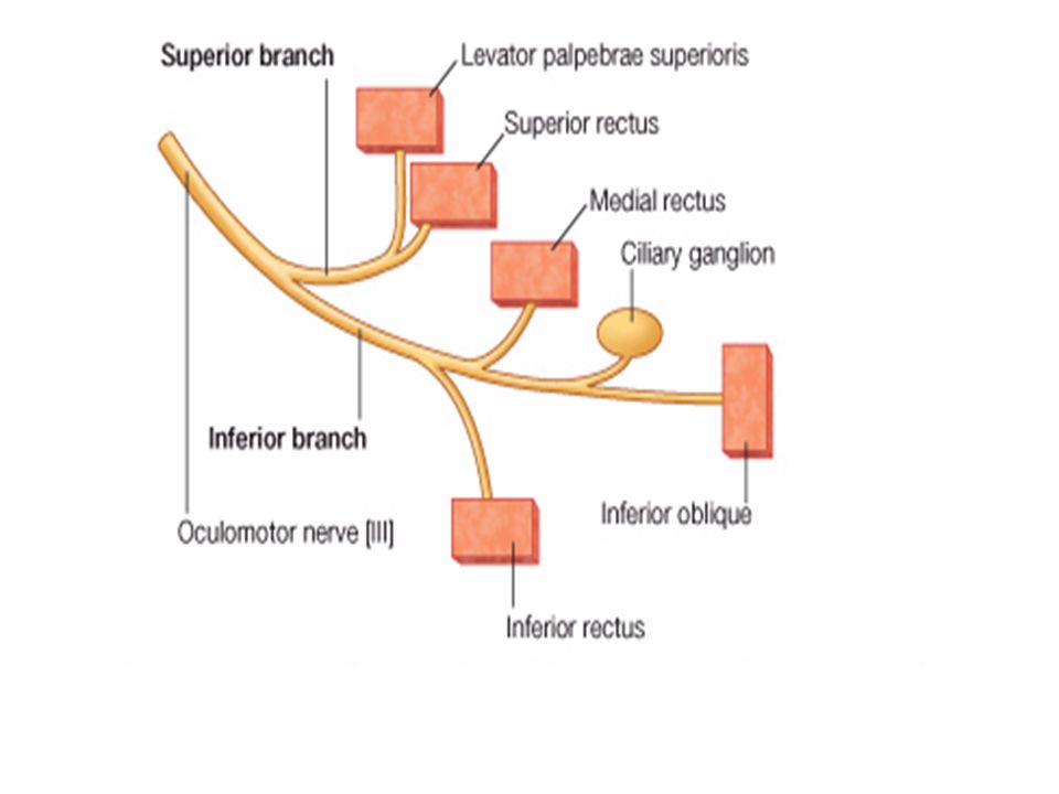
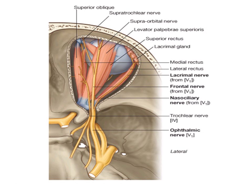
Branches of the Oculomotor Nerve:
1. Superior division: which supplies the: a. Levator palpebrea superior. b. Superior rectus muscles. 2. Inferior division: which supplies the: a. Medial rectus. b. Inferior rectus. c. Inferior oblique muscles. d. Sphencter pupillae muscle. e. Ciliary muscle.

48
Lesion of the Oculomotor Nerve:
Ptosis: paresis of the levator palpebrea superior muscle. Diplopia: occurs only on elevation of eye lid. Squint: divergent paralytic. Mydriasis: dilated fixed pupil. Loss of light and accommodation reflexes.

49
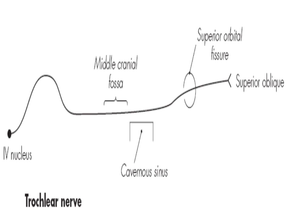
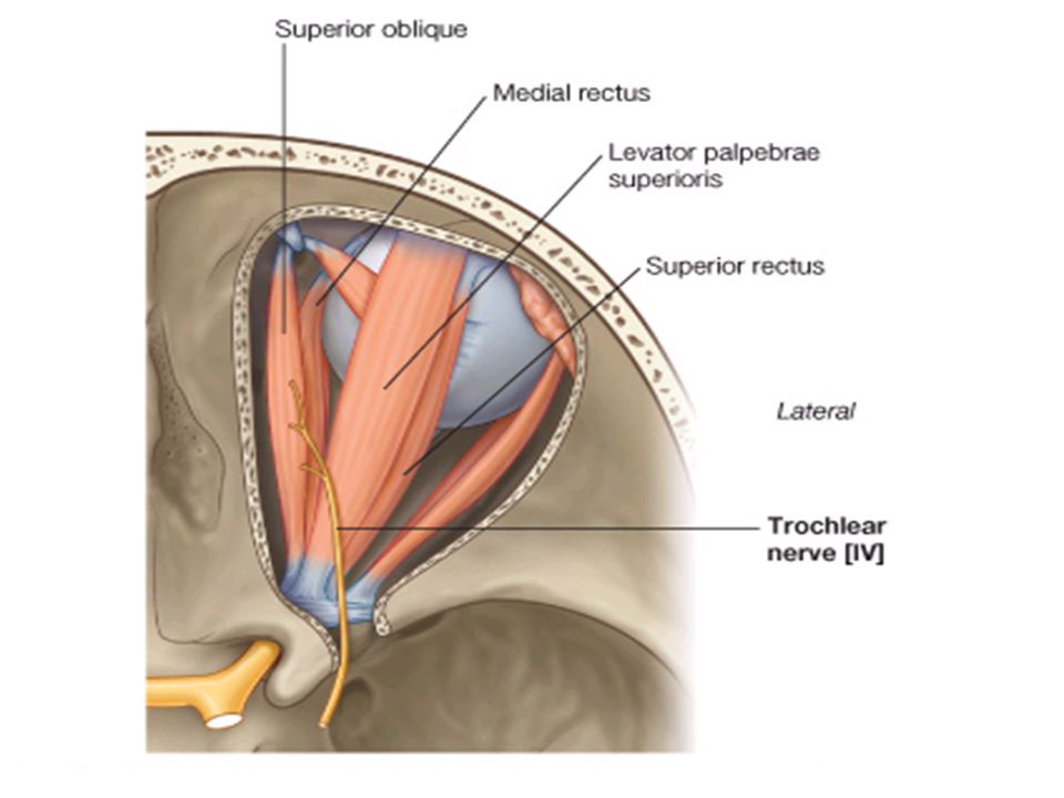
The Trochlear Nerve (IV)
The trochlear nerve is so called because superior oblique muscle (which it supplies) is arranged as a pulley (Latin: trochlea – pulley). Function: motor nerve → movement of the eye ball → supplies the superior oblique muscles. Site of the nucleus: it lies in the tegmentum of the midbrain.
 is arranged as a pulley (Latin: trochlea – pulley). Function: motor nerve → movement of the eye ball → supplies the superior oblique muscles. Site of the nucleus: it lies in the tegmentum of the midbrain.")
50
The Trochlear Nerve It is the smallest cranial nerve.
The trochlear nerve is purely a motor nerve. The trochlear nerve emerges from the posterior surface of the midbrain. The nerve travels in the lateral wall of the cavernous sinus and then enters the orbit via the superior orbital fissure of the sphenoid bone where it supplies the superior oblique muscle of the eye that controls downward and lateral movement of the eyeball.

58
Lesion of the Trochlear Nerve
Diplopia on looking down and out. Limitation of movement during examination for eye movement down and out.

59
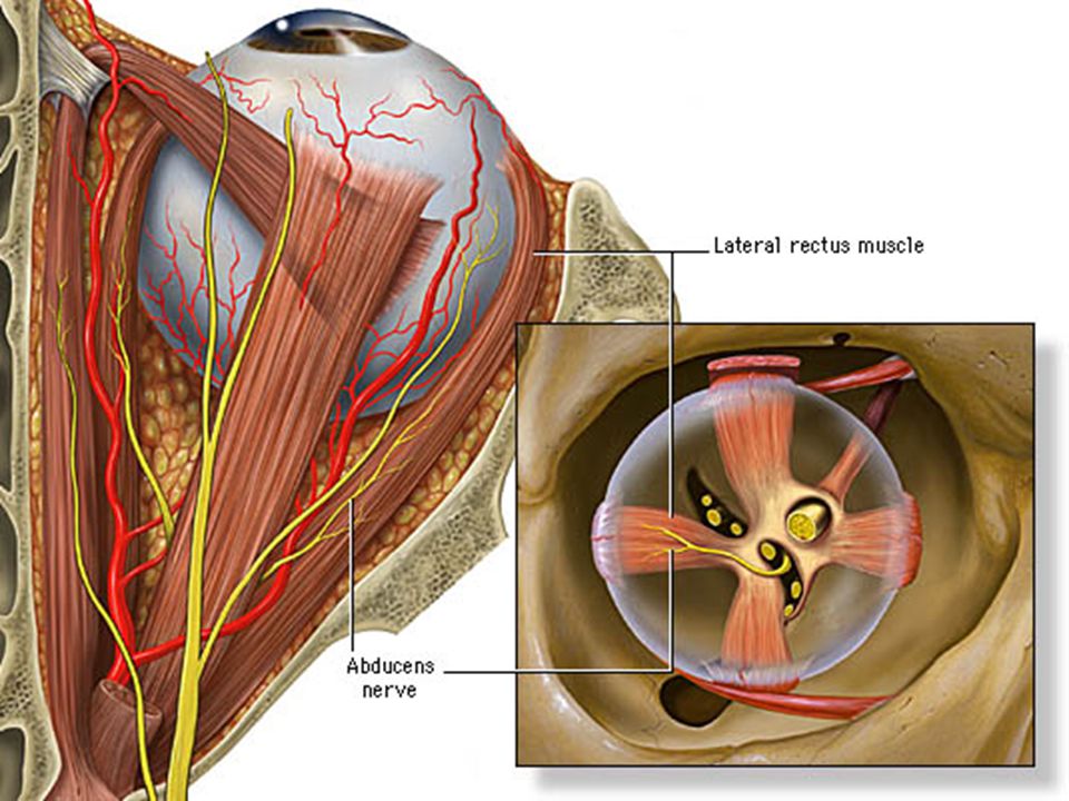
The Abducent Nerve (VI)
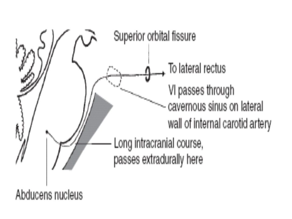
Abducent nerve: (Latin for "abduction"). The abducent nerve is so called because lateral rectus muscle (which it supplies) abducts the eyeball. Function: motor nerve → movement of the eye ball → supplies the lateral recti muscles (abduction). Site of the nucleus: it lies in the lower part of the ventral surface of pons.
. The abducent nerve is so called because lateral rectus muscle (which it supplies) abducts the eyeball. Function: motor nerve → movement of the eye ball → supplies the lateral recti muscles (abduction). Site of the nucleus: it lies in the lower part of the ventral surface of pons.")
60
The Abducent Nerve The abducent nerve is purely a motor nerve. The abducent nerve emerges from the lower border of the pons (between the pons and medulla oblongata). The abducent nerve travels the medial wall of the cavernous sinus and then enters the orbit via the superior orbital fissure of the sphenoid bone where it supplies the lateral rectus muscle of the eye that controls abduction of the eye.
. The abducent nerve travels the medial wall of the cavernous sinus and then enters the orbit via the superior orbital fissure of the sphenoid bone where it supplies the lateral rectus muscle of the eye that controls abduction of the eye.")
66
Lesion of the Abducent Nerve
Diplopia on looking out wards. Limitation during examination of eye movement in the outward direction. Convergent paralytic squint.

67
Thank You

Similar presentations


 and passes through foramina of skull There are 12 pairs of cranial nerves They have both name.>")


>")


 Prepared and presented by: Dr. Iyad Mousa Hussein,>")


>")