Download presentation
Presentation is loading. Please wait.

1
Boerhaave Syndrome Preethi Yeturu and Erik Mikaitis MS IV

2
Background Transmural perforation of the esophagus Distinguished from Mallory-Weiss syndrome (non-transmural tear) Diagnosis is difficult because often no classic symptoms are present
 Diagnosis is difficult because often no classic symptoms are present")
3
History First described in 1724 by Hermann Boerhaave His patient, Baron van Wassenaer, would eat large meals and induce vomiting by ingesting ipecac so that he could immediately have another large meal After vomiting, he began having severe chest pain & dyspnea and died 18 hours later At autopsy, Boerhaave found olive oil and roast duck in the left pleural cavity.

4
Pathophysiology Rupture is caused by a sudden rise in intraluminal esophageal pressure produced during vomiting. Neuromuscular incoordination results in failure of the cricopharyngeus muscle to relax. Most common location of the tear is the left posterolateral wall of the lower third of the esophagus. (2 nd most common is subdiaphragmatic or upper thoracic area)
.")
5
Causes Commonly associated with: Alcoholism Alcoholism Bulimia Bulimia Overindulgence in food and drink Overindulgence in food and drink

6
Epidemiology Rare but most lethal perforation of the GI tract Most studies report a 100% mortality within 7 days without surgery Only a 70% overall survival with surgery However, the syndrome is very rare: Only 16 cases reported from 1958-1973 Only 16 cases reported from 1958-1973 A 1980 review cited only 300 cases in literature worldwide A 1980 review cited only 300 cases in literature worldwide

7
Epidemiology Accounts for 15% of all traumatic ruptures or perforations of the esophagus Other 85% of ruptures are from iatrogenic perforation (Not Boerhaave’s syndrome)
")
8
Mortality and Morbidity The overall mortality of 30% is due to: Subsequent infection Subsequent infection Mediastinitis Mediastinitis Pneumonitis Pneumonitis Pericarditis Pericarditis Empyema Empyema

9
Epidemiology There is equal distribution of Boerhaave’s throughout all races Male-to-female ratio ranges from 2:1 to 5:1 Most frequently seen in patients aged 50- 70 years 80% of patients are middle aged men

10
Diagnosis

11
Clinical History Repeated episodes of retching and vomiting Sudden onset of chest pain in lower thorax and upper abdomen Pain may radiate to the back or to the left shoulder Swallowing can aggravate the pain

12
Clinical History Hematemesis is not seen after rupture (which helps distinguish from Mallory- Weiss) Swallowing may precipitate cough SOB is common due to pleuritic pain or pleural effusion
 Swallowing may precipitate cough SOB is common due to pleuritic pain or pleural effusion")
13
Physical Exam Mackler triad is the classic presentation, including: Vomiting Vomiting Lower thoracic pain Lower thoracic pain Subcutaneous emphysema Subcutaneous emphysema Presentation may depend on: Location of tear Location of tear Cervical tear may have neck of upper chest pain Mid to lower esophagus tear may have interscapular or epigastric pain. Cause of the injury Cause of the injury Time since the perforation Time since the perforation

14
Physical Exam Pleural effusion is common Subcutaneous emphysema is very helpful It is seen in 28-66% of patients It is seen in 28-66% of patients Typically found later Typically found later Tachypnea and abdominal rigidity are other classic findings Pneumomediastinum is an important finding May cause crackling on chest auscultation (Hamman crunch) May cause crackling on chest auscultation (Hamman crunch) Heard coincident with each heartbeat (can be mistaken for pericarial friction rub) Heard coincident with each heartbeat (can be mistaken for pericarial friction rub) Found in 20% of cases Found in 20% of cases
 May cause crackling on chest auscultation (Hamman crunch) Heard coincident with each heartbeat (can be mistaken for pericarial friction rub) Heard coincident with each heartbeat (can be mistaken for pericarial friction rub) Found in 20% of cases Found in 20% of cases")
15
Later stages of Illness Can manifest as signs of infection and sepsis Symptoms may include fever, hemodynamic instability, progressive obtundation Diagnosis at later stages is more difficult as septic complications begin to dominate the clinical picture

16
Workup Laboratory Studies Findings are non-specific Findings are non-specific May present with leukocytosis with left shift May present with leukocytosis with left shift 50% of patients have hematocrit over 50%. 50% of patients have hematocrit over 50%. Thought to be due to fluid loss into pleural spaces and tissues Thought to be due to fluid loss into pleural spaces and tissues

17
Thoracentesis If patient presents with pleural effusion: Pleural fluid can aid in diagnosis Pleural fluid can aid in diagnosis May find undigested food particles and gastric juices May find undigested food particles and gastric juices If no gross particles are found, cytology can confirm its presence If no gross particles are found, cytology can confirm its presence pH of the fluid will be less than 6 and amylase will be elevated pH of the fluid will be less than 6 and amylase will be elevated Squamous cells from saliva may be found Squamous cells from saliva may be found

18
Imaging Studies Upright Chest X-ray 90% of patients have an abnormality after perforation 90% of patients have an abnormality after perforation Most common finding is a left unilateral effusion Most common finding is a left unilateral effusion May have: May have:PneumothoraxHydropneumothoraxPneumomediastinum Subcutaneous emphysema Mediastinal widening V-sign of Naclerio V-sign of Naclerio Streaks of air that dissect the planes behind the heart and form a ‘V’. Fairly specific, but very insensitive Found of 20% of patients

22
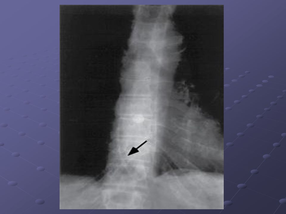
Imaging Studies Esophagram Helps confirm the diagnosis Helps confirm the diagnosis Shows extravasation of contrast Shows extravasation of contrast Outlines the length of the perforation and its location (aids in decision of surgical approach, thoracic vs. abdominal) Outlines the length of the perforation and its location (aids in decision of surgical approach, thoracic vs. abdominal) Initially use water soluble contrast (Gastrografin) which has 90% sensitivity Initially use water soluble contrast (Gastrografin) which has 90% sensitivity Barium is associated with severe medistinitis Barium is associated with severe medistinitis If study is negative but suspicion remains high, try left and right lateral decubitus images If study is negative but suspicion remains high, try left and right lateral decubitus images
 Outlines the length of the perforation and its location (aids in decision of surgical approach, thoracic vs. abdominal) Initially use water soluble contrast (Gastrografin) which has 90% sensitivity Initially use water soluble contrast (Gastrografin) which has 90% sensitivity Barium is associated with severe medistinitis Barium is associated with severe medistinitis If study is negative but suspicion remains high, try left and right lateral decubitus images If study is negative but suspicion remains high, try left and right lateral decubitus images.")
25
Imaging Studies CT Scan Can reveal decisive criteria for diagnosis Can reveal decisive criteria for diagnosis Helpful in patients too ill to tolerate esophagrams Helpful in patients too ill to tolerate esophagrams Shows localized collections of fluid Shows localized collections of fluid Visualizes adjacent structures to help narrow the differential diagnoses. Visualizes adjacent structures to help narrow the differential diagnoses. Can demonstrate periesophageal air tracks Can demonstrate periesophageal air tracks It may not precisely localize the site of perforation It may not precisely localize the site of perforation

27
Procedures Endoscopy Not commonly used Not commonly used Carries a risk of increasing the size and extent of the perforation as well as pushing more air through the perforation Carries a risk of increasing the size and extent of the perforation as well as pushing more air through the perforation More useful in thoracic esophagus More useful in thoracic esophagus May be useful when perforation is suspected but not proven May be useful when perforation is suspected but not proven

28
Treatment

29
Medical care Therapy includes: IV volume resuscitation IV volume resuscitation Broad-spectrum aantibiotics Broad-spectrum aantibiotics Prompt surgical intervention Prompt surgical intervention Conservative vs aggressive treatment depends on: Time delay Time delay Extent of perforation Extent of perforation Overall medical condition Overall medical condition

30
Medical management Conservative management may be appropriate if: The disruption is well contained within the mediastinum The disruption is well contained within the mediastinum The cavity should be drained back into the esophagus The cavity should be drained back into the esophagus Few symptoms Few symptoms Clinical sepsis should be minimal Clinical sepsis should be minimal

31
Conservative Management Consists of the following: IVF IVF Antibiotics (Primaxin) Antibiotics (Primaxin) NGT on suction NGT on suction Keep patient NPO Keep patient NPO Drainage with tube thoracostomy Drainage with tube thoracostomy Early us of nutritional supplements (via jejunostomy tube) Early us of nutritional supplements (via jejunostomy tube)
 Antibiotics (Primaxin) NGT on suction NGT on suction Keep patient NPO Keep patient NPO Drainage with tube thoracostomy Drainage with tube thoracostomy Early us of nutritional supplements (via jejunostomy tube) Early us of nutritional supplements (via jejunostomy tube)")
32
Surgical Care First successful surgical repair in 1947 Goals in surgery: Direct repair of the rupture Direct repair of the rupture Adequate drainage of the mediastinum and pleural cavity Adequate drainage of the mediastinum and pleural cavity Left thoracotomy is the preferred approach Omental flap may be used to support the primary closure Grastrostomy and jejunostomy tubes are placed for drainage and nutrition respectively

33
Surgical Care Alternatives to primary repair: Creation of an esophageal diversion through the use of a loop or end-cervical esophagostomy Creation of an esophageal diversion through the use of a loop or end-cervical esophagostomy T-tubes result in the formation of a controlled fistula and cause a drainage of esophageal secretions and refluxed gastric materials T-tubes result in the formation of a controlled fistula and cause a drainage of esophageal secretions and refluxed gastric materials Primary repair can be considered for perforations as old as 72 hours Primary repair can be considered for perforations as old as 72 hours

34
Surgical Care

35
Late complications Empyema Esophagotrachael fistula esophagobronchial fistula

36
Consultations Thoracic or general surgery as soon as diagnosis is suspected Infectious disease for antimicrobial therapy

37
Complications Esophageal rupture which may lead to: Septicemia Septicemia Pneumomediastinum Pneumomediastinum Medistinitis Medistinitis Pleural effusion Pleural effusion Empyema Empyema Subcutaneous emphysema Subcutaneous emphysema

38
Other complications A rupture extending into the pleura will cause a hydropneumothorax ARDS

39
Prognosis Directly related to early recognition and appropriate intervention Early intervention allows for prompt surgical repair Patients who undergo repair within 24 hours have a 70-75% survival. Repair at 24-48 hours, survival drops to 35-50% At more than 48 hours, survival is 10%

40
Questions?

Similar presentations


, FCCP>")


. -Cytological tests (>")

