Download presentation
Presentation is loading. Please wait.

1
Common Shoulder Disorders
Abdulaziz Al-Ahaideb Canadian Board of Orthopedics Associate Professor Knee and Shoulder surgeon د عبدالعزيز الأحيدب

2
Common shoulder disorders
Basic shoulder anatomy Impingement syndrome Rotator cuff pathology Adhesive capsulitis Glenohumeral osteoarthritis Recurrent shoulder dislocations Acromioclavicular separation

3
Shoulder Anatomy The greatest range of motion body.

4
Shoulder Anatomy: Bony Anatomy
Humerus Scapula Glenoid Acromion Coracoid Scapular body Clavicle Sternum

5
Joints Glenohumeral joint: the main joint Acromioclavicular (AC) joint
Sternoclavicular (SC) joint Scapulothoracic joint
 joint. Scapulothoracic joint.")
6
Shoulder Anatomy: Rotator Cuff Muscles
Depress humeral head against glenoid

7
Shoulder anatomy: Rotator cuff muscles
Supraspinatus: Abduction Infraspinatus: External rotation Teres Minor: Subscapularis: Internal rotation

8
Muscles Deltoid: largest, strongest muscle of the shoulder.

9
Pectoralis Major Deltoid Long Head of Biceps

10
Shoulder (Anterior View)
")
11
LABRUM Cartilage ring around the glenoid. Deepens the socket of the G-H Joint

12
Subacromial bursa Between the acromion and the rotator cuff tendons.
Protects the acromion and the rotator cuff from grinding against each other.

13
Common shoulder disorders
Basic shoulder anatomy Impingement syndrome Rotator cuff pathology Adhesive capsulitis Glenohumeral osteoarthritis Recurrent shoulder dislocations Acromioclavicular separation

14
Impingement Syndrome Describes a condition in which the supraspinatus and bursa are pinched as they pass between the head of humerus (greater tuberosity) and the lateral aspect of the acromion
 and the lateral aspect of the acromion.")
15
Risk factors Age: over 40 years Overhead activities
Bursitis and supraspinatus tendinitis Acromial shape: type II & III acromion AC arthritis or AC joint osteophytes

17
Symptoms Pain in the acromial area when the arm is flexed and internally rotated Inability to use the overhead position. The pain may result from subacromial bursitis or rotator cuff tendinitis Pain when sleeping on the affected side.. Pain will often become worse at night, as the subacromial bursa becomes hyperemic after a day of activity Decreased range of motion especially abduction Weakness

18
Differential diagnosis
Rotator cuff tears Calcific tendinitis Biceps tendinitis Cervical radiculopathy Acromioclavicular arthritis Glenohumeral instability Glenohumeral osteoarthritis.

19
Physical examination Atrophy of rotator cuff muscles.
Decreased range of motion (esp. internal rotation & adduction) Weakness in flexion and external rotation. Pain on resisted abduction and external rotation. Pain on “impingement tests”..
 Weakness in flexion and external rotation. Pain on resisted abduction and external rotation. Pain on impingement tests ..")
20
Impingement tests Neer’s impingement test:
passive elevation of the internally rotated arm in the sagittal plane (shoulder passive forward flexion). Hawkins’ impingement test: with the elbow flexed to 90 degrees, the shoulder passively flexed to 90 degrees and internally rotated.
. Hawkins’ impingement test: with the elbow flexed to 90 degrees, the shoulder passively flexed to 90 degrees and internally rotated.")
21
Neer’s test Hawkins test

22
Radiological findings
Plain X-rays: Acromial spurs AC joint osteophytes Subacromial sclerosis Greater tuberosity cyst MRI: To confirm the diagnosis and rule out rotator cuff tear

24
Supraspinatous outlet view
Type of acromion: I : flat II : curved III: hooked

25
Management Conservative treatment: Operative: Always start with it
Indicated when conservative measures fail

26
Conservative treatment
Avoid painful and overhead activities Physiotherapy: Stretching and range of motion exercises Strengthening exercises NSAIDs Steroid injection into the subacromial space

27
Operative treatment The goal of surgery is to remove the impingement and create more subacromial space for the rotator cuff Indicated if there is no improvement after 6 months of conservative treatment The anterolateral edge of the acromion is removed Open (called: Acromioplasty) or arthroscopic technique (called subacromial decompression) Success rate 70-90%
 or arthroscopic technique (called subacromial decompression) Success rate 70-90%")
28
Common shoulder disorders
Basic shoulder anatomy Impingement syndrome Rotator cuff pathology Adhesive capsulitis Glenohumeral osteoarthritis Recurrent shoulder dislocations Acromioclavicular separation

29
Rotator cuff

30
Rotator cuff muscles Supraspinatus: Infraspinatus: Subscapularis:
Initiation of abduction + external rotation Infraspinatus: External rotation Subscapularis: Internal rotation Teres Minor:

31
Function of rotator cuff muscles
Keep the humeral head centered on the glenoid regardless of the arm’s position in space. Generally work to depress the humeral head while powerful deltoid contracts

32
Causes of rotator cuff tears
Intrinsic factors: Vascular Degenerative ( age-related) Extrinsic factors: Impingement Acromial spurs AC joint osteophytes Repetitive use Traumatic (e.g. a fall or trying to catch or lift a heavy object)
 Extrinsic factors: Impingement. Acromial spurs. AC joint osteophytes. Repetitive use. Traumatic (e.g. a fall or trying to catch or lift a heavy object)")
33
Diagnosis History Physical examination X-rays MRI

34
Symptoms Pain radiating to deltoid insertion or biceps
Insidious progression of pain Night pain Popping noises Weakness Could be asymptomatic

35
Signs Painful arc Restricted internal rotation Subacromial crepitus
60 degrees degrees elevation Drop arm test Restricted internal rotation Subacromial crepitus Weak RC muscles Inability to lift the shoulder Palpation of cuff defect and wasting

36
Imaging Plain radiographs AP + lateral view Supraspinatus outlet view
AC - joint view Ultrasonography Arthrogram MRI MRI - arthrogram Diagnosis of the tear + impingement Size of tendon retraction Atrophy and fatty degenerationreplacement

38
Management Goals of treatment Elimination of pain
Restoration of function Full range of motion Prevention of progression or recurrence

39
Modalities of management
Degenerative type: (always start with conservative) Rest Physio NSAIDs Steroid injection If no improvement of 6 months, surgical repair (open or arthroscopic) is indicated Traumatic type: (acute surgical repair)
 Rest. Physio. NSAIDs. Steroid injection. If no improvement of 6 months, surgical repair (open or arthroscopic) is indicated. Traumatic type: (acute surgical repair)")
40
Operative treatment Depends on: Patient factors: - Activity level
- Expectations - Needs Pain or weakness - Interferes with work, sports, activities of daily living. - Unresponsive to appropriate non-operative treatment

41
Operative treatment Wide spectrum of tears: Partial Complete Small
Large Massive (irreparable)
")
42
Operative treatment Surgical Modalities
1. Rotator cuff tendon debridement 2. Subacromial decompression - open - arthroscopic 3. Rotator cuff tendon repair - completely surgically open repair - arthroscopic assisted mini-open repair - completely arthroscopic repair 4. Reverse shoulder arthroplasty

43
Operative treatment Rotator cuff repair
- Active, normal acromion, >50% tear depth Arthroscopic subacromial decompression Sedentary, hooked acromion, <50% tear depth Between these extremes gray zone

44
Operative treatment DECISION MAKING
Factors Rotator Cuff Repair Subacromial decompression Age Young Old Patient’s activities Active sedentary Depth of RC tear > 50% < 50% Acromial morphology flat or curved Hooked Normal AC joint AC joint osteophytes MRI of RC Normal Fatty degeneration Muscle structure Tendon retraction Patient’s preference

45
SUBACROMIAL DECOMPRESSION
Operative treatment SUBACROMIAL DECOMPRESSION Open Arthroscopic Skin incision Big Small (a few portals) Cost High Low Return to work Late Early Rehabilitation & ROM Late Early correlation of other Difficult Feasible Abnormalities Surgical experience & learning centre Easy Very demanding Deltoid detachment Needed Not needed
 Cost High Low. Return to work Late Early. Rehabilitation & ROM Late Early. correlation of other Difficult Feasible. Abnormalities. Surgical experience. & learning centre Easy Very demanding. Deltoid detachment Needed Not needed.")
46
Complications of RC tears
Complete tears: if not treated chronic pain and loss of motion and with time becomes irreparable rotator cuff arthropathy Complications of surgery: not improving, stiffness, infection, RSD

47
Common shoulder disorders
Basic shoulder anatomy Impingement syndrome Rotator cuff pathology Adhesive capsulitis Glenohumeral osteoarthritis Recurrent shoulder dislocations Acromioclavicular separation

48
Adhesive Capsulitis Also called “frozen shoulder”
It is characterized by pain and restriction of all movements of the shoulder (global stiffness) Usually self limiting (typically begins gradually, worsens over time and then resolves but may take >2 years to resolve)
 Usually self limiting (typically begins gradually, worsens over time and then resolves but may take >2 years to resolve)")
49
Adhesive Capsulitis Risk factors: DM (esp. insulin dependent)
Following injury or surgery to the shoulder Hypo and Hyperthyroidism High cholesterol

50
Adhesive Capsulitis Diagnosis: Stages: Mainly clinical
X-rays and MRI to rule out other pathologies Stages: Pain (freezing stage) Stiffness (frozen stage) Resolution (thawing stage)
 Stiffness (frozen stage) Resolution (thawing stage)")
51
Adhesive Capsulitis Treatment Resolves if untreated over 2-4 years
Physiotherapy Pain and anti-inflammatory medications Steroid injections Manipulation under anesthesia Arthroscopic capsular release

52
Common shoulder disorders
Basic shoulder anatomy Impingement syndrome Rotator cuff pathology Adhesive capsulitis Glenohumeral osteoarthritis Recurrent shoulder dislocations Acromioclavicular separation

53
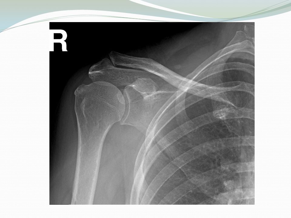
Glenohumeral osteoarthritis

54
Glenohumeral osteoarthritis
Mainly wear and tear Could be a result of RA, AVN or malunited fractures

55
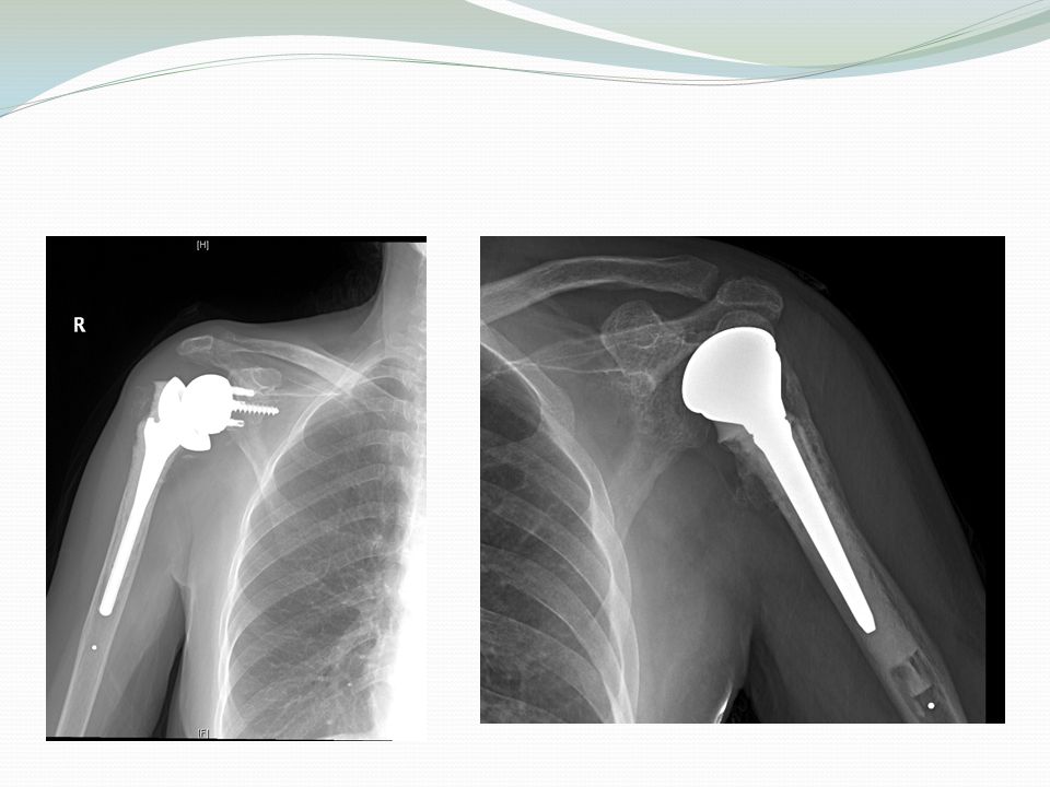
Glenohumeral osteoarthritis
The management starts with conservative measures. If it fails to relieve the pain, the best management for elderly patient is arthroplasty (hemi or total shoulder replacement) If associated with irreparable rotator cuff tear for reverse shoulder replacement.
 If associated with irreparable rotator cuff tear for reverse shoulder replacement.")
57
AC arthritis

58
Causes of AC Arthritis Degenerative osteoarthritis.( wear and tear in old aged people) Rheumatoid Arthritis . Gouty Arthritis. Septic Arthritis. Atraumatic distal claivcle osteolysis in weight lifters.

59
Signs and Symptoms Pain , which worsens with movement and progressively worsens.( the patient may suffer a night pain which is a sign of arthritis) It is commonly associated with impingement syndrome Positive across the chest adduction Diagnosis: Clinical and by x-rays

60
Across the chest adduction test

61
AC osteoarthritis Non-surgical Treatment
Rest , avoid weightlifting and push-ups Pain medications and NSAID to reduce pain and inflammation

62
Common shoulder disorders
Basic shoulder anatomy Impingement syndrome Rotator cuff pathology Adhesive capsulitis Glenohumeral osteoarthritis Recurrent shoulder dislocations Acromioclavicular separation

63
Glenohumeral Joint Most common dislocated joint Lacks bony stability
Composed of: Fibrous capsule Ligaments Surrounding muscles Glenoid labrum

64
Dislocation of the Shoulder
According to the direction: Mostly Anterior > 95 % of dislocations Posterior Dislocation occurs < 5 % True Inferior dislocation (luxatio erecta) occurs < 1% According to the mechanism: Traumatic Non traumatic dislocation may present as Multi directional dislocation due to generalized ligamentous laxity. It may become painless habitual
 occurs < 1% According to the mechanism: Traumatic. Non traumatic dislocation may present as Multi directional dislocation due to generalized ligamentous laxity. It may become painless habitual.")
65
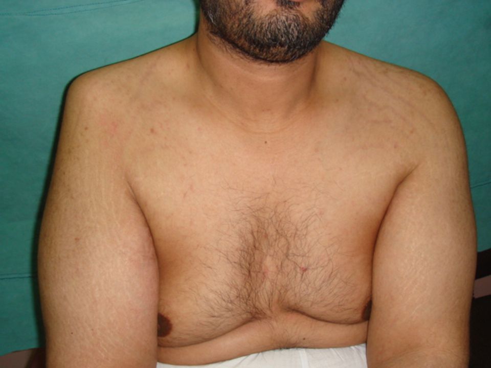
Anterior Shoulder dislocation
Usually also inferior Bankart’s Lesion

66
Mechanism of anterior shoulder dislocation
Usually Indirect fall on Abducted and extended shoulder May be direct when there is a blow on the shoulder from behind

67
Clinical picture of acute anterior shoulder dislocation
Patient is in severe pain Holds the injured limb with other hand close to the trunk The shoulder is abducted and the elbow is kept flexed There is loss of the normal contour of the shoulder

68
Clinical picture Loss of the contour of the shoulder may appear as a step Anterior bulge of head of humerus may be visible or palpable A gap can be palpated above the dislocated head of the humerus

70
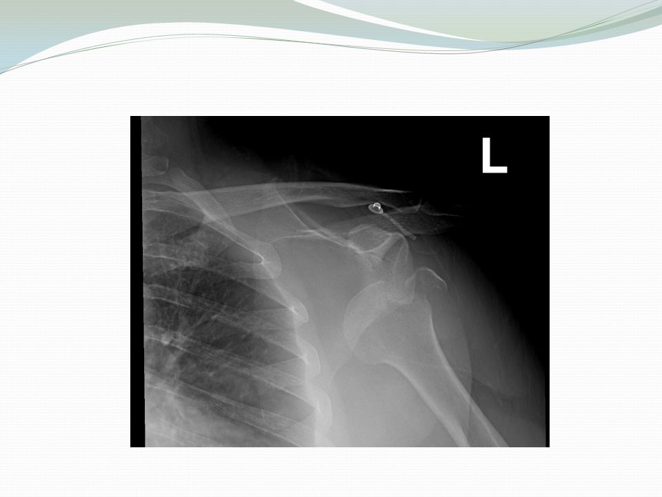
X-ray anterior shoulder dislocation

71
Associated injuries of anterior Shoulder Dislocation
Injury to the neuro vascular bundle in axilla Injury of the Axillary Nerve ( Usually stretching leading to temporary neuropraxia ) Associated fracture
 Associated fracture.")
73
Axillary Nerve Injury It is a branch from posterior cord of Brachial plexus It hooks close round neck of humerus from posterior to anterior It pierces the deep surface of deltoid and supply it and the part of skin over it

74
Axillary nerve injury

75
Management of Anterior Shoulder Dislocation
Is an Emergency It should be reduced in less than 24 hours or there may be Avascular Necrosis of head of humerus Following reduction the shoulder should be immobilized strapped to the trunk for 3-4 weeks and rested in a collar and cuff

76
Methods of Reduction of anterior shoulder Dislocation
Hippocrates Method ( A form of anesthesia or pain abolishing is required ) Stimpson’s technique ( some sedation and analgesia are used but No anesthesia is required ) Kocher’s technique is the method used in hospitals under general anesthesia and muscle relaxation
 Stimpson’s technique ( some sedation and analgesia are used but No anesthesia is required ) Kocher’s technique is the method used in hospitals under general anesthesia and muscle relaxation.")
77
Hippocrates Method

78
Stimpson’s technique

79
Kocher’s Technique

80
Complications of anterior Shoulder Dislocation : Early
Neurovascular injury ( rare ) Axillary nerve injury Associated Fracture of neck of humerus or greater or lesser tuberosities
 Axillary nerve injury. Associated Fracture of neck of humerus or greater or lesser tuberosities.")
81
Complications of anterior shoulder Dislocation : Late
Avascular necrosis of the head of the Humerus (high risk with delayed reduction) Recurrent shoulder dislocations (the commonest complication) , treated by Bankart repair (either arthroscopic or open)
 Recurrent shoulder dislocations (the commonest complication) , treated by Bankart repair (either arthroscopic or open)")
82
Bankart lesion

83
Bankart lesion

84
Arthroscopic Bankart repair

85
Common shoulder disorders
Basic shoulder anatomy Impingement syndrome Rotator cuff pathology Adhesive capsulitis Glenohumeral osteoarthritis Recurrent shoulder dislocations Acromioclavicular separation

86
Acromioclavicular Joint
The AC joint is different from joints like the knee or ankle, because it doesn't need to move very much. The AC joint only needs to be flexible enough for the shoulder to move freely. The AC joint just shifts a bit as the shoulder moves.

87
The AcromioClavicular joint is stabilized by three ligaments
2 CC ligaments Conoid trapezoid AC ligament

88
Acromioclavicular separation
Mechanisms of Injury: Fall on the tip of the unprotected shoulder. Fall on the outstretched hand. Downward force on the acromion from above.

89
Acromioclavicular separation
Rockwood Classification: type I: sprain of joint with out a complete tear of either ligament type II: tear of AC ligaments with intact coracoclavicular ligaments; will not show marked elevation of lateral end of clavicle type III: both AC & CC ligaments are torn type IV: distal clavicle is dislocated posteriorly into trapezial fascia type V: distal clavicle is dislocated inferiorly

90
type III: both AC & CC ligaments are torn

91
Type III

93
Thank you

Similar presentations