Download presentation
Presentation is loading. Please wait.

1
ANATOMY OF PHARYNX By-Dr. kcsudeep

2
PHARYNX IN GENERAL It is a conical fibromuscular tube forming upper part of air and food passage. 12-14 cm long. Extends from base of skull to lower border of cricoid cartilage. Width is 3.5 cm and it becomes narrow at pharyngo-oesophageal junction.narrowest part of GIT.

4
STRUCTURE OF PHARYNGEAL WALL
IT CONSIST OF 4 LAYERS: Mucous membrane Pharyngeal aponeurosis(pharyngobasilar fascia) Muscular coat Buccopharyngeal fascia
 Muscular coat. Buccopharyngeal fascia.")
6
Muscular coat consist of two layers of muscles with 3 muscles in each layer.
External layer superior, middle, inferior constrictor muscle Internal layer stylopharyngeus, salpingopharyngeus, palatopharyngeus.

7
KILLIAN’S DEHISCENCE Inferior constrictor has two parts ;thyropharyngeus with oblique fibres and cricopharyngeus with transverse fibres. Between these two parts exists a potential gap’killian’s dehiscence’ or “gate way of tears”. Perforation can occur during oesophagoscopy.

8
WALDEYER’S RING Scattered throughout the pharynx in its subepithelial layer is lymphoid tissue which is aggregated at places to form masses, collectively called waldeyer’s ring. The masses are: Nasopharyngeal tonsil or adenoids. Palatine tonsils. Lingual tonsils. Tubal tonsils. Lateral pharyngeal bands. Nodules (in post. Pharyngeal wall)
")
10
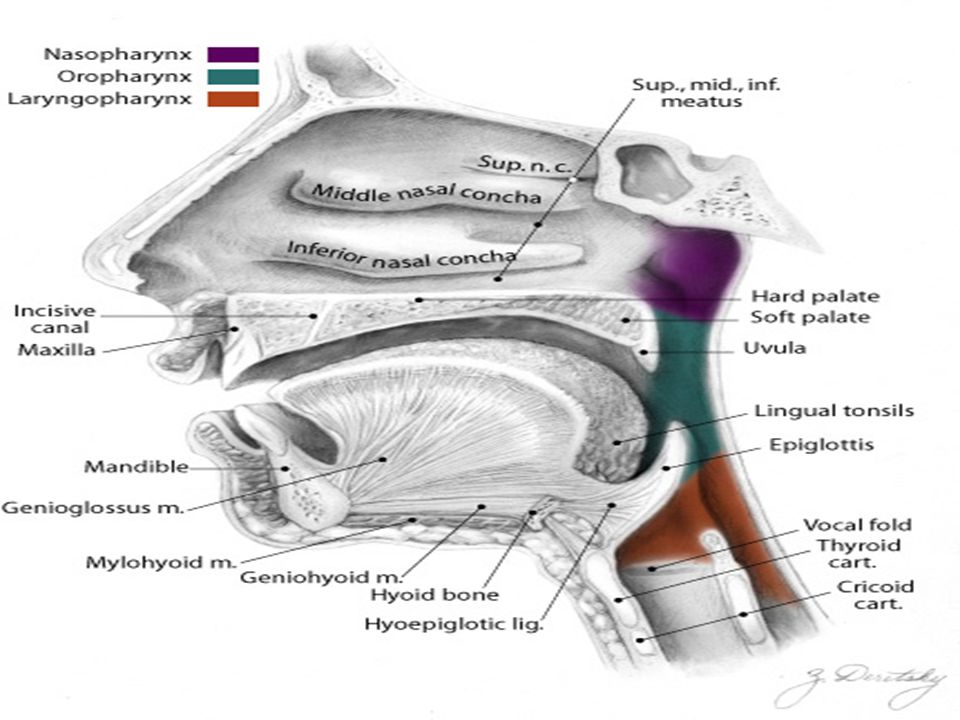
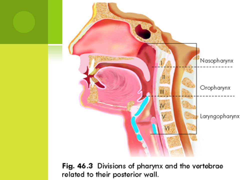
DIVISION OF PHARYNX Nasopharynx. Oropharynx.
Hypopharynx or laryngopharynx. NASOPHARYNX: Roof, posterior wall, floor, anterior wall and lateral wall. Each lateral wall opening of eustchaintube.Above and behind is elevation called torus tubarius . Above and behind tubal elevation is a recess called “fossa of Rosenmuller”-> commonest site for carcinoma.

12
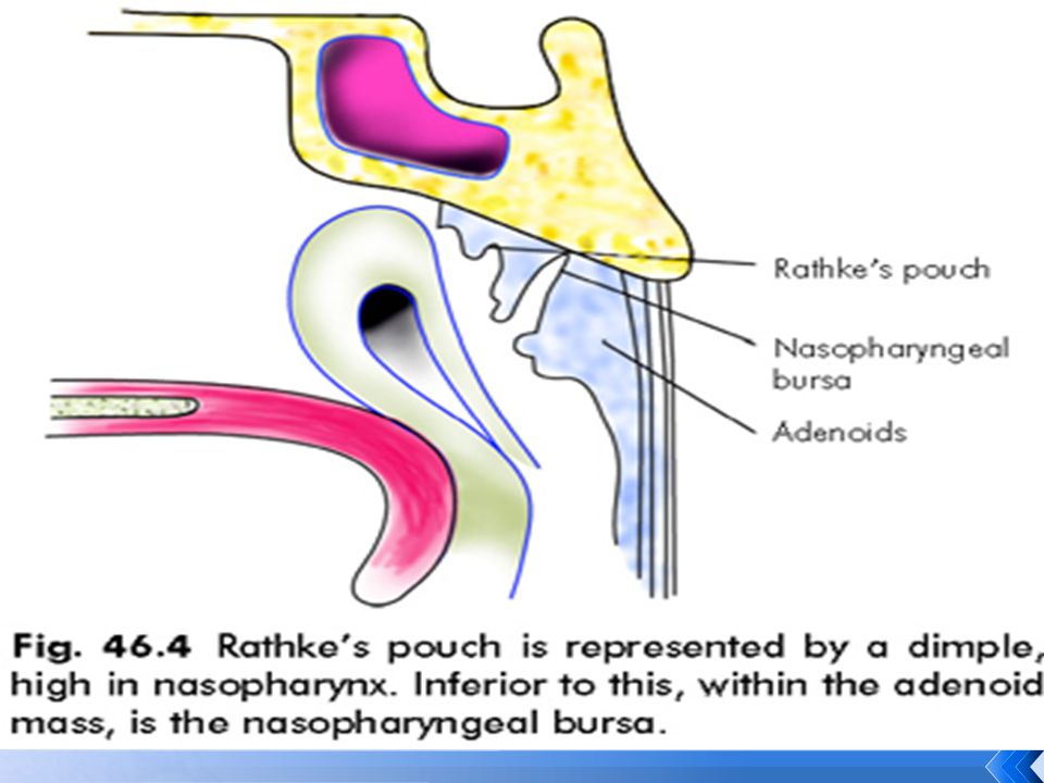
NASOPHARYNGEAL TONSIL
It is a subepithelial collection of lymphoid tissue at junction of roof and posterior wall of nasopharynx and causes the overlying mucous membrane to be thrown into radiating folds . It increases upto 6 yrs and gradually atrophies.

14
ACUTE AND CHRONIC PHARYNGITIS
ACUTE PHARYNGITIS: Aetiology Viral Bacterial_ grp. A Beta hemolytic streptococci. Fungal.

15
CLINICAL FEATURES: Milder infections:
Discomfort in throat, malaise and low grade fever. Pharynx is congested but no lymphadenopathy. Moderate and severe infections: Pain in throat, dysphagia, headache, malaise, high fever. Pharynx shows erythema exudate , enlargement of tonsils and lymphoid follicles.

16
Very severe cases: Oedema of soft palate and uvula with enlargement of cervical lymphnodes. Viral infections are mild and accompanied by rhinorrhoea and hoarsness. DIAGNOSIS: Culture of throat swab.

17
TREATMENT: General measures:
Bed rest , plenty of fluids, warm saline gargles and analgesics form main stay of treatment. Specific treatment: Streptocoal pharyngitis—penicillin G , for 10 days. benzathine pn. Erythromycin mg/kg daily for 10 days. For diptheria – antitoxin with pn.

18
VIRAL INFECTIONS CAUSING PHARYNGITITS
Herpangina: caused by group A coxsackie virus mostly affects child . characteristics feature include : fever sore throat and vesicular eruption on soft palate and pillars. Infectious mononucleosis: caused by EBV .affects older child and young adult . characterised by fever, sore throat,exudative pharyngitis ,lymphadenopathy, splenomegaly and hepatitis

19
Fungal pharyngitis: candida infection of oropharynx
Fungal pharyngitis: candida infection of oropharynx . Nystatin is drug of choice. CHRONIC PHARYNGITIS: It is chronic inflammatory condition of pharynx. Hypertrophy of mucosa, seromucinous glands, subepithelial lymphoid follicles and even muscular coat. Chronic pharyngitis is of two types: 1)Chronic catarrhal pharyngitis 2)chronic hypertrophic (granular)pharyngitis.
Chronic catarrhal pharyngitis. 2)chronic hypertrophic (granular)pharyngitis.")
20
AETIOLOGY: 1) Persistent infection in near by structures.
2) Mouth breathing. 3)Chronic irritants. 4)Environmental pollution. SYMPTOMS: Discomfort or pain in throat. Foreign body sensation in throat. Tiredness of voice. Cough.
 Mouth breathing. 3)Chronic irritants. 4)Environmental pollution. SYMPTOMS: Discomfort or pain in throat. Foreign body sensation in throat. Tiredness of voice. Cough.")
21
SIGNS: Chronic catarrhal pharyngitis: Congestion of posterior pharyngeal wall with engorgement of vessels ; faucial pillars may be thickened. increased mucus secretion which may cover pharyngeal mucosa. Chronic hypertrophic(granular) pharyngitis: 1)Pharyngeal wall appears thick and oedematous with congested mucosa and dilated vessels.
 pharyngitis: 1)Pharyngeal wall appears thick and oedematous with congested mucosa and dilated vessels.")
23
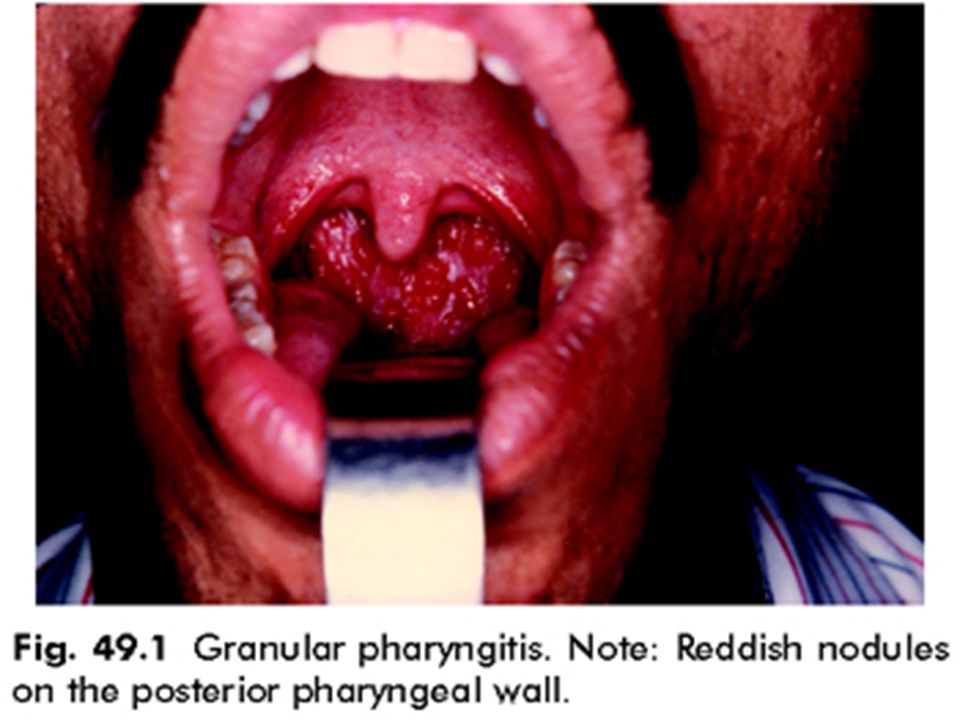
2) posterior pharyngeal wall may be studded with reddish nodules.
3)Lateral pharyngeal bands become hypertrophied. 4)Uvula may be elongated and oedematous. TREATMENT: Causative factor should be eradicated. Voice rest may be required. Warm saline gargles Mandle’s paint (consists of iodine 1.25 grams, potassium iodine 2.5 grams, water 2.5 ml )may be applied to pharyngeal mucosa. Cautery of lymphoid granules by silver nitrate.
Lateral pharyngeal bands become hypertrophied. 4)Uvula may be elongated and oedematous. TREATMENT: Causative factor should be eradicated. Voice rest may be required. Warm saline gargles. Mandle’s paint (consists of iodine 1.25 grams, potassium iodine 2.5 grams, water 2.5 ml )may be applied to pharyngeal mucosa. Cautery of lymphoid granules by silver nitrate.")
24
THANK YOU !!

Similar presentations